Retinal angiospasm is an emergency condition in ophthalmology, which is characterized by disruption of metabolic processes in cells due to cessation of blood flow. Like any emergency form of the disease, vasospasm requires surgical intervention by a specialist. Observation of people who have suffered this condition allows us to draw the following conclusions:
- disruption of the cardiovascular system is one of the causes of vasospasm, as evidenced by 91.2% of cases;
- in particular, atherosclerosis and arterial hypertension lead to retinal vasospasm, 60% of cases prove this connection;
- in 25-30% of cases it is not possible to determine what caused the vasospasm;
- this emergency condition usually occurs in people over 40 years of age;
- the number of sick men is twice as high as women.
What are the causes of this condition?
The causes of retinal circulatory disorders have not been fully elucidated. In childhood, the reduction of the middle layer of blood vessels occurs due to the development of the peripheral nervous system. In adults, the causes of the disease include the following:
- Hypertension leads to a sharp reflex contraction of blood vessels, this also applies to the eye area.
- Diabetes. This disease causes diabetic retinopathy. The latter condition is accompanied by episodes of vasospasm.
- Atherosclerosis leads to increased blood pressure and impaired blood flow through the vascular system of organs and tissues.
- Bad habits. Smoking and alcohol abuse can lead to retinal vasospasm, since nicotine and ethyl alcohol are substances that lead to vasoconstriction, disrupting the blood supply to the retina.
- Intoxication is often observed in people who have been in unhealthy conditions for a long time. In particular, compounds of carbon disulfide and lead increase the tension of the vascular wall, which changes the diameter of the lumen of blood vessels, leading to impaired blood flow.
The mechanism of the onset of the disease can be described as follows: an increased concentration of calcium ions and a decrease in potassium and sodium ions lead to changes in the smooth muscle layer. As a result, the lumen of the vessels narrows, slowing down blood flow. Long-term intoxication leads to an increase in the tone of the sympathetic nervous system, which leads to vasospasm. Observations of insulin-dependent patients indicate an increase in the degree of rigidity of their vascular walls. This fact also leads to retinal vasospasm.
What complications can vasospasm of the retina lead to?
Retinal angiospasm is an emergency condition in ophthalmology, which is characterized by disruption of metabolic processes in cells due to cessation of blood flow.
Like any emergency form of the disease, vasospasm requires surgical intervention by a specialist. Observation of people who have suffered from this condition allows us to draw the following conclusions: • dysfunction of the cardiovascular system is one of the causes of vasospasm, this is evidenced in 91.2% of cases; • in particular, atherosclerosis and arterial hypertension lead to retinal vasospasm, 60% of cases prove this connection; • in 25-30% of cases it is not possible to determine what caused the vasospasm; • this emergency condition usually occurs in people over 40 years of age; • the number of sick men is twice as high as women. What are the causes of this condition? The causes of retinal circulatory disorders have not been fully elucidated. In childhood, the reduction of the middle layer of blood vessels occurs due to the development of the peripheral nervous system. In an adult, the causes of the disease include the following: 1. Hypertension leads to a sharp reflex contraction of blood vessels, this also applies to the eye area. 2. Diabetes mellitus. This disease causes diabetic retinopathy. The latter condition is accompanied by episodes of vasospasm. 3. Atherosclerosis leads to increased blood pressure and impaired blood flow through the vascular system of organs and tissues. 4. Bad habits. Smoking and alcohol abuse can lead to retinal vasospasm, since nicotine and ethyl alcohol are substances that lead to vasoconstriction, disrupting the blood supply to the retina. 5. Intoxication is often observed in people who spend a long time in conditions harmful to health. In particular, compounds of carbon disulfide and lead increase the tension of the vascular wall, which changes the diameter of the lumen of blood vessels, leading to impaired blood flow. The mechanism of the onset of the disease can be described as follows: an increased concentration of calcium ions and a decrease in potassium and sodium ions lead to changes in the smooth muscle layer. As a result, the lumen of the vessels narrows, slowing down blood flow. Long-term intoxication leads to an increase in the tone of the sympathetic nervous system, which leads to vasospasm. Observations of insulin-dependent patients indicate an increase in the degree of rigidity of their vascular walls. This fact also leads to retinal vasospasm. Symptoms Retinal vasospasm typically affects both eyes, and in rare cases, only one. Typical complaints of patients are blurred vision and the appearance of flickering dots before the eyes. If vasospasm is short-term, the symptoms go away. Such a spasm may also be accompanied by a distortion of visual perception, similar to meta- and photomorphopsia. Some patients complain of discomfort in the orbital area of the affected organ of vision. During the spasm, the patient may experience headache and dizziness, pulsation in the temples. After the attack, the general condition returns to normal, visual function is restored. If the resulting vasospasm is prolonged, the consequence may be an irreversible loss of visual acuity. Complications Prolonged spasm can lead not only to a decrease in visual acuity, but also to its complete loss. The frequency of attacks causes severe discomfort and disability due to the fact that patients lack the ability to anticipate the period of development of the next fragment. The progressive form of the disease entails an increase in intraocular pressure. Over time, the patient develops secondary ophthalmopyrtension and develops ocular migraine. Rapid attacks are tolerated without complications. Diagnosis The disease is diagnosed based on medical history and examination; in some cases, additional diagnostic methods are used. During vasospasm, such phenomena as overflow of blood vessels and swelling of the conjunctiva are present; after the end of the attack, changes in the anterior area of the eyes are absent upon visual examination. When diagnosing vasospasm of the retina, the following methods are used: - Ophthalmoscopy. A sharply narrowed central artery and its branches of small diameter are visible upon examination. Venous arteries are filled with blood. There is swelling of the optic nerve and its pale pink color. — Non-contact tonometry. Diagnosis by this method is based on the presence of increased intraocular pressure during vasospasm. After the symptoms disappear, the indicator returns to normal. If this phenomenon is not observed, it is necessary to refer to electronic tonography of the eye. — Antiography of the retina. Provides an opportunity to examine the dynamics in the retinal vessels using fluorescein circulation analysis. In case of disease, the blood-retinal barrier does not allow contrast to pass through. — Optical coherence tomography of the retina. The scan reveals a sharp enlargement of the macula (macula). The foveal fossa is leveled, the retinal reaction is reduced. The curve has an atypical straight appearance. Treatment Etiotropic treatment has not been developed. Pathogenetic therapy is aimed at dilating spasmodic vessels to restore retinal blood flow in the ischemic area. It is extremely important to stop the symptoms of vasospasm in a timely manner, since prolonged disruption of microcirculation entails complete or partial loss of vision. For this reason, immediately after examining the fundus, infusions of plasma substitutes and antispasmodics are prescribed. The next stage of treatment is performing electrophoresis with vasodilators (drotaverine) and peripheral vasodilators (bendazole). In order to reduce intraocular pressure, saluretics and carbonic anhydrase inhibitors are used. If there is no effect, an irrigation system is installed in the retrobulbar space. Instillation of solutions of beta-adrenergic receptor blockers (timolol, pilocarpine hydrochloride) is indicated. If accompanied by an increase in systemic blood pressure, drugs are administered intramuscularly. In order to stimulate the regeneration of the receptor apparatus, peptide bioregulators are used. If the pathology is severe, retrobulbar administration of M-anticholinergics (atropine) is additionally indicated. Prognosis and preventive measures The prognosis for vasospasm is influenced by the nature of the attack. A short-term narrowing of the small branches of the central nervous system of the eyeball passes without a trace. Vasospastic reactions lasting 15 minutes or more, when irreversible changes in the retina occur, are considered unfavorable in terms of prognosis. Specific preventive measures are currently unknown. Nonspecific prevention consists of monitoring blood pressure and blood glucose levels, taking statins for atherosclerosis, and using personal protective equipment when working with pesticides in production conditions.
Symptoms
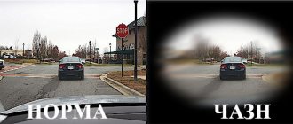
Retinal vasospasm typically affects both eyes, and in rare cases, only one. Characteristic complaints of patients are blurred vision and the appearance of flickering dots before the eyes. If vasospasm is short-term, the symptoms go away. Such a spasm may also be accompanied by a distortion of visual perception, similar to meta- and photomorphopsia. Some patients complain of discomfort in the orbital area of the affected organ of vision. During the spasm, the patient may experience headache and dizziness, pulsation in the temples. After the attack, the general condition returns to normal, visual function is restored. If the resulting vasospasm is prolonged, the consequence may be irreversible loss of visual acuity.
Diagnostic measures
The presence of a specific pathology, as well as its exact cause, can only be determined during a face-to-face consultation with a specialist. During it, the doctor will ask several general questions regarding lifestyle and chronic diseases, study the medical history in detail, conduct some functional tests, and clarify the presence of similar pathologies in close relatives. As part of the examination, the doctor will ask you about the frequency and intensity of your symptoms, monitor your clinical picture, and identify the suspected etiology of the disease.
If suspicions are partially confirmed, you will be prescribed simple tests:
Rheovasography (RVG) is a non-invasive functional method for assessing pulse blood supply to the extremities, as well as the tone, elasticity and patency of peripheral vessels using a specific device;
Measurement of the brachial-ankle index - a one-time determination of the level of blood pressure in the area of the shoulders and ankles (normally it is the same);
Biochemical blood test (cholesterol content), and other tests to detect cardiac dysfunction.
For a more in-depth study of the course of the disease, the following measures are taken:
1. Duplex scanning of arteries and veins;
2. Angiography using a contrast agent;
3. Magnetic resonance angiography;
4. Multislice computed tomography of the lower extremities;
5. Functional tests.
Complications
Prolonged spasm can lead not only to a decrease in visual acuity, but also to its complete loss. The frequency of attacks causes severe discomfort and disability due to the fact that patients lack the ability to anticipate the period of development of the next fragment. The progressive form of the disease entails an increase in intraocular pressure. Over time, the patient develops secondary ophthalmopyrtension and develops ocular migraine. Rapid attacks are tolerated without complications.
Symptoms of vasospasm of cerebral vessels
Previously, the disease occurred only in elderly patients. In recent years, pathology has become “younger”. It is facilitated by the unfavorable environmental component that prevails in large cities and industrial zones, and the presence of toxic elements in the air. The latter penetrate the respiratory system and are transported directly to the brain, having a negative effect on the blood vessels.
Angiospasm is a fairly powerful and long-term compression of blood vessels. Spasm occurs due to incorrect blood circulation and supply of nutrients. This disease requires immediate treatment, since untimely assistance contributes to the development of complications, such as stroke.
You should consult a doctor if you have the following symptoms:
- Painful sensations in the occipital and temporal region;
- Frequent dizziness;
- High pressure;
- Tinnitus that occurs when a person is in a calm or active state;
- Loss of coordination, speech impairment.
Diagnostics
The disease is diagnosed based on medical history and examination; in some cases, additional diagnostic methods are used. During vasospasm, such phenomena as overflow of blood vessels and swelling of the conjunctiva are present; after the end of the attack, changes in the anterior area of the eyes are absent upon visual examination.
When diagnosing retinal vasospasm, the following methods are used:
— Ophthalmoscopy. A sharply narrowed central artery and its branches of small diameter are visible upon examination. Venous arteries are filled with blood. There is swelling of the optic nerve and its pale pink color.
— Non-contact tonometry. Diagnosis by this method is based on the presence of increased intraocular pressure during vasospasm. After the symptoms disappear, the indicator returns to normal. If this phenomenon is not observed, it is necessary to refer to electronic tonography of the eye.
— Antiography of the retina. Provides an opportunity to examine the dynamics in the retinal vessels using fluorescein circulation analysis. In case of disease, the blood-retinal barrier does not allow contrast to pass through.
— Optical coherence tomography of the retina. The scan reveals a sharp enlargement of the macula (macula). The foveal fossa is leveled, the retinal reaction is reduced. The curve has an atypical straight appearance.
Popular questions about vascular spasms of the extremities
What causes spasms?
The cause of vascular spasms in the extremities is a number of diseases such as angiospastic syndrome, obliterating atherosclerosis and endarteritis, diabetes mellitus. Also, spasms of the blood vessels of the arms and legs occur as a reaction to prolonged exposure to cold, nicotine intoxication or frostbite.
How long to treat vascular spasms of the extremities?
Some medications for spasms of blood vessels are recommended for use throughout life, others - as part of a therapeutic course, which depends on the degree of the disease.
Which doctor treats vascular spasms?
After identifying symptoms associated with vascular spasms of the extremities, you must consult an angiologist or phlebologist.
Treatment
Etiotropic treatment has not been developed. Pathogenetic therapy is aimed at dilating spasmodic vessels to restore retinal blood flow in the ischemic area. It is extremely important to stop the symptoms of vasospasm in a timely manner, since prolonged disruption of microcirculation entails complete or partial loss of vision. For this reason, immediately after examining the fundus, infusions of plasma substitutes and antispasmodics are prescribed. The next stage of treatment is performing electrophoresis with vasodilators (drotaverine) and peripheral vasodilators (bendazole).
In order to reduce intraocular pressure, saluretics and carbonic anhydrase inhibitors are used. If there is no effect, an irrigation system is installed in the retrobulbar space. Instillation of solutions of beta-adrenergic receptor blockers (timolol, pilocarpine hydrochloride) is indicated. If accompanied by an increase in systemic blood pressure, drugs are administered intramuscularly. In order to stimulate the regeneration of the receptor apparatus, peptide bioregulators are used. If the pathology is severe, retrobulbar administration of M-anticholinergics (atropine) is additionally indicated.
Does vasospasm occur in children?
A little person does not know how to talk about his feelings.
He is afraid of everything unusual. Angiospasm in infants is caused by violations of the mother's regimen during pregnancy (smoking, drinking alcohol), difficult childbirth, and the use of vacuum extraction. Even moderate trauma to the cerebral vessels causes changes in the baby’s behavior: increased anxiety, crying, poor appetite, sleep disturbance, decreased weight gain.
Clinical signs of vasospasm depend on the area of the lesion, the location of the narrowed vessels, and the possibilities for the development of collateral blood supply. With age, the child develops its own protective mechanism, and the tone of the vascular wall is restored.
In adolescence, a breakdown is possible due to school overload and hormonal imbalance. Angiospasm of cerebral vessels is manifested by increased fatigue in class, short-term headaches with dizziness, and decreased vision. A diagnosis of neurocirculatory dystonia is usually made.
You can learn in detail about how vasospasm of peripheral and central vessels manifests itself in adults on our website in the article “Symptoms of vascular spasms.”
Forecast and preventive measures
The prognosis for vasospasm is influenced by the nature of the attack. A short-term narrowing of the small branches of the central nervous system of the eyeball passes without a trace. Vasospastic reactions lasting 15 minutes or more, when irreversible changes in the retina occur, are considered unfavorable in terms of prognosis. Specific preventive measures are currently unknown. Nonspecific prevention consists of monitoring blood pressure and blood glucose levels, taking statins for atherosclerosis, and using personal protective equipment when working with pesticides in production conditions.
Prevention
To prevent spasms of blood vessels in the extremities (arms and legs), it is necessary to eliminate risk factors as much as possible:
- Adhere to a healthy lifestyle (balance your diet, quit bad habits, exercise).
- Monitor your own weight and general health.
- People over 40 years of age should monitor blood glucose and cholesterol levels, as well as blood pressure.
- If your heredity is aggravated by diseases such as diabetes, heart attack, stroke, do screening at least 4 times a year.
- Avoid hypothermia and injury.