Functions
Every second, many signals pass through our brain. The process does not stop even in sleep. The body needs to perceive the world around it, make movements, ensure the functioning of the heart, respiratory, digestive, genitourinary systems, etc. Two main groups of neurons are involved in organizing all this activity - sensory and motor.
When we touch cold or hot and feel the temperature of the object, this is the merit of the sensitive cells. They instantly transmit information received from the periphery of the body. This ensures reflex activity.
Neurons form our entire central nervous system. Their main tasks:
- get information;
- transmit it through the nervous system.
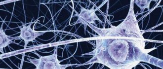
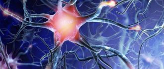
These unique cells are capable of instantly transmitting electrical impulses.
To ensure the process of life, the body must process a huge amount of information that comes to it from the outside world, and respond to any sign of changing environmental conditions. To make this process as efficient as possible, neurons are divided according to their functions into:
- Sensitive (afferent) are our guides to the world around us. They are the ones who perceive information from the outside, from the senses, and transmit it to the central nervous system. The peculiarity is that thanks to their contact activity, we feel temperature, pain, pressure, and have other feelings. Sensitive cells of narrow specialization transmit taste and smell.
- Motor (motor, efferent, motor neurons). Motor neurons transmit information through electrical impulses from the central nervous system to muscle groups and glands.
- Intermediate (associative, intercalary, intercalary). Now let’s take a closer look at what function interneurons perform, why they are needed, and what is their difference. They are located between sensory and motor neurons. Interneurons transmit nerve impulses from sensory fibers to motor fibers. They provide “communication” between efferent and afferent nerve cells. They should be treated as a kind of natural “extenders,” long cavities that help transmit a signal from a sensory neuron to a motor one. Without their participation this would not have been possible. This is their function.
The receptors themselves are cells of the skin, muscles, internal organs, and joints specially designated for this function. Receptors can begin in the cells of the epidermis and mucous membrane. They are able to accurately capture the smallest changes, both outside the body and inside it. Such changes may be physical or chemical. Then they are instantly transformed into special bioelectric impulses and sent directly to sensory neurons. This is how the signal travels from the periphery to the center of the body, where the brain deciphers its meaning.
Impulses from the organ to the brain are carried out by all three groups of neurons - motor, sensory and intermediate. The human nervous system consists of these groups of cells. This structure allows you to respond to signals from the outside world. They provide reflex activity of the body.
If a person ceases to feel taste, smell, hearing and vision decrease, this may indicate disorders in the central nervous system. Depending on which sense organs are affected, a neurologist can determine in which part of the brain the problems have arisen.
There are two groups of functions of the nervous system:
1) Somatic. This is conscious control of the skeletal muscles.
2) Vegetative (autonomous). This is control of internal organs uncontrolled by consciousness. The operation of this system occurs even if a person is in a state of sleep.
Types and characteristics of neurons
Nerve cells called neurons receive, send, and conduct bioelectrical signals. There are efferent (motor) neurons - these are components of the central nervous system that redirect signals to executive organs, for example, skeletal muscles. Afferent (sensitive) neurons are cells that perceive external and internal stimuli, which ensures the body’s connection with the external environment and reactions to changes in the functional activity of internal organs.
Intercalary cells provide interconnections within the overall neural network. Neurons of all types (sensitive, efferent, associative) are functional units that support the activity of the nervous system; they are found in all tissues of the body, where they play the role of connecting links between receptor (perceiving irritating stimuli) and effector organs that respond to irritating stimuli.
Effector organs include muscles and glands, and receptor organs include sensory organs. The significance of the signals transmitted varies significantly depending on the type of cell and its role in the functioning of the central nervous system. For example, sensory neurons that perceive impulses from the external environment transmit signals from skin receptors and sensory organs in the direction of the brain; motor neurons redirect commands generated in the brain, causing contraction of skeletal muscles and initiating movement.
Despite the different meanings of bioelectric impulses, their nature is the same and consists in changing the electrical potential in the area of the plasma membrane of the nerve cell. The mechanism of propagation of nerve impulses is based on the ability of an electrical disturbance that appears in one place of the cell to be transmitted to other areas. In the absence of factors that enhance the signal, the pulses fade away as they move away from the source of excitation.
Sensory, also known as sensitive, is an afferent neuron that conducts impulses from distal parts of the body to the central parts of the central nervous system. For example, sensory fibers form fibers that extend from the light-sensitive cells of the organs of vision. Signals leave the retina, traveling along millions of axons belonging to the structures of the basal ganglia, towards a region of the visual cortex.
The sensory neuron, together with the executive (motor) neurons, forms a simple reflex arc.
For example, the knee reflex is an unconditioned stretch reflex reaction that occurs as a result of the activity of a similar reflex arc. A reaction in the form of uncontrolled extension of the lower leg occurs when there is a mechanical impact on the tendon of the femoral muscle, which lies under the patella. Reaction mechanism:
- Mechanical effects on the neuromuscular spindles located in the hip extensor muscle.
- An increase in the intensity of nerve signals in the endings entwining the neuromuscular spindles due to their stretching.
- Transmission of impulses to sensory neurons located in the spinal ganglia through dendrites extending from the femoral nerve.
- Transmission of impulses from sensory cells to alpha motor neurons located in the anterior horns within the boundaries of the spinal cord.
- Signal transmission from alpha motor neurons to contractile muscle fibers of the femoral muscle.
The knee reflex mechanism involves interneurons that transmit inhibitory impulses to motor neurons of the flexor muscles, and other interneurons, for example, Renshaw cells. The knee reflex mechanism also involves gamma motor neurons, which regulate the intensity of spindle stretch.
In the spinal cord, formed by gray matter, there are three types of neurons - motor, intercalary, and autonomic. Moreover, the vegetative ones are located in the visceral (related to the internal organs) nuclei. These cells interact with afferent (ascending pathways that transmit impulses from peripheral receptors to the central zones of the central nervous system) fibers responsible for general visceral sensitivity.
Visceral afferents conduct nerve signals (usually painful or reflex sensations) from internal organs, elements of the circulatory system, glands to the corresponding zones of the central nervous system. Visceral afferents are part of the autonomic nervous system. Reflex arcs within the autonomic department of the central nervous system differ in structure from the arcs of the somatic department.
Efferent components (descending pathways that transmit impulses from the cortical and subcortical zones of the brain to peripheral areas) are formed by two types of neurons - intercalary and effector (motor). The intercalary ones are located in the nuclei belonging to the vegetative part of the central nervous system. The name “intercalary” is due to its location between the sensory and motor neurons.
Sensitive
A sensory neuron is a component of the nervous system that transmits information to the brain about stimuli affecting a specific area of the body. Examples of irritants include factors: sunlight, mechanical impact (impact, touch), the action of a chemical substance. Sensory neurons are located in the ganglia of the brain - the spinal cord and brain.
The connection formed with a sensitive neuron can provoke excitation or inhibition, which is sent along nerve fibers to the cortical parts of the brain. As the level of sensory pathways increases, the transmitted information is processed to identify important features. Sensitive neurons belong to pseudounipolar neurons - their axon and dendrites extend from the body together, subsequently separate and are located in the spinal cord, brain (axon) and in the peripheral parts of the body (dendrites).
Insert
Interneurons transmit converted nerve impulses obtained as a result of processing sensory information received from various sources, for example, from the organs of vision and skin receptors. As a result, the processed information becomes the initial data for the formation of adequate motor commands.
Motor
Motor nerve cells are of two types - large and small. In the first case we are talking about α-motoneurons, in the second – about γ-motoneurons. Alpha motor neurons are present in the basal ganglia of lateral (closer to the lateral plane) and medial (closer to the median plane) localization. These are the largest cells present in nervous tissue.
Their axons interact with striated fibers contained in skeletal muscles. As a result, synapses (sites where nerve signals are transmitted) are formed. The axons of alpha motor neurons interconnect with intercalary analogues, also known as Renshaw cells, leading to the formation of collateral pathways and inhibitory synapses in the spinal cord.
Gamma motor neurons are part of the neuromuscular spindle, which is a complex receptor consisting of nerve endings (afferent, efferent). The main function of neuromuscular spindles is to regulate the force and rate of contraction or stretching of skeletal muscles.
Structure
Sensory neurons are most often unipolar. This means that they are equipped with only one bifurcating process. It leaves the cell body (soma) and simultaneously performs the functions of both an axon and a dendrite. The axon is the input, and the dendrite of the sensory neuron is the output. After excitation of sensitive sensory cells, a bioelectric signal passes along the axon and dendrite.
There are also bipolar nerve cells that have two processes, respectively. They can be found, for example, in the retina and structures of the inner ear.
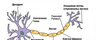
The body of the sensitive cell is shaped like a spindle. 1, and more often 2 processes (central and peripheral) extend from the body.
Peripheral in its shape is very similar to a thick long stick. It reaches the surface of the mucous membrane or skin. This process is similar to the dendrite of nerve cells.
The second, opposite process extends from the opposite part of the cell body and is shaped like a thin thread covered with swellings (they are called varicosities). This is an analogue of the nerve process of a neuron. This process is directed to a specific part of the central nervous system and branches out this way.
Sensitive cells are also called peripheral. Their peculiarity is that they are located directly behind the peripheral nervous system and the central nervous system, but without them the operation of these systems is unthinkable. For example, olfactory cells are located in the epithelium of the nasal mucosa.
Structure and functions
The intercalary cell consists of a body from which a single axon and dendrites extend. The dendrites of intercalary cells are often short. Their axons variably pass within the boundaries of the spinal cord from the posterior horns to the anterior ones (close the arc at the level of the spinal cord segment) or spread to other levels of brain structures - spinal, brain.
One of the functions of interneurons is inhibition of the intensity of certain signals. For example, interneurons of the neocortex (the new cortex responsible for higher mental functions - sensory perception, conscious thinking, voluntary motor activity, speech) selectively reduce the intensity of some signals coming from the thalamus to prevent the need to be distracted by extraneous, unimportant stimuli. If the impulse provoked by an external stimulus is not strong enough, it may fade before reaching the cortex of the brain.
The area of influence of intercalary cells is limited by individual structural features - the length of axonal processes, the number of collateral branches. Typically, intercalary ones are equipped with axons with terminals (the terminal section represented by the synaptic ending - the place of contact with other cells) ending within the same center, which determines integration within the group.
Interneurons close reflex arcs, they perceive excitation from afferent nerve structures, process data and transmit them to motor neurons. Associative cells play a leading role in the formation of neural networks, where the storage period of incoming and processed information is extended.
How do they work
The function of a sensitive neuron is to receive a signal from special receptors located on the periphery of the body and determine its characteristics. The impulses are perceived by the peripheral processes of sensory neurons, then they are transmitted to their body, and then along the central processes they follow directly to the central nervous system.
The dendrites of sensory neurons connect to various receptors, and their axons connect to other neurons (interneurons). For a nerve impulse, the simplest path is the following - it must pass through three neurons: sensory, intercalary, motor.
The most typical example of the passage of an impulse is when a neurologist knocks on the knee joint with a hammer. In this case, a simple reflex is immediately triggered: the knee tendon, after a blow to it, sets in motion the muscle that is attached to it; Sensitive cells from the muscle transmit the signal through sensory neurons directly to the spinal cord. There, sensory neurons make contact with motor neurons, and they send impulses back to the muscle, causing it to contract, and the leg straightens.
By the way, in each section of the spinal cord (cervical, thoracic, lumbar, sacral, coccygeal) there is a pair of roots: sensory posterior, motor anterior. They form a single trunk. Each of these pairs controls its own specific part of the body and sends a centrifugal signal about what to do next, how to position a limb, torso, what to do to the gland, etc.
Sensory neurons take part in the work of the reflex arc. It consists of 5 elements:
- Receptor. Converts irritation into a nerve impulse.
- The impulse along the neuron follows from the receptor in the central nervous system.
- The interneuron, which is located in the brain, transmits a signal from the sensory neuron to the executive one.
- The motor (executive) neuron conducts the main impulse from the brain to the organ.
- An (executive) organ is a muscle, gland, etc. It reacts to the received signal by contraction, secretion, etc.
INTRODUCTION TO COGNITIVE NEUROSCIENCE. Chapter 3. Neurons and connections between them.
INTRODUCTION TO COGNITIVE NEUROSCIENCE. From the textbook BRAIN. COGNITION. INTELLIGENCE. E-book https://t.me/kudaidem/1879
Chapter 3. Neurons and connections between them.
What do we know about the processes occurring at the neuronal level? Can we now construct a consistent theory regarding events at this level?
The main cells of the brain are neurons, highly conserved from an evolutionary point of view . They have persisted relatively unchanged for many hundreds of millions of years, and even very different animal species have the same types of neurons . From many points of view, neurons are no different from other cells, but there is one thing that sets them apart from the rest: a specialization in electrochemical signaling, thanks to which they are able to receive an incoming signal at the dendrites and send an electrochemical signal along the axon. The entire brain can be considered as a highly complex structure consisting of interconnected neurons.
(Fig. 3.1).
Dendrites and axons are extensions of the neuron body; one neuron can have up to ten thousand dendrites and one axon.
(Fig. 3.2 and 3.3).
The action potential (AP) travels along the axon much more slowly than the electric current in a computer, but our brain performs many tasks much better than modern computers. Currently, computers are far behind humans in tasks of perception, language communication, semantic memory, motor control and creativity.
Neuroscience focuses on the connections and interactions of neurons. It is convenient to start considering such connections with a generalized neuron .
Classic neurons connect via synapses, which can be excitatory or inhibitory.
(Fig. 3.6)
Neuron activity is mediated by dozens of factors—the sleep-wake cycle, the availability of neurotransmitter precursors, and many others. All of these factors influence the probability of a signal passing between two neurons and can be represented as synaptic weights. Thus, the entire diversity of neurons can be successfully represented in the form of an integrative neuron, and all methods of interneuron communication can be successfully represented in the form of the probability of signal transmission between neurons.
There are at least six major neurotransmitters and at least thirty "less important" ones, mostly neuropeptides .
Even the dendrites of a single cell appear to be capable of information processing . There is also evidence that neuroglia , the supporting tissue of the nervous system, can also take part in information processing.
It is now known that stem cells exist in some parts of the adult brain. The formation of new synapses occurs throughout life; To form new synapses, dendritic processes can form within a few minutes.
(Fig. 3.7).
1.3. Information processing by neurons.
Artificial neural networks have been used to model many brain functions—recognizing image elements, controlling robots, learning, and improving functioning based on experience.
In many cases, such networks performed tasks better than computer programs based on logic and mathematics.
They also help us understand the principles of operation of real neural networks in the brain.
Neural networks help us understand the functioning of the nervous system.
Thus, artificial neural networks can serve as models for studying real structures in the brain.
We will limit ourselves to considering synapses of only two types - excitatory (increasing the probability of the passage of AP (Action Potential) on the postsynaptic neuron) and inhibitory (reducing this probability).
Glutamate, the most common neurotransmitter in the central nervous system, is an excitatory.
GABA (gamma aminobutyric acid) is the most common inhibitory neurotransmitter .
Arrays of neurons, often referred to as maps, are common throughout the nervous system.
2.1. Simplified case: receptors, pathways and circuits.
Each sensory nerve may contain several parallel channels, each carrying slightly different information. Thus, the visual tract has a channel for transmitting color, called small-cell, and a channel for transmitting the shape and size of an object, called large-cell.
In the same way, somatosensory pathways combine channels for transmitting touch, pressure, pain and some others.
Most sensory fibers terminate in the thalamus, where they transmit signals to neurons that terminate in the cortex.
(Fig. 3.10 and 3.11)
Thus, in most signaling pathways there are feedback loops, such as in a neural network with two or more layers.
From this point of view, the brain appears to be a system of arrays and networks influencing each other .
A neuron array is a two-dimensional network of neurons .
When arrays correspond to the spatial organization of a particular structure, they are called maps.
Both temporal and spatial coding take place in the brain, along with many other ways of encoding and processing information.
Spatial maps are the most visual form of spatial coding.
(Fig. 3.15 and 3.16).
Somatosensory information
such as touch and pain is also processed by cortical maps. Other senses, such as hearing, taste, and smell, are much less associated with receptor location, but the auditory cortex has a map-like wedge-shaped region.
Thus, even information from sense organs not associated with space is processed by arrays and maps of neurons.
Our brain organizes huge amounts of incoming information to reflect the position of surrounding objects. The motor cortex, as you might guess, also looks like a disproportionate map of the body's skeletal muscles.
The main question regarding sensory technology today is how high-level processing of perceived information occurs. And the neural network model provides one possible answer.
The brain continually adjusts motor systems based on sensory information and adapts sensory systems through motor activity.
Sensory systems can be thought of as hierarchical systems, consisting of lower-order hierarchical systems, starting with receptors and gradually moving to increasingly complex objects.
Motor systems can be represented as a hierarchical structure of the opposite direction, ending in motoneurons .
There is a continuous exchange of information between the two systems in a cycle from perception to action, from the lowest to the highest levels of planning, thinking and analysis of possible developments.
(Fig. 3.20 Hierarchical system from the field of architecture).
In the diagram of a hierarchical information processing system, each array of neurons is called a map; cards exist at different levels, and the signal can go up, down, and to another card at the same level.
When considering the electrical activity of tens of billions of neurons, the brain inevitably begins to seem like a huge orchestra, rather than a single instrument. Over hundreds of millions of years of evolution, neurons with a variety of types of temporal and spatial coding have appeared in the brain (block 3.1).
Information paths have many choice points from which it can be directed along several different paths or transferred to a higher or lower level.
If we return to the step pyramid, then such a branched path is similar to a person’s path to the top: he can reach it in a direct or roundabout way.
The visual picture of the world is subject to constant changes. However, the brain still processes such changes. An animal cannot afford not to notice a predator hiding in the grass just because it is sunset or because a shadow falls on it.
In order to survive, we had to have a superior visual system.
For example, a cat stalking a prey can examine a tree with only one eye, while the other eye remains passive. This leads to the phenomenon of binocular competition —competition between visual inputs from different eyes.
Many animals receive completely different inputs from different eyes - animals such as rabbits and deer have no areas of visual field overlap at all, so for them the phenomenon of binocular competition is impossible.
The brain constantly has some expectations about the external conditions it encounters. When going down the stairs in the dark, we expect that there will be a step under our feet.
When analyzing ambiguously interpreted objects, expectation determines the choice of the most acceptable interpretation option. Many words in a language have more than one meaning, so even as you read this you are forced to deal with ambiguities. The brain relies not only on incoming information - it has many reasons for choosing one option or another , based on predicting the outcome and expectation.
Selective attention allows us to dynamically change our sensory preferences, and long-term memory increases the strength of the synapses responsible for accurate perception.
Many scientists believe that the entire cortex, along with associated areas such as the thalamus, should be considered a single functional unit. It is often called the thalamocortical system .
(Plastic brain).
One of the main properties of animal behavior is the ability to adapt.
The main property of the brain , therefore, is adaptability . However, what changes in the structure of the brain itself lead to such adaptability?
For these purposes, methods for visualizing brain structures, which have received intensive development in the past two decades, are much better suited.
Although most imaging techniques are domain-specific, thereby emphasizing functional separation rather than integration, attempts have been made to study learning as a systemic process involving global changes in brain structure and function.
The development of magnetic resonance imaging technology has made it possible to begin to study changes in the structural components of communication - white matter tracts - under the influence of learning.
Learning to juggle has been shown to cause changes in both the gray and white matter of the brain.
These results were truly revolutionary, since for many years it was believed that the structure of the brain is unchanged .
Such discoveries, which allow us to consider the brain as a functionally and structurally labile organ , are without a doubt a step forward in our understanding of the learning process.
(4.0. Adaptation and training of neuron arrays).
The most well-known rule for training neural networks, expressed in the slogan “ neurons that fire together, wire together.”
Neurons that fire together wire together.
(Hebb Training).
Donald Hebb postulated in 1949 that assemblies of neurons can learn by strengthening connections between neurons that fire simultaneously when stimulated .
The basis of learning and memory is the effectiveness of synaptic connections.
There are many ways to influence the efficiency of synaptic transmission. Thus, two neurons can form more synapses, more neurotransmitter can be produced in the synapses themselves, and the receptors of the postsynaptic neuron can become more effective.
two types of changes involved in learning ; they can be thought of as increased excitation and increased inhibition .
A long-term increase in the excitability of a single neuron is called long-term potentiation .
A long-term decline is long-term depression . Both events take place in the hippocampus.
Visually, Hebbian learning can be represented as thickening of the lines between network nodes, as in a simple collection of cells.
Models with a third, hidden layer allow the neural network to change the strength of connections.
A classic three-layer forward network with a hidden layer and tunable interaction strength can be trained efficiently by mapping the neural network output to the desired output and adjusting the strength of the connections to achieve the desired result.
The process is called backpropagation and is in many ways similar to negative feedback .
Networks of this type are the most common today.
In a self-organizing auto-associative network, the output is matched to the input.
This strategy is useful when recognizing patterns, such as the sound of a familiar voice.
Self-organizing systems are used in nature to solve many problems.
The organisms themselves and their nervous systems can be considered as self-organizing systems.
The self-organizing network is able to cope with the fundamental problem of recognizing human faces.
A person learns to respond to normal, undeformed faces very early in life and soon becomes able to distinguish familiar from unfamiliar faces.
The problem solved by the network is much simpler than that solved by humans, since in the model only the formation of the circuit occurs.
The network is able to learn to predict the location of the mouth at the bottom of the picture and the two eyes at the top.
4.2. Darwinian approach in the nervous system: the cells and synapses best adapted to the task survive.
Neural Darwinism suggests that neurons develop and connect to each other according to Darwinian principles.
Selectionism is an effective way of adaptation.
The selection of neurons leads to the formation of long-lived neural aggregates that perform tasks of adaptation, learning, pattern recognition, and the like.
Neural networks are characterized by a high level of parallelism (which means the ability to perform many different calculations at the same time) and distribution (the ability to process information in different places using different mechanisms).
This indicates the greater proximity of neural networks to biological methods of information processing.
Neural networks are quite easy to translate into mathematical expressions.
Neural networks are capable of processing symbolic information, and symbols can be translated into neural networks.
Neural network learning occurs as the network recognizes the input and cuts off alternative options.
There are many ways to coordinate the work of neurons . One of them is large-scale rhythms , which coordinate the work of large groups of neurons in the same way that a conductor coordinates the playing of a symphony orchestra. If a large mass of neurons is activated simultaneously, then their activity, as a rule, sums up.
Current evidence favors much faster gamma and theta correlations at the frequencies at which the brain does most of this work.
Encephalogram rhythms are now considered to signal different but coordinated processes.
For example, high-density gamma rhythms are thought to be associated with conscious visual perception and the process of solving simple equivalence problems.
Alpha rhythms are traditionally associated with the absence of tasks requiring focused attention, while theta rhythms are currently believed to control the hippocampal region and frontal cortex during long-term memory retrieval. Delta rhythms - signals of deep sleep - group fast neuronal activity in order to consolidate the received data.
When designing an aircraft, engineers build some functional redundancy into its design in case critical systems fail. So, if one engine fails, most of the aircraft will be able to reach the runway on the remaining ones.
Humans and animals also have a certain amount of functional redundancy .
This rule also applies to the brain. The brain is able to work even after receiving very significant damage.
6.0. Conclusion.
Lateral inhibition is a common strategy for highlighting differences between two homogeneous signal regions, such as dark spots on a light background.
Cells in sensory systems have so-called receptive fields that are tuned to certain input parameters, such as line orientation, color, movement, shape and type of object. As the level of visual maps increases, their resolution decreases, while the ability to integrate information increases.
Because sensory and motor systems are studied separately, the brain appears to be a huge sensorimotor organ that allows continuous high-level interactions between input and output.
Spatial arrays of neurons make spatial coding possible, but we should not forget that the nervous system also has temporal coding. The fundamental rhythms of the encephalogram are believed to be responsible for the temporal coordination of the activity of large groups of neurons.
Recent research suggests that the gamma rhythm is responsible for the integration of sensory information into conscious sensations, and the theta rhythm is responsible for retrieving information from long-term memory.
Test assignments for this chapter.
1. Describe the main functions of an integrative neuron.
2. What is lateral inhibition and what role does it play in sensory systems?
3. How can sensory and motor systems be viewed as hierarchical structures?
4. Describe the role of bidirectional interactions in brain function.
5. What is the Darwinian approach to the nervous system and what aspects of brain processes does it address?
6. Name the three most common properties of sensory systems.
Conclusion
The biology of the human body is very thought out and perfect. Thanks to the activity of many sensitive neurons, we can interact with this wonderful world and respond to it. Our body is very receptive, the development of its receptors and sensitive nerve cells has reached the highest level. Thanks to such a thoughtful organization of the central nervous system, our senses can perceive and transmit the smallest shades of taste, smell, tactile sensations, sound, and color.
We often believe that the main thing in our consciousness and the functioning of the body is the cortex and hemispheres of the brain. At the same time, we forget what enormous capabilities the spinal cord provides. It is the functioning of the spinal cord that ensures the receipt of signals from all receptors.
It is difficult to name the limit of these possibilities. Our body is very plastic. The more a person develops, the more opportunities are placed at his disposal. This simple principle allows us to quickly adapt to changes in the world around us.