The medical term “vascular genesis” arose from the religious doctrine of genesis (origin, origin). The meaning is tied to the causes and mechanisms of development of diseases of the arteries and veins of the brain.
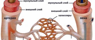
Through arterial vessels, blood comes from the carotid and vertebral arteries. And veins form a system for the outflow of waste waste from cells and intercellular space.
Any vascular disorders entail more or less severe changes in the functioning of the brain (diffuse and focal).
At-risk groups
Typically, focal changes in the white matter of the brain of a dystrophic nature most often occur in old age. Most lesions appear during life and as a result of hypoxia and ischemia. People who lead a sedentary and unhealthy lifestyle are also susceptible to the disease. Genetic predisposition also plays a role. The risk group includes people suffering from high or low blood pressure, diabetes, rheumatism, obesity and atherosclerosis. In addition, emotional individuals prone to stress are at risk of developing pathology.
You may be interested: Inhalations with Miramistin in a nebulizer
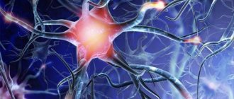
The white matter of the brain coordinates all human activity. It is what connects different parts of the nervous system. White matter is necessary for the two hemispheres to work together.
Classification
Depending on the type of brain cells that degenerated and gave rise to pathogenesis, the following types of tumors are distinguished:
- astrocytomas are the most common type, their share in the general population is about 50%;
- oligodendrogliomas – the share in the general population of tumors of this type is up to 10%;
- Ependymomas are the rarest forms (occurrence less than 7%).
According to the WHO classification (which is generally accepted), neoplasia is ranked depending on the degree of malignancy.
Benign gliomas
These are tumors of the first degree of malignancy, for example, astrocytomas: giant cell, pilocytic, juvenile subependymal. They are benign because they grow slowly, have no signs of cancer, and are easy to treat. The prognosis for life with benign glioma is favorable. After tumor removal, patients live 10 years or longer.
Low grade glioma
Second degree of malignancy. It is also classified as a benign neoplasm, but with a borderline degree of malignancy. The growth of pathological tissue is slow (low-grade), they are well differentiated, as a rule, there is only one sign of cancer (cell atypia). Tumors of this type can degenerate into cancer and easily progress to the third and fourth degree of malignancy. These include diffuse and fibrillary astrocytomas. Treatment is complex: surgery to remove atypical tissues, additionally radio and chemotherapy.
Malignant gliomas
These include gliomas of grade 3 and 4:
- Third degree of malignancy . There are all signs of malignancy, except for tissue necrosis. Tissues lose clear differentiation, tumor growth accelerates (high-grade), the boundaries are unclear, and growth into nearby tissues is typical. The most striking example is anaplastic astrocytoma, which most often develops in middle-aged and older people. Treatment is hampered by the lack of clear boundaries of the tumor; life expectancy depends on how much atypical tissue is removed, as well as the stage of carcinogenesis at the beginning of treatment.
- The most dangerous is the fourth degree of malignancy (glioblastoma). It develops between the ages of 40 and 70 years. In this case, all the signs of malignancy are present, including necrosis. The tumor grows quickly, penetrates other tissues and has no clear boundaries. This significantly complicates therapy. The prognosis is unfavorable.
Neoplasia is divided into two types, depending on growth characteristics:
- Tumors of nodular growth . As a rule, these are benign neoplasms with clear contours. They are characterized by formation anywhere in the brain and the presence of cysts. Examples: pleomorphic xanthoastrocytoma and piloid astrocytoma.
- Diffuse type formations . There are no obvious boundaries, the size can reach significant sizes, neoplasia grows into adjacent brain tissue, which complicates the removal of dysplasia. These are often malignant tumors, such as glioblastoma or anaplastic astrocytoma, or those that can quickly degenerate.
Causes
You may be interested in: The act of defecation: mechanism, regularity, causes of violations
Focal changes in the brain substance of a dystrophic nature occur in a number of diseases of different origins:
- Changes in vascular origin: atherosclerosis, migraine, hypertension, etc.
- Inflammatory diseases. Multiple sclerosis, Behcet's disease, Sjögren's disease, inflammatory bowel disease (Crohn's disease).
- Infectious diseases. HIV, syphilis, borreliosis.
- Intoxication and metabolic disorders, carbon monoxide poisoning, B12 deficiency.
- Traumatic processes associated with radiation therapy.
- Congenital diseases caused by metabolic disorders.
The occurrence of pathology is caused by impaired blood supply in any part of the brain. It is fraught with loss of function of brain tissue. The more blood flow has decreased, the more noticeable the consequences. An example is damage to the spinal or cerebral blood flow. Such violations progress slowly, but entail serious consequences.
Stages
There are several stages that indicate the development of a disease associated with insufficient nutrition of the brain.
The dynamics may be different, as it is influenced by several factors, such as heredity, lifestyle, environmental conditions, and so on. At the first stage of the disease, people often have headaches, irritability, forgetfulness and sleep disturbances. In the second stage, memory deteriorates more severely; a person can sleep during the day, but at night sleep is disturbed. Obsessive thoughts also appear, the patient begins to think about the same problem. The gait becomes unsteady. Uncoordinated movements appear. Performance decreases. At the last stage of the disease, dementia sets in, the person stops recognizing relatives and finding his way around on the street.
Signs
You may be interested in:Hot spring "Avan" in Tyumen: detailed information
The signs of focal changes in the brain substance of a dystrophic nature are also different. With focal changes, not the entire brain suffers, but only its individual parts. Tissue degeneration occurs when there is insufficient supply of nutrients necessary for the normal functioning of the body’s nervous systems. We are talking about proteins - the building material of the human body. Proteins break down into amino acids, which, in turn, stimulate the nervous system. It also requires fats and carbohydrates - the main sources of energy needed by every living creature.
Of the vitamins, the brain needs B1 (activates its work), B3 (provides energy at the intracellular level), B6 (it is difficult to imagine metabolic processes without it, in addition, it is also a kind of antidepressant), B12 (promotes memory preservation and helps to stay alert) . All these vitamins can be obtained in sufficient quantities by creating the right diet.
Pathologies
Focal changes in the substance of the brain of a dystrophic nature most often take the form of pathologies such as:
- A cyst is a small cavity that is filled with fluid. It often interferes with the normal functioning of neighboring areas of the brain, as it compresses blood vessels. Cysts are divided into intracerebral (cerebral) and arachnoid. The latter appears in the meninges. Its occurrence is promoted by the accumulation of cerebrospinal fluid and inflammatory processes. Cerebral occurs in place of dead brain tissue.
- Necrotic state of tissue - appears when the supply of important nutrients to areas of the brain for any reason deteriorates. Dead cells form so-called dead zones and are not regenerated.
- Hematomas and brain scars occur after severe trauma or concussion. Foci of this type lead to structural damage.
Symptoms
The clinical picture of gliotic foci of vascular origin is determined by the localization of the replaced tissue. The modified tissue does not cause gross disturbances, however, in the presence of large-scale foci, gliosis “reduces” the general background of life, worsening its quality.
Localization:
Frontal lobes
Leads to a general decrease in cognitive abilities: the pace of thinking slows down, control over one’s behavior is partially lost. Patients have difficulty learning new information and skills. Causal relationships are more difficult to establish. The patient is slower to think.
With deep lesions of gliosis, complex motor patterns are forgotten: patients forget how to tie shoelaces or how to play a musical instrument. The vocabulary becomes poor: sentences are monotonous, there are few or no synonymous words in speech.
The emotional-volitional sphere is upset. Emotions “dull”: all feelings lose their expression and color. Motivation decreases: the desire to explore the world around us is lost.
Temporal, parietal and occipital region
Hearing, speech and vision are affected. The perception of complex compositions is impaired. The sense of rhythm is disrupted. Visual accuracy deteriorates. The threshold of general sensitivity increases: the senses of tactile touch lose their sharpness. Memory deteriorates.
Single supratentorial foci of gliosis of vascular origin
The presence of foci in the cerebellar structures forms a picture of coordination disorder. Gait is disturbed. It is called “drunk” walking: balance is disturbed, the patient spreads his legs wide apart to maintain balance and not fall.
Limbs are shaking. This happens at rest and during movement. Individual fingers also tremble. Vision is impaired. Nystagmus appears - a synchronous rotation of the eyeballs to one side with a frequency of 60 movements per minute.
Muscle tone is impaired towards weakening. At the same time, tendon reflexes decrease. Muscles decrease in size. The synchronicity of the work of the flexor and extensor muscles is disrupted. Handwriting becomes disordered: the patient’s letters are difficult to read and spell out.
The clinical picture of single supratentorial foci of gliosis of vascular origin also affects speech disorder. It loses its smoothness and becomes scanned. For example, a person speaks slowly and in syllables: “mo-lo-ko.” At the same time, the speech rhythm is observed.
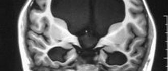
Diagnostics
The complete picture of focal changes in the brain substance of a dystrophic nature is determined using an MRI study. This procedure allows you to see even small areas of transformation in the white matter. And they, in turn, lead to cancer and stroke.
Focal dystrophic lesions come in different sizes and differ in location. Based on this, the examination may show some types of disorders.
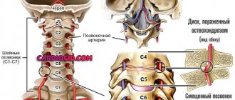
In the cerebral hemisphere, blockage of vital arteries is usually diagnosed due to abnormal development of the embryo or acquired atherosclerotic plaques. A herniated cervical spine is also detected.
Changes in the white matter of the brain indicate hypertension and congenital developmental anomalies. In other cases, numerous areas of brain pathology may indicate a pre-stroke condition, senile dementia, or epilepsy.
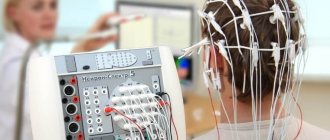
Sometimes doctors perform tests on a patient to detect the presence of cognitive impairment. That is, cognitive dysfunction. Such as orientation in space and time, understanding of external processes, the ability to remember information, drawing, writing, reading, etc.
Focal changes in the brain substance of a dystrophic nature can develop in three ways:
- In the first case, the disease is remitting in nature. Symptoms increase gradually, the condition worsens, and brain productivity decreases. But from time to time, remissions occur—temporary improvements in health, after which the patient becomes worse again.
- Progressive focal changes in the brain substance of a dyscirculatory dystrophic nature develop very quickly. Each stage of the disease takes no more than two years, which is considered a short period for organic brain lesions.
- Typically, the deterioration of a person suffering from focal changes lasts for many years, eventually leading to dementia.
It should be remembered that single focal changes in the brain substance of a dystrophic nature often appear in young people, and single damage to the white matter in an elderly person is considered normal. Structural disorders of the cerebral arteries of the atherosclerotic type appear in 50% of patients over 50 years of age. For the most part, hypertensive patients suffer from this. Therefore, you need to show the MRI result to a neurologist so that he can determine the severity of the disorders in the brain by comparing the MRI result and the clinical picture of the disease.
How do vascular lesions of the brain manifest clinically?
Diseases of the brain of vascular origin, depending on the degree of damage to the arterial bed, can cause reversible (transient) symptoms or, using the body’s compensatory capabilities, form clinical manifestations that indicate the localization of the source of maximum destruction.
In ischemic brain disease, initial changes in nerve cells cause subtle disturbances in higher cortical functions:
- psyche;
- functioning of the sense organs;
- coordination of movements;
- vegetatives (sweating, tachycardia).
They are detected in cases of nervous overstrain, excitement, and stressful situations. Then the disorders take on a dyscirculatory character.
Headache may worsen with head movements or bending
The most common manifestations are:
- headaches - very intense and dull, localized in the back of the head, eyebrows or throughout the head;
- feeling of “noise in the head or ears”;
- dizziness;
- sensation of blood pulsating in the head;
- photophobia;
- nausea and vomiting;
- weakness in the left, right or all limbs;
- chilliness of hands and feet;
- difficulty speaking;
- visual impairment;
- memory disorder;
- insomnia.
Objective signs are:
- paresis and muscle paralysis;
- asymmetry of nasolabial folds;
- “sailing” breathing;
- pathological reflexes in the arms and legs.
In case of venous insufficiency, the patient additionally experiences:
- cyanotic tint of lips, nose, ears, cheeks;
- swollen lower eyelids;
- focal symptoms are less pronounced.
Headaches occur after night, when tilting the head (outflow worsens).
In severe cases:
- the patient is unconscious (cerebral coma);
- breathing is hoarse due to paresis of the respiratory muscles and vocal cords, arrhythmic;
- the face is purple and puffy (with a cerebral hemorrhage);
- focal symptoms depend on the location of the hematoma.
Diet
In the early stages of this disease, it is enough to reconsider your lifestyle and diet, choosing a more gentle regimen and diet. In the diet, it is advised to reduce the consumption of animal fats, and it is better to completely replace them with vegetable ones. You should eat fish and seafood instead of fatty meat, and reduce the amount of salt in your diet. Fresh vegetables and fruits will be of great benefit.
Treatment
There are a huge number of focal anomalies, so each has its own cause. Treatment of brain pathologies is based on the destruction of those factors that led to the appearance of lesions in brain tissue. In addition to eliminating the underlying disease, the doctor may also prescribe vitamins and medications to help combat the deterioration of cerebral blood flow.
The treatment process depends directly on what somatic problems in the body led to the occurrence of lesions in the brain. For infections, for example, antibiotics are taken; for injuries, diuretics, decongestants, and anticonvulsants are taken. If the damage to the brain tissue was caused by a circulatory disorder, then nootropics and coagulants are prescribed.
Source
Mental changes of vascular nature
Often patients with vascular disorders of the brain present complaints of a special nature, indicating changes in the psyche. Preserved criticality is replaced by its loss, then the change in the character of the sick person can be judged by the reviews of employees at work and relatives.
The most typical:
- sleep disturbances (short-term, superficial);
- constant fatigue;
- intolerance to bright light, loud sounds, smells;
- increased irritability;
- memory loss;
- anxiety, suspiciousness.
In case of head injury, an X-ray examination of the skull is required.