The physiology of the cerebral venous system is currently still poorly understood. Therefore, only experienced phlebologists and neuropathologists know what venous discirculation is and how to treat it. Although in fact, under such a complex term lies an ordinary violation of venous blood flow. In a healthy person at rest, the average speed of venous blood movement is approximately 220 mm/min, and in those suffering from dyscirculation it decreases to 47 mm/min. Knowledge of the anatomy of the circulatory system of the brain will help you independently determine the symptoms caused by dyscirculation, as well as take preventive measures in advance.
The mechanism of occurrence of venous discirculation
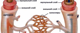
Cerebral veins can be divided into 2 subtypes: superficial and deep. The veins that are located in the soft membrane (superficial) are designed to drain blood from the cerebral cortex, and those that are located in the central parts of the hemispheres (deep veins) serve to drain blood from the white matter. The above vessels carry blood to the superior and inferior longitudinal sinuses. From these collectors, blood is pumped into the internal jugular vein, and then flows from the brain through the vertebral vein system.
This rather simplified description of the complex route of blood outflow allows us to understand why for such a long time doctors cannot determine the true causes of cerebrovascular accidents.
At the moment, doctors have learned that cerebral venous discirculation occurs due to pathological processes in the cavity between the membranes of the brain or in the cervical and spinal plexus. In 75% of cases, these pathological processes are cervical osteochondrosis or atherosclerotic plaques.
Classification
The pathological process can be divided according to the severity of the clinical picture and course. This is an integral, unifying method of typing; it should also be called staging of the disease.
Based on the system of characteristics, the following forms are called:
- First stage. As a rule, it occurs without pronounced manifestations. The silent phase of the pathological process. It is detected at this moment only by chance, because the symptomatic complex is scanty or absent altogether. Main problem. Only at such a moment can you completely eliminate organic disorders, restore blood flow and get rid of the problem. As it progresses, this is no longer possible.
- Second stage. The symptoms are obvious. The person still retains the ability to work and can take care of himself in everyday life, but great difficulties arise. You may need outside help. Without treatment, disruption of the cerebral venous outflow rapidly progresses. Structural changes are visible using imaging methods.
- Third stage. Accompanied by severe neurological abnormalities. Hemodynamics are significantly worse than they should be. Urgent medical attention is required. Unfortunately, at this stage it will no longer be possible to radically restore the patient’s original or at least acceptable condition. The only goal is partial improvement, as much as possible. The outcome is a stroke or other dangerous complications.
Staging is carried out based on the results of instrumental studies. The basic techniques are determined by the situation.
Causes of obstruction of blood flow from the brain
It is quite difficult to determine exactly what exactly provoked the disruption of the normal outflow of blood from the brain, because more than one year may pass after the event that provoked the blockage. The main causes of venous discirculation may be:
- pulmonary and heart failure;
- compression of extracranial veins;
- jugular vein thrombosis;
- brain tumors;
- traumatic brain injury;
- cerebral edema;
- systemic diseases (lupus erythematosus, Wegener's granulomatosis, Behcet's syndrome).
Discirculation can be provoked by either one disease or a complex of several unpleasant symptoms. For example, a mutation in the prothrombin protein in combination with the use of contraceptives in the form of pills increases the risk of developing dysgemia (another name for venous discirculation).
Development mechanism
Understanding what exactly provokes venous discirculation is not always easy. There can be many reasons. In total, there are three key factors that are involved in pathogenesis.
Among them:
- Mechanical compression. The vessels are compressed, venous outflow is difficult, this means that blood cannot move through them normally, and the result is a rapid progression of organic changes.
When it comes to the brain, the main type of mechanical obstruction is a neoplasm. Cyst or tumor.
It is also possible to localize the root cause in the neck area. In this case, there is compression of the vessels by osteophytes (bone outgrowths), hernias or displaced vertebrae, usually on one side, which causes an asymmetry of the venous outflow.
- The second possible reason is a hormonal factor. It is relatively rare. It is observed as a result of physical overload, severe stress or psycho-emotional stress.
The main substances are cortisol, adrenaline and others. There is also an increase in pressure in the veins and arteries, which disrupts the rate of blood outflow.
- Finally, a significant factor in the development of the disease is a change in the rheological properties of the blood. First of all, its fluidity. The intensity of movement of liquid tissue decreases. It is possible to change the further scheme only with the use of medications and timely treatment.
There are some other mechanisms, but the essence always remains the same: the outflow of venous blood is hampered, and the quality of nutrition of cerebral structures decreases.
Hence the neurological deficit, which is manifested by symptoms characteristic of encephalopathy, a transient ischemic attack. In a word - chronic malnutrition of the brain.
In the absence of therapy, progression is rapid. It takes from a couple of months to several years for dangerous complications to develop. Depends on many factors.
Attention:
The average duration before the onset of a stroke is about 3-5 years, give or take, this is an average figure.
Risk factors
In addition to the above diseases, disruption of venous blood flow can provoke an unhealthy lifestyle. If you find yourself with at least one of the risk factors listed below, you need to make an appointment with a neurologist to discuss measures to prevent dysgemia.
High blood pressure and a sedentary lifestyle are the first step to dysgemia
The following deviations should alert you:
- presence of diabetes mellitus;
- high blood pressure;
- obesity degree 2 or higher;
- high cholesterol;
- high triglyceride levels;
- passive lifestyle.
Causes
Factors in the development of the pathological process are diverse and complex. Doctors know them quite well; diagnostic tasks include identifying provoking moments that are associated with the onset of the disorder.
- Arterial hypertension. Stable or periodic increase in pressure. The numbers are different. A drop in blood pressure levels has the same negative effect. In both cases, there is a change in the tone of all blood vessels, and the nutrition of the cerebral structures is poor.
- Cardiac (heart) failure. Regardless of phase. Stages 2-3 are especially difficult. The reason lies in the low contractility of the myocardium and the inability to provide adequate nutrition to brain tissue. Read more about chronic heart failure in this article.
- Tumors of cerebral structures. As stated earlier. Cysts, directly solid neoplasms. They compress tissues, provoke an increase in intracranial pressure and worsen venous outflow.
- Osteochondrosis of the cervical spine. A common reason.
- Endocrine diseases. From excess production of thyroid hormones to problems with the adrenal glands, pituitary gland and other structures. Also, the reason becomes relevant at peak moments: puberty, pregnancy, attenuation of the reproductive system (menopause).
- Traumatic brain injuries. Especially when hematomas form.
- Blood diseases, blood flow disorders.
The list of problems is wide. Patients who smoke, regularly consume alcohol, caffeine-based drinks, and people with little physical activity are at risk.
The issue is resolved within the framework of prevention, primary and secondary.
Symptoms of pathology
Dysgemia is almost always accompanied by periodic dull headaches, sometimes with nausea and vomiting. Less commonly, a disturbance of consciousness occurs, after which focal symptoms appear:
- numbness of the limbs;
- severe aphasia;
- isolated epileptic seizures;
- impaired vascular-platelet hemostasis.
Signs of venous discirculation may appear irregularly and last for several minutes. If the disease is not treated, then unpleasant symptoms can constantly bother the patient.
Only a doctor can help prevent the development of severe dysgemia
The most serious symptoms that occur if the disorder is ignored are:
- dizziness;
- blurred vision;
- sudden loss of consciousness;
- tingling in the neck, especially on the left;
- moderate hypoxia;
- sudden reflex movements;
- constant drowsiness.
Symptoms and signs of obstructed venous outflow
The first clinical symptoms of impaired venous outflow with cervical osteochondrosis appear 2-3 months after the onset of development of pathological changes in the vascular bed. In this case, a developed clinical picture is already present and the venous vessels are quite deformed. It is recommended to visit a doctor earlier. Therefore, it is important to know the first signs of obstructed venous outflow, which can occur literally 2-3 days after the development of the disorder.
These symptoms of obstructed venous outflow include a feeling of fullness in the back of the head and temples in the first hours after waking up. These pains are dull and indistinct in nature and may be accompanied by an attack of nausea, lack of appetite, general weakness and weakness. If such manifestations appear, it is necessary to consult a vertebrologist as soon as possible.
In the future, as the disease develops, symptoms of obstructed venous outflow of the head may manifest themselves in the form of:
- regularly occurring obsessive noise in the ears (this can be the splashing of waves, the sound of the surf, humming, buzzing, etc.);
- decreased hearing and visual acuity on the affected side or symmetrically with prolapse of the vena cava;
- gradual development of problems with falling asleep and insomnia, accompanied by obsessive thoughts at night and a feeling of enormous fatigue in the morning;
- development of depressed mood, sometimes with suicidal thoughts;
- decreased mental performance, memory, ability to concentrate;
- high degree of dependence on meteorological conditions and magnetic storms;
- bluish tint of the skin in the neck, collar area and face;
- pronounced swelling of the face and infraorbital areas in the morning;
- a dull, pressing headache in the back of the head and temples (may occur in the morning, after an emotional shock, heavy physical activity or prolonged tension in the neck muscles);
- dizziness, which at first are orthostatic in nature (when standing up suddenly), then turn into a constantly present state of lightheadedness, often leading to fainting.
With prolonged obstruction of the venous outflow of blood from the structures of the brain, muscle weakness and partial numbness of the upper extremities develop. Problems with memory, ability to perform mental work, concentration, etc. quickly begin. Neuroses and psychoses may develop, and the patient's mood is constantly depressed. Approximately 20% of patients develop a syndrome of persistent increased intracranial pressure over time. This condition can lead to regular epileptic seizures.
What does ignoring the problem lead to?
Ignoring symptoms for a long time results in oxygen and glucose not reaching the brain. This can lead to neurological problems. Lack of treatment can provoke more severe conditions.
Stroke
If any tumor blocks the flow of blood in the carotid artery, a heart attack or stroke may occur. As a result, some brain tissue may die. The death of even a small amount of tissue can affect speech, coordination, and memory. The severity of the consequences of a stroke depends on how much tissue has died and how quickly the outflow of venous blood has been restored. Some patients are able to fully recover, but most of those affected suffer irreversible changes.
Brain hemorrhage
With chronic problems with cerebral venous circulation, bleeding in the cranial cavity may occur. This occurs when the walls of the arteries weaken and burst. Even minor bleeding puts pressure on the brain, which can lead to loss of consciousness.
Hypoxia
Hypoxia occurs when completely or partially blocked venous drainage prevents oxygen from reaching the brain. Individuals with hypoxia often feel lethargic and dizzy. If the vessels are not promptly unblocked, coma and death may occur.
Dyscirculatory encephalopathy (including atherosclerotic origin)
Discirculatory hypertensive encephalopathy is a painful syndrome that is caused by a violation of venous blood flow. With minor dyscirculation, encephalopathy develops very slowly and is practically asymptomatic. The syndrome quickly disappears when the original causes of dysgemia are eliminated. But with a prolonged lack of oxygen or as a result of a complete blockage of venous outflow, brain death can occur (just 6 minutes after the complete cessation of blood flow).
Violation of venous circulation in the brain
The brain is a complex structure, its normal functioning depends on the state of blood circulation. In addition to the need to deliver oxygen and glucose to the nervous tissue, the outflow of venous blood and the removal of toxins with it, the result of cell activity, is important. If this process is disrupted, chronic venous insufficiency of the brain is formed.
A peculiarity of the vessels of the brain is the course of the veins: it does not coincide with the direction of the arteries, and a network is formed that is independent of them. If the outflow of blood through one of the vessels is impaired, venous blood is directed to the other, and compensatory expansion occurs. A prolonged decrease in tone leads to vascular atrophy, they collapse, and the risk of thrombosis increases. Dilated vessels contribute to the development of venous circulation insufficiency, the functioning of the valves is disrupted, they do not close tightly, and the direction of blood flow is disrupted.
Stages of the pathological process
The following stages are distinguished during cerebral venous insufficiency:
- latent: no clinical symptoms, no complaints;
- cerebral venous dystonia: some symptoms are observed: headaches, weakness;
- venous encephalopathy: severe symptoms caused by organic lesions are observed, venous outflow is impaired in all areas of the brain, there is a high risk of hemorrhages from dilated vessels.
Classification
There are chronic and acute variants of venous circulation in the brain. Chronic include venous congestion and venous encephalopathy, acute include venous hemorrhage, thrombosis of the veins and venous sinuses, thrombophlebitis.
Causes and risk factors
Insufficiency of cerebral venous circulation can be caused by diseases or individual characteristics of the patient. The most common causes of pathology:
- neoplasms in brain tissue can cause disruption of venous outflow;
- head injuries that impair blood circulation to the brain;
- injuries during childbirth;
- hematomas formed as a result of stroke, atherosclerosis, bruises and other causes contribute to the formation of tissue edema, which complicates the outflow of blood from the affected area;
- blood clots and embolisms narrow the lumen of the vessel or completely close it, preventing the movement of blood;
- diseases of the spinal column, in which deformed sections of the canals compress the vessels and disrupt blood flow, also cause venous insufficiency;
- vascular features: hereditary predisposition and impaired development of veins can provoke the development of a violation of the outflow of venous blood.
Circulatory disorders can be physiological and occur when coughing, sneezing, or physical strain. Such short-term deviations do not cause significant harm to health.
One-time attacks of cerebral circulatory disorders do not cause serious consequences for the body. However, prolonged blood stagnation can contribute to the development of serious consequences. The following risk factors increase the likelihood of cerebral venous insufficiency:
- frequent stress;
- smoking;
- alcohol abuse;
- prolonged dry cough;
- professional singing;
- hypertension;
- heart failure;
- reading in the wrong position
- professional swimming;
- frequent wearing of clothes that compress the neck;
- chronic rhinitis:
- work in high-altitude, underwater, underground professions;
- office work that involves staying in a position with the head tilted or turned;
- frequent physical activity of high intensity
Clinical manifestations
Disturbances of venous circulation, as a rule, are genetically determined. Currently, the role of the initial tone of the veins in the formation of venous discirculation is undeniable. Constitutional and hereditary factors are key for the development of venous dyshemia. Patients with a family “venous” history usually have several typical manifestations of constitutional venous insufficiency - varicose veins or phlebothrombosis of the lower extremities, hemorrhoids, varicocele, impaired venous outflow from the cranial cavity, esophageal varicose veins. Pregnancy is often the trigger.
Typical complaints:
- morning or afternoon headache of varying intensity;
- dizziness, depending on changes in body position;
- noise in the head or ears;
- visual disturbances (decreased visual acuity, photopsia);
- “tight collar” symptom - increased symptoms when wearing tight collars or ties;
- “high pillow” symptom - increased symptoms when sleeping with a low headboard;
- sleep disorders;
- feeling of discomfort, “fatigue” in the eyes in the morning (symptom of “sand in the eyes”);
- pastiness of the face and eyelids in the morning (with a pale, purple-cyanotic tint);
- mild nasal congestion (outside the symptoms of acute respiratory infections);
- darkening of the eyes, fainting;
- numbness of the limbs.
Signs of impaired cerebral venous blood flow are related to weather conditions. Headaches are poorly controlled by analgesics; often some relief is brought only by a change in body position - in a horizontal position, venous blood flow is redirected along collaterals - bypassing the affected vessel.
The patient’s psyche changes in such a way that minor experiences can lead to neuroses. Tearfulness increases, the patient often breaks into a scream. Mania and depression are observed. Severe damage leads to psychosis, accompanied by hallucinations and delusions; this can make the patient dangerous to himself and others.
Diagnostics:
- radiography determines the strengthening of the pattern of the veins of the skull, which indicates the presence of a pathological process;
- angiography is a contrast method for diagnosing blood stagnation, determining the patency of blood vessels;
- computed tomography and magnetic resonance imaging make it possible to accurately determine the presence of a pathological process in the brain, as well as in surrounding tissues;
- ultrasound examination of the veins of the brain and neck;
- rheoencephalography is a functional diagnostic method that assesses the condition of blood vessels;
- An increased level of pressure in the ulnar vein allows one to suspect abnormalities in the vessels of the brain.
Therapy is complex and includes several areas
- drug treatment;
- non-drug treatment: physiotherapy, massage, physical therapy;
- surgery.
Drug therapy
The following drugs are used to normalize cerebral circulation:
- venotonics strengthen the vascular wall, reduce permeability, have an analgesic effect, and eliminate inflammation;
- diuretics to eliminate swelling;
- neuroprotectors improve nutrition and metabolism of the brain;
- anticoagulants to thin the blood and prevent thrombosis;
- vitamin therapy (vitamins B and PP).
Non-drug treatment
There are a number of non-drug therapies that are effective as an additional treatment method and improve vascular tone. However, before treating impaired cerebral venous outflow with their help, it is necessary to assess individual risks and contraindications: in some cases, such procedures can lead to the opposite effect and worsen the patient’s condition.
Applicable:
- head and neck massage;
- oxygen therapy;
- foot baths;
- physical therapy: breathing exercises, neck exercises, yoga classes.
Intern of the ophthalmology department Gurlo A.I.
Head of the department ophthalmological department Rudova
Diagnostic methods
If the patient complains of several of the above symptoms, then all the doctor’s efforts will be aimed at identifying and treating the cause of the dyscirculation. This involves a physical examination and a medical history. To confirm the violation of venous outflow, several studies are prescribed with visualization of veins in the brain and vertebrobasilar region.
Complete blood count
It is prescribed to detect antinuclear antibodies and determine the erythrocyte sedimentation rate. If the results of the analysis confirm the presence of antibodies and a reduced ESR, then an additional study is prescribed to determine complement components and the level of antibodies to anti-deoxyribonucleic acid. The results of the above tests will reveal that the cause of dysgemia is systemic lupus erythematosus or Wegener's granulomatosis.
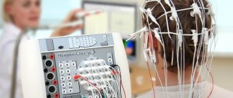
Electroencephalogram (EEG)
An electroencephalogram with impaired outflow of venous blood may be normal. But this study is strongly recommended after unilateral thalamic infarction. A slowdown in the basic alpha rhythm indirectly indicates coordination abnormalities and problems with blood outflow.
An EEG can help a doctor identify venous dyscirculation
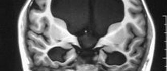
Computed tomography (CT)
CT is an important imaging modality and is often indicated for the initial diagnosis of dysgemia. In the tomograph image, you can see whether any neoplasm or thrombosis is the cause of dyshemia.
CT angiography
CT angiography is also prescribed to visualize the cerebral venous system. Only angiography can indicate the absence of flow in the venous channels.
Contrast magnetic resonance imaging
Contrast magnetic resonance imaging is an excellent method for visualizing blood flow in the great cerebral veins. It is prescribed if angiography does not reveal any disturbances in the outflow of venous blood into the VBB.
Prevention
To prevent circulatory problems in the brain:
- Get rid of bad habits.
- Avoid stress, overexertion, and overtime.
- Eat right.
- Do not subject your body to excessive physical activity, but also do not lead a sedentary lifestyle.
- Treat chronic diseases in a timely manner, especially for cervical osteochondrosis, which people often do not give due importance. If your neck hurts, immediately go to a neurologist.
- Monitor your blood pressure, especially if you are over 40. Buy an automatic electronic blood pressure monitor and measure your blood pressure 2 times a day. This will take at most 5 minutes a day, but in this way you will eliminate hypertension and hypotension. If you notice that your blood pressure changes, immediately go to a therapist, and then to a cardiologist.
- Once every six months to a year, undergo a preventive examination with a therapist, cardiologist, or neurologist.
How to treat venous discirculation?
The doctor may recommend several different treatment methods, depending on the identified causes of the disease. But most patients will be advised to make changes to their daily lifestyle, namely:
- stop smoking and drinking alcohol;
- perform simple physical exercises daily;
- follow a diet to lower cholesterol levels;
- Monitor your blood sugar and blood pressure daily.
As for the drug treatment of patients with venous discirculation, specific therapy is prescribed, which includes taking anticoagulants or thrombolytics (depending on the medical history). But the use of systemic anticoagulation as primary treatment is recommended for all patients without exception (even for a child and in the presence of intracranial hemorrhage).
Drug therapy is the most effective method of treating dysgemia
Most often, drugs containing heparin are prescribed. When administered intravenously, its action begins immediately, which is very important for patients with acute dyshemia.
Enoxaparin sodium is a low molecular weight heparin and is prescribed if it is necessary to restore venous outflow to patients suffering from allergic reactions, or for prophylaxis. The main advantage of enoxaparin is the possibility of intermittent administration of the drug, which allows the patient not to go to the hospital, but to take advantage of outpatient treatment.
Warfarin is prescribed to patients with bleeding disorders for whom heparin and enoxaparin are strictly contraindicated. The drug has a slight effect on coagulation activity, but the therapeutic effect can only be seen after a few days. Therefore, such treatment is not prescribed in the acute stages of dyscirculation. The dose of the administered drug must be carefully monitored by a doctor, so use at home is excluded. Higher doses are administered at the beginning of treatment in order to speed up the time to restore normal outflow, but at the same time, this tactic leads to an increased risk of bleeding. Treatment with warfarin should be continued for 3-6 months to obtain lasting results.
Surgical intervention to get rid of dyscirculation is prescribed in extreme cases
If the disturbances in the venous system are too severe, the doctor may recommend surgery to quickly improve the flow of blood from the brain. But surgery is prescribed only if medical methods do not work.
Types of surgical operations recommended for dysgemia:
- endarterectomy (removal of the inner lining of the affected artery);
- bypass surgery: a new blood vessel is placed near the narrowing of the vein to create a new route for blood flow;
- angioplasty: a balloon catheter is inserted into a narrow section of the artery to widen the walls and improve blood flow.
Symptoms of the first stage
At the first stage, when discirculation is just beginning, there is no symptomatic complex at all, or the list of disorders is so small that it has no characteristic features. It is possible to develop a minimal list of manifestations.
Among them:
- Mild episodic headache. Sluggish, makes itself felt intermittently. Absent most of the time.
- Weakness, constant desire to sleep, regardless of the amount of rest per day. This is the most typical sign of venous discirculation of the brain, which is caused by adaptation mechanisms against the background of a reduced volume of nutrition.
- Occasional fainting is rare. Or conditions preceding syncopation.
The first stage lasts up to several years. It is possible to identify it, but only if you look for it purposefully.
That is, diagnosing a disorder at the initial stage is a matter of chance. We are talking about great luck, because there is every chance of restoring the normal state of affairs and completely eradicating the disease.
Second phase
Already much more dangerous. The clinic of stagnation of venous blood is pronounced, accompanied by a group of ghosts:
- Headache. Intense. Continues for several hours, then spontaneously subsides and disappears. The nature of the unpleasant sensation is pressing, bursting. Accompanied by a group of other signs. The manifestation is especially noticeable in the morning, also in the evening, after heavy emotional, mental stress, visual tension and other negative stress factors affecting the state of the body.
- Cyanosis of the nasolabial triangle, facial skin. Not always. The symptom appears at a moment of nervous tension.
- Numbness of fingers, toes. Dermal cover. Feels like tingling, decreased sensitivity. Temporary disturbances in motor activity are possible.
- Paresthesias are intermittent and paroxysmal. The intensity is maximum at the peak of the attack.
- Tinnitus. It develops against the background of a decrease in the quality of blood flow in the brain. It intensifies when changing the position of the body in space.
- Flashing spots in the eyes, decreased visual acuity, photopsia or sharp flashes of light (in fact, these are the simplest hallucinations against the background of irritation of the cerebral cortex, especially the occipital lobe).
- Dizziness. Accompanied by impaired coordination and motor activity. The patient also loses the ability to move normally, and his gait is unsteady.
Signs do not always develop; there is generally a connection with a trigger factor: overwork, a sharp change in ambient temperature, and other factors.
Third stage
Accompanied by pronounced changes in well-being. Neurological deficits reach a peak and precede stroke.
And if the first signs of venous discirculation are not noticeable either to the person himself or often to doctors, everything is obvious here.
- Behavioral disorders. The sufferer becomes apathetic. Depressed, reacts sluggishly to surrounding stimuli and external irritants. This condition turns out to be prevalent, and the patient is almost always in this condition.
- Cognitive dysfunction. It manifests itself as a decrease in memory, speed of thinking, and attention. It is impossible to solve applied problems. Psychological tests are resolved at first at a slower rate, and then cease to be performed at all, because the mental sphere suffers significantly.
- Instead of sensory disturbances in the limbs, paresis and even complete paralysis of the arms/legs occur, depending on the situation. This is an alarming sign indicating structural changes in the brain, irreversible disorders.
- Speech problems. She becomes inarticulate. The reason lies in the mechanical factor. More dangerous options are also possible, for example, aphasia associated with destruction of Wernicke’s, Broca’s areas, temporal or frontal lobes.
- Changes in coordination and orientation in space. It even gets to the point where the patient is unable to move independently, falls, and does not understand his own body and how to control it. A typical sign of destruction of the extrapyramidal system, in particular the cerebellum. The frontal lobe is also affected.
Cerebral venous dyscirculation is accompanied by focal neurological symptoms most of the time, the signs are almost always identical. Only the list and severity of manifestations differ.
A special, isolated case is venous discirculation in the VBB (vertebrobasilar region). It is localized in the neck and occipital lobe of the brain.
Disruption of nutrition and outflow in these structures leads mainly to damage to the occipital lobe and extrapyramidal system. Vision and motor activity suffer.
In severe cases, irreversible eye damage is possible and even probable. A stroke can lead to complete blindness; such situations often occur in clinical practice.
The prospects for restoring visual function are more than vague. Therefore, you should not take it to the limit; you should immediately consult a doctor.
Forecasts for venous discirculation
The prognosis and speed of recovery will depend on several factors.
Success in treating the underlying disease that caused dysgemia
For example, the survival prognosis for dysgemia may be quite negative if the patient has had a stroke or thrombosis. But if the cause of the disease is hypertension or diabetes, the prognosis will be much better.
Presence of hypoxia
The prognosis will be poor if venous discirculation previously led to hypoxia. Even after eliminating dysgemia, sudden loss of consciousness or problems with the musculoskeletal system are possible.
Treatment of venous outflow disorders in cervical osteochondrosis
The basis for the treatment of impaired venous outflow in cervical osteochondrosis is the principle of complete restoration of the patency and functionality of the damaged blood vessel. To do this you need:
- restore the normal structure of the intervertebral disc (height and shock-absorbing capacity);
- ensure elasticity of muscles, ligaments and tendons to prevent the risk of re-compression;
- restore the free outflow of venous fluid;
- improve the condition of brain structures.
In our manual therapy clinic, if the patient has difficulty venous outflow from the head, treatment begins with individual recommendations. The doctor talks about how you need to change your lifestyle, organize your sleeping and working space, develop a diet and physical activity regimen in order to completely restore the circulation of venous fluid and eliminate the risk of relapse of cervical osteochondrosis. An individual course of treatment is then developed.
It may include:
- traction traction of the spinal column - allows you to restore the physiological gaps between the vertebral bodies and start the process of regeneration of the fibrous ring of the cartilage disc;
- osteopathy and massage to restore microcirculation of blood and lymphatic fluid, improve the condition of all soft tissues;
- physical therapy and kinesiotherapy to increase the tone of the muscles of the neck and collar area, restore the process of diffuse nutrition of the cartilage tissue of the intervertebral discs;
- laser exposure and other methods of physiotherapy;
- reflexology (acupuncture, pharmacopuncture) to quickly relieve symptoms and start the regeneration processes of all damaged tissues.
If you need to undergo a course of treatment and restore the obstructed outflow of venous blood due to cervical osteochondrosis, then make an appointment right now for a free appointment with a vertebrologist in our manual therapy clinic. The doctor will conduct an examination, make a diagnosis and tell you about the prospects for using manual therapy methods in your case of illness.
Diagnostics
It is carried out in an outpatient or inpatient (much less often) setting. Requires the participation of a neurologist as the main specialist. If necessary, other doctors are also involved.
The basis consists of the following activities:
- Oral interview with a person. To identify all the symptoms and create a clinical picture. This is the basis for developing hypotheses regarding diagnosis.
- Anamnesis collection. From past illnesses to family history of pathologies, in order to understand what could have caused the disorders. These are routine primary techniques, but they set the vector for further examination.
- Measurement of blood pressure and heart rate.
- Duplex scanning of the vessels of the neck and brain. A basis that allows you to provide comprehensive information about the functional state of blood flow in the named structures.
- MRI if organic lesions are suspected. At advanced stages, it plays a role in determining the severity of the disorder.
- Electrocardiography and ECHO-CG as methods for identifying problems with the heart and blood vessels.
- Phlebography as a more specific study. It is carried out for the purpose of targeted visualization of the condition of the veins.
Usually this is enough. As needed, specialists check basic reflexes and assess the concentration of specific hormones. As part of identifying the causes of the violation.