Somatic nervous system
(from the Greek soma - body) - part of the human nervous system, which is a set of afferent (sensitive) and efferent (motor) nerve fibers innervating muscles (in vertebrates - skeletal), skin, joints. The somatic system is the part of the peripheral nervous system that carries motor (motor) and sensory (feeling) information to and from the central nervous system. This system consists of nerves attached to the skin, sensory organs and all the muscles of the skeleton. It is responsible for almost all conscious muscle movements, as well as for processing sensory information received through external stimuli: vision, hearing and touch. The name somatic nervous system comes from the Greek word “soma” (body). The somatic nervous system contains two main types of neurons: sensory (afferent) neurons, which carry information from the nerves to the central nervous system, and motor (efferent) neurons, which carry information throughout the body from the brain and spinal cord to muscle tissue.
The neurons of the somatic nervous system extend from the central nervous system directly to the muscles and receptors. The body of the neuron is located in the central nervous system, and the axons extend further until they reach the skin, sensory organs, or muscles. Electrochemical impulses travel through axons from the brain to the spinal cord. The somatic nervous system also includes reflex arcs that are responsible for unconscious actions (reflexes). With the help of reflex arcs, muscles move without signals from the brain. This happens when nerve pathways connect directly to the spinal cord. Some examples of reflex arcs are when you quickly remove your hand from a hot pan or unconsciously raise your leg when the doctor taps your knee.
Structure of the autonomic nervous system
There are also differences in the structure of the two parts of the ANS.
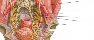
The centers of its sympathetic part are located in the thoracic and lumbar parts of the spinal cord, and the centers of the parasympathetic part are in the brain stem and sacral part of the spinal cord (see figure). The highest centers that regulate and coordinate the work of both parts of the ANS are the hypothalamus and the cortex of the frontal and parietal lobes of the cerebral hemispheres. Autonomic nerve fibers emerge from the brain and spinal cord as part of the cranial and spinal nerves and are directed to the autonomic ganglia. The ganglia of the sympathetic part of the ANS are located near the spine, and the parasympathetic part is located in the walls of the internal organs or near them. Therefore, the preganglionic and postganglionic sympathetic fibers are almost the same length, and the parasympathetic preganglionic fiber is much longer than the postganglionic fiber. After passing through the ganglion, autonomic fibers, as a rule, are directed to the innervated organ along with blood vessels, forming plexuses in the form of a network on the vessel wall.
The paravertebral ganglia of the sympathetic part of the ANS are combined into two chains, which are located symmetrically on both sides of the spinal column and are called sympathetic trunks. In each sympathetic trunk, consisting of 20–25 ganglia, the cervical, thoracic, lumbar, sacral and coccygeal sections are distinguished.
- Additional points
- Reflex as the basic principle of the nervous system
- What is the autonomic part of the nervous system?
- Autonomic (autonomic) nervous system. Functions of the autonomic nervous system.
- Role
- What is the somatic nervous system?
- Cells of the brain and spinal cord
- 6. Central Nervous System (CNS)
- What controls the autonomic nervous system?
- How does the somatic system work?
- How does the ANS work?
- What are the types of disease
- 3. Characteristics of the Central Nervous System
From the 3 cervical ganglia of the sympathetic trunk, nerves arise that regulate the activity of the organs of the head and neck, as well as the heart. These nerves form plexuses on the wall of the carotid arteries and, together with their branches, reach the lacrimal gland and salivary glands, glands of the mucous membrane of the oral and nasal cavities, the larynx, pharynx and the muscle that dilates the pupil. The cardiac nerves, arising from the cervical ganglia, descend into the chest cavity and form a plexus on the surface of the heart.
From 10–12 thoracic ganglia of the sympathetic trunk, nerves depart to the organs of the thoracic cavity (heart, esophagus, lungs), as well as large and small splanchnic nerves, heading into the abdominal cavity to the ganglia of the celiac (solar) plexus. The solar plexus is formed by autonomic ganglia and numerous nerves and is located in front of the abdominal aorta on the sides of its large branches. The celiac plexus supplies the innervation of the abdominal organs - the stomach, small intestine, liver, kidneys, and pancreas.
From the 4 lumbar ganglia of the sympathetic trunk depart the nerves involved in the formation of the celiac plexus and other autonomic plexuses of the abdominal cavity, which provide sympathetic innervation to the intestines and blood vessels.
The sacrococcygeal section of the sympathetic trunk consists of four sacral ganglia and one unpaired coccygeal ganglia lying on the inner surface of the sacrum and coccyx. Their branches participate in the formation of the vegetative plexuses of the pelvis, which provide sympathetic innervation to the organs and vessels of the pelvis (rectum, bladder, internal genital organs), as well as the external genitalia.
The nerve fibers of the parasympathetic part of the ANS leave the brain as part of the III, VII, IX and X cranial nerves (in total, 12 pairs of cranial nerves depart from the brain), and from the spinal cord as part of the II–IV sacral nerves. The parasympathetic ganglia in the head area are located near the glands. Postganglionic fibers are sent to the organs of the head along the branches of the trigeminal nerve (V cranial nerve). Parasympathetic innervation is received by the lacrimal and salivary glands, glands of the mucous membrane of the oral and nasal cavities, as well as the muscle that constricts the pupil and the ciliary muscle (provides accommodation - adapting the eye to seeing objects at different distances).
The largest number of parasympathetic fibers passes through the vagus nerve (X cranial nerve). The branches of the vagus nerve innervate the internal organs of the neck, chest and abdominal cavities - the larynx, trachea, bronchi, lungs, heart, esophagus, stomach, liver, spleen, kidneys and most of the intestines. In the chest and abdominal cavities, the branches of the vagus nerve are part of the autonomic plexuses (in particular, the celiac plexus) and together with them reach the innervated organs. The pelvic organs receive parasympathetic innervation from the splanchnic pelvic nerves emerging from the sacral spinal cord. Parasympathetic ganglia are located in or near the wall of the organ.
Structure and types of the nervous system: structural classification
To simplify the structure of the nervous system, in medicine there are several classification options depending on the structure and functions performed. Thus, anatomically, the human nervous system can be divided into 2 broad groups:
- central (CNS), formed by the brain and spinal cord;
- peripheral (PNS), represented by nerve ganglia, endings and nerves themselves.
The basis of this classification is extremely simple: the central nervous system is a kind of connecting link in which the analysis of the incoming impulse and further regulation of the activity of organs and systems is carried out. And the PNS serves to transport the received signal from the receptors to the CNS and the subsequent activator, but from the CNS to the cells and tissues that will perform a specific action.
central nervous system
The central nervous system is a key component of the nervous system, because it is here that the main reflexes are formed. It consists of the spinal cord and brain, each of which is reliably protected from external influences by bone structures. Such thoughtful protection is necessary, since each part of the central nervous system performs vital functions, without which it is impossible to maintain health.
Spinal cord
This structure is contained within the spinal column. It is responsible for the simplest reflexes and involuntary reactions of the body to stimuli.
In addition, spinal cord neurons coordinate the activity of muscle tissue, which regulates defense mechanisms. For example, upon feeling an extremely hot temperature, a person involuntarily withdraws his palm, thereby protecting himself from a thermal burn. This is a typical reaction controlled by the spinal cord.
Brain
The human brain consists of several sections, each of which performs a number of physiological and psychological functions:
- The medulla oblongata is responsible for the vital functions of the body - digestion, respiration, blood movement through the vessels, etc. In addition, the nucleus of the vagus nerve is located here, which regulates the autonomic balance and psycho-emotional reaction. If the nucleus of the vagus nerve sends active impulses, a person’s vitality decreases, he becomes apathetic, melancholic and depressed. If the activity of impulses emanating from the core decreases, the psychological perception of the world changes to a more active and positive one.
- The cerebellum regulates the precision and coordination of movements.
- The midbrain is the main coordinator of muscle reflexes and tone. In addition, neurons regulated by this part of the central nervous system contribute to the adaptation of the sensory organs to external stimuli (for example, pupil accommodation at dusk).
- The diencephalon is formed by the thalamus and hypothalamus. The thalamus is the most important organ that analyzes incoming information. The hypothalamus regulates the emotional background and metabolic processes; there are centers responsible for the sensation of hunger, thirst, fatigue, thermoregulation, and sexual activity. Thanks to this, not only physiological processes are coordinated, but also many human habits, for example, the tendency to overeat, the perception of cold, etc.
- Cerebral cortex. The cerebral cortex is a key link in mental functions, including consciousness, speech, perception of information and its subsequent comprehension. The frontal lobe regulates motor activity, the parietal lobe is responsible for bodily sensations, the temporal lobe controls hearing, speech and other higher functions, and the occipital lobe contains the centers of visual perception.
Peripheral nervous system
The PNS provides interconnection between organs, tissues, cells and the central nervous system. Structurally, it is represented by the following morphofunctional units:
- Nerve fibers, which, depending on the functions performed, are motor, sensory and mixed. Motor nerves transmit information from the central nervous system to muscle fibers, sensitive nerves, on the contrary, help to perceive information received through the senses and transmit it to the central nervous system, and mixed nerves are involved to one degree or another in both processes.
- Nerve endings, which are also motor and sensory. Their function is no different from fiber structures with the only nuance - the nerve endings begin or, conversely, end the chain of impulses from the organs to the central nervous system and back.
- Nerve ganglia, or ganglia, are clusters of neurons outside the central nervous system. The spinal ganglia are responsible for transmitting information received from the external environment, and the autonomic ganglia are responsible for transmitting information about the state and activity of the internal organs and resources of the body.
In addition, all peripheral nerves are classified depending on their anatomical features. Based on this characteristic, there are 12 pairs of cranial nerves that coordinate the activity of the head and neck, and 31 pairs of spinal nerves that are responsible for the torso, upper and lower extremities, as well as internal organs located in the abdominal and thoracic cavities.
Cranial nerves originate from the brain. The basis of their activity is the perception of sensory impulses, as well as partial participation in respiratory, digestive and cardiac activity. The function of each pair of cranial nerves is presented in more detail in the table.
| No. | Name | Function |
| I | Olfactory | Responsible for the perception of various odors, transmitting nerve impulses from the olfactory organ to the corresponding center of the brain. |
| II | Visual | Regulates the perception of visual data by delivering impulses from the retina. |
| III | Oculomotor | Coordinates the movement of the eyeballs. |
| IV | Block | Along with the oculomotor pair of nerves, it takes part in the coordinated movement of the eyes. |
| V | Trigeminal | Responsible for sensory perception of the facial area, and also participates in the act of chewing food in the oral cavity. |
| VI | Abductor | Another nerve that regulates the movements of the eyeballs. |
| VII | Facial | The nerve that coordinates facial contractions of the facial muscles. In addition, this pair is also responsible for taste perception, transmitting signals from the papillae of the tongue to the brain center. |
| VIII | vestibulocochlear | This pair is responsible for the perception of sounds and the ability to maintain balance. |
| IX | Glossopharyngeal | Regulates the normal activity of the pharyngeal muscles and partially transmits taste sensations to the brain center. |
| X | Wandering | One of the most significant cranial nerves, the functionality of which determines the activity of internal organs located in the neck, chest and abdominal wall. These include the pharynx, larynx, lungs, heart muscle and digestive tract organs. |
| XI | Dorsal | Responsible for contractions of muscle fibers of the cervical and shoulder regions. |
| XII | Sublingual | Coordinates the activity of the tongue and partially forms speech skills. |
The activity of the spinal nerves is classified much more simply - each specific pair or complex of pairs is responsible for its assigned area of the body with the same name:
- cervical - 8 pairs,
- infants - 12 pairs,
- lumbar and sacral - 5 pairs respectively,
- coccygeal - 1 pair.
Each representative of this group belongs to mixed nerves, formed by two roots: sensory and motor. That is why the spinal nerves can both perceive an irritating effect, transmitting an impulse along the chain, and intensify activity in response to a message from the central nervous system.
Additional points
It is also worth noting that there are distinctive features in terms of morphology. As noted above, fibers may have a myelin sheath (in the somatic system) or not (in the autonomic system). There is also a difference in size: for somatic fibers it is in the range of 12-14 microns, and for autonomous fibers it is up to 7 microns. And this directly affects the speed of nerve impulses, that is, the smaller the unit, the longer it will transmit signals, and vice versa. This characteristic also affects the degree of excitability, so autonomous fibers require greater force when irritated.
In general, not only the somatic, but also the entire nervous system has many nuances on which the full functionality of the entire organism depends.
Reflex as the basic principle of the nervous system
I. M. Sechenov in 1863 in his work “Reflexes of the Brain” he developed the idea that the reflex is the basic principle of the functioning of not only the spinal cord, but also the brain.
A reflex is the body’s response to irritation with the participation of the central nervous system. Reflexes are divided into: 1) unconditioned reflexes: innate (hereditary) reactions of the body to irritations carried out with the participation of the spinal cord or brain stem; 2) conditioned reflexes: acquired on the basis of unconditioned reflexes temporary reactions of the body, carried out with the obligatory participation of the cerebral cortex, which form the basis of higher nervous activity.
Each reflex has its own reflex arc - this is the path along which excitation passes from the receptor to the effector (executive organ).
The reflex arc is represented by a chain of neurons that provide the perception of irritation, the transformation of the energy of irritation into a nerve impulse, the conduction of a nerve impulse to the nerve centers, the processing of incoming information and the implementation of a response.
Any reflex arc consists of 5 components
1. A receptor is a specialized cell designed to perceive a stimulus (sound, light, chemical, etc.).2. Afferent pathway, which is represented by afferent neurons.3. The area of the central nervous system represented by the spinal cord or brain; 4. The efferent pathway consists of the axons of efferent neurons extending beyond the CNS.5. An effector is a working organ (muscle, gland, etc.).
The simplest reflex arc includes 2 neurons and is called monosynaptic (based on the number of synapses). A more complex one is represented by 3 neurons and is called three-neuron or disynaptic. However, most reflex arcs include a large number of interneurons and are called polysynaptic.
Reflex arcs can pass through the spinal cord only (for example, withdrawing a hand when touching a hot object) or through the brain only (for example, closing the eyelids when a stream of air is directed at the face), or through both the spinal cord and the brain.
Reflex arcs are closed into reflex rings using feedback connections. The concept of feedback and its functional role was indicated by Bell in 1826. He wrote that two-way connections are established between the muscle and the central nervous system. With the help of feedback, signals about the functional state of the effector are sent to the central nervous system.
The morphological basis of feedback is the receptors located in the effector and the afferent neurons associated with them. Thanks to feedback afferent connections, fine regulation of the effector’s work and an adequate response of the body to environmental changes are carried out.
Peripheral nervous system
In order to ensure the relationship between the central nervous system and organs, there is a peripheral part of the nervous system.
The peripheral nervous system includes nerve endings, neurons, and nerves. The main function of the PNS is to manage and control the skeletal muscles, regulate the functioning of all organs, and maintain homeostasis. That is, after the brain sends a signal to the spinal cord, the corresponding part of it sends a synaptic signal through the axons of nerve cells to the desired organ. This can be either an exciting signal (for example, muscle contraction) or a relaxing one.
The PNS provides two-way communication between a person and his environment: he can not only perceive signals, but also respond to them with the help of movements and facial expressions.
What is the autonomic part of the nervous system?
Despite the enormous influence of the autonomic system on the body of every person as a biological unit, in fact, no one can say that they are able to feel its work every second. When functioning properly, people simply feel healthy.
Important Cheat sheet on the subject of dysarthria and cerebral palsy - file n1.doc
This is the main goal of the vegetative segment - the creation inside the body of an apparatus that would connect all organs and tissues into a single conglomerate to preserve a person as an integral natural unit. For example, when the temperature of the external environment increases, the activity of the respiratory, cardiovascular and metabolic systems is immediately adjusted. By interacting, they create comfortable conditions for the functioning of the brain and liquid tissues - prevention of dehydration.
In addition, the vegetative department controls digestive, urinary and reproductive functions. Not a single internal structure is left without double supervision - for example, some impulses slow down the pulse rate, while others increase the heart rate. This is the advantage of the human body over the plant or animal world.
Essentially, throughout evolution, the vegetative divisions have allowed humans to adapt to changing external conditions and the survival of the human race. Under new circumstances, the cardiovascular and respiratory systems, as well as digestion, provided internal tissues with nutrients. This guaranteed the safety of the individual. Subsequently, the innervation became more complex and modified. Ultimately, a modern person does not carry out a single type of activity without autonomic regulation, albeit at an unconscious level.
Autonomic (autonomic) nervous system. Functions of the autonomic nervous system.
Above, a fundamental qualitative difference was noted in the structure, development and function of non-striated (smooth) and striated (skeletal) muscles. Skeletal muscles are involved in the body's response to external influences and respond to changes in the environment with quick and appropriate movements. Smooth muscles, embedded in the viscera and blood vessels, work slowly but rhythmically, ensuring the flow of life processes in the body. These functional differences
are associated with differences in innervation: skeletal muscles receive motor impulses from the animal, somatic part of the nervous system, smooth muscles - from the autonomic one.
Autonomic nervous system
controls the activities of all organs involved in the implementation of plant functions of the body (nutrition, respiration, excretion, reproduction, circulation of fluids), and also carries out trophic innervation (I. P. Pavlov).
Trophic function of the autonomic nervous system
determines the nutrition of tissues and organs in relation to the function they perform in certain environmental conditions (
adaptation-trophic function
).
It is known that changes in the state of higher nervous activity affect the function of internal organs and, conversely, changes in the internal environment of the body affect the functional state of the central nervous system. Autonomic nervous system
strengthens or weakens
the function of
specifically working organs.
This regulation is tonic in nature, so the autonomic nervous system changes the tone of the organ. Since the same nerve fiber is capable of acting only in one direction and cannot simultaneously increase and decrease tone, accordingly, the autonomic nervous system is divided into two sections, or parts: sympathetic and parasympathetic - pars sympathica and pars parasympathica
.
Sympathetic department
in its main functions it is trophic. It enhances oxidative processes, consumption of nutrients, increased breathing, increased heart activity, and increased oxygen supply to the muscles.
The role of the parasympathetic department
protective: constriction of the pupil in strong light, inhibition of cardiac activity, emptying of the abdominal organs.
Comparing the area of distribution of sympathetic and parasympathetic innervation
, it is possible, firstly, to detect the predominant importance of one particular vegetative department.
The bladder, for example, receives mainly parasympathetic innervation, and transection of the sympathetic nerves does not significantly change its function; Only the sweat glands, hair muscles of the skin, spleen, and adrenal glands receive sympathetic innervation. Secondly, in organs with dual autonomic innervation, interaction between the sympathetic and parasympathetic nerves is observed in the form of a certain antagonism. Thus, irritation of the sympathetic nerves causes dilation of the pupil, constriction of blood vessels, acceleration of heart contractions, inhibition of intestinal motility; irritation of the parasympathetic nerves
leads to constriction of the pupil, dilation of blood vessels, slowing of the heartbeat, and increased peristalsis.
However, the so-called antagonism of the sympathetic and parasympathetic parts
should not be understood statically, as a opposition between their functions.
These parts interact, the relationship between them changes dynamically at different phases of the function of a particular organ; they can act both antagonistically and synergistically
.
Antagonism and synergism
- two sides of a single process. The normal functions of our body are ensured by the coordinated action of these two parts of the autonomic nervous system. This coordination and regulation of functions is carried out by the cerebral cortex. The reticular formation is also involved in this regulation.
Autonomy of the autonomic nervous system
is not absolute and manifests itself only in local reactions of short reflex arcs.
autonomic nervous system
proposed by the PNA is not accurate, which explains the retention of the old, more correct and logical term “
autonomic nervous system
”.
The division of the autonomic nervous system
into sympathetic and parasympathetic divisions is carried out mainly on the basis of physiological and pharmacological data, but there are also morphological differences due to the structure and development of these divisions of the nervous system.
Vegetatics
Classifying the nervous systems of people according to their structure is not the only way to divide its departments. The autonomic nervous system, which directly controls the organs of all systems, is of enormous importance. A person cannot consciously control the activity of the vegetative system, but information about how it works sometimes helps to correct well-being in case of vegetative disorders, for example, with a common disease - VSD (vegetative-vascular dystonia).
Role
It is difficult to overestimate the role of the somatic system in the full functioning of the human body. After all, it regulates a lot of conscious muscle contractions - from facial expressions to fine motor skills of the fingers.
Due to the fact that the somatic system does not respond in a timely manner to external irritating factors, a person gets the opportunity to control his body and keep it intact. In general, this structure, with the help of reflex arcs, regulates the work of every, even the smallest, skeletal muscle - through impulses from motor neurons.
You can imagine the role of the somatic system using the example of any extreme situation:
- sense organs perceive and transmit information about changes in the external environment;
- the corresponding neuron of the nerve fiber transmits the signal to the sensory neuron in the spinal ganglia;
- the impulse travels through the entire chain of neurons to the motor neuron;
- the required muscle group is activated.
It is the speed of reaction to the threat that directly determines the preservation of human bodies as units of living matter. The role of somatic regulation is not simply limited to processing the information received and reacting to it - the system is involved in the regulation of voluntary movements by the consciousness. For example, move your foot from a broken board slightly to the side to avoid falling, or move your body higher/lower and regain your balance.
What is the somatic nervous system?
Translated from Greek, "soma" means "body". It follows from this that the somatic nervous system is the structure of the human body that controls all actions performed by the body. The work of the somatic nervous system is subject to the will of man. This is ensured due to the presence of afferent (responsible for sensitivity) and efferent (motor) fibers. At the same time, the system itself exchanges information between the structures of the body and the central nervous system.
Nerve fibers of this part of the nervous system are present in almost every part of the body. The somatic nervous system controls the activity of muscle fibers, the processing of sensory information received from peripheral fibers, which is perceived by the organs of touch, hearing, and vision.
Where is the somatic nervous system located?
The human somatic nervous system includes two types of neurons: sensory and motor. The former are engaged in delivering the received information from nerve endings to the central nervous system. Motor ones deliver impulses throughout the body, from the central parts, spinal cord and brain - to muscle tissue.
The cell bodies of the neurons of the somatic system are located in the central part of the nervous system. At the same time, their axons stretch throughout the body until they reach muscle structures, sensory organs or skin. In addition, the somatic system also contains reflex arcs that ensure the development of unconscious actions and reflexes. With their help, muscle fibers contract even in the absence of signals from the brain, since the nerve pathways are directly connected to the spinal cord.
Divisions of the somatic nervous system
The somatic nervous system has two divisions:
- spinal nerves;
- cranial nerves.
Important Anxiety-neurotic personality disorder: a syndrome or a separate diagnosis?
The first belong to peripheral nerve fibers. They are responsible for transmitting information received from the sensory organs to the spinal cord and motor commands from the spinal cord. The cranial nerves deliver the received information to the brain stem and, in parallel, in the opposite direction.
These nerve fibers innervate the eyes, nose, tongue, mouth, ears, transmitting information from the organs of hearing, taste and smell to the brain. In total, the somatic nervous system has 12 pairs of cranial nerves, 31 pairs of spinal nerves, which, due to the large number of branches, form a dense nerve network.
Organs of the nervous system
The classification of the nervous system is, first of all, its physical structure. It consists of:
- brain;
- spinal cord;
- nerves;
- ganglia and nerve endings.
The brain is the most important organ that regulates the activity of all organs, and in which stimuli (commands) are formed that are sent to the cells of internal organs and muscles.
The brain consists of several sections, each of which is “responsible” for certain functions.
| Brain region | Main functions |
| Medulla oblongata and pons | Making decisions about launching reactions that regulate the most important vital functions: breathing, heart and blood vessels, digestion and wakefulness. |
| Cerebellum | Automation of movements: maintaining balance, moving in space, voluntary movements (for example, writing). |
| Midbrain | Reaction to stimuli, attention to what is happening. |
| Diencephalon | Regulation of the endocrine system, “filtering” of signals entering the brain. |
| Cortex | Smell, short-term memory, speech, thinking process, will and initiative. |
The brain actively exchanges signals with the spinal cord, which is located along the entire length of the spine, consisting of 31 fragments - vertebrae. The spine consists of four sections, each of which controls a specific “floor” of the body:
- cervical: neck, arms and diaphragm;
- thoracic: organs of the peritoneum and chest;
- lumbar: legs;
- sacrococcygeal: pelvis.
Thus, the signal from the nervous system from the brain enters the corresponding part of the spinal cord, and from there to the necessary organs, cells, and tissues. And the path from the spinal cord to specific nerve endings lies along the nerves, or, more precisely, along the axons of neurons in the form of short electrical impulses.
Cells of the brain and spinal cord
The brain and spinal cord are made up of cells whose names and characteristics are determined by their functions. Cells characteristic only of the nervous system are neurons and neuroglia.
Neurons –
These are the workhorses of the nervous system. They send and receive signals to and from the brain through a network of interconnections so numerous and complex that it is completely impossible to count them or chart them completely. At best, we can roughly say that the brain contains hundreds of billions of neurons and many times more connections between them.
Brain tumors arising from neurons or their precursors include embryonal tumors (previously called primitive neuroectodermal tumors - PNETs)
, such as
medulloblastomas
and
pineoblastomas
.
The second type of brain cells are called neuroglia. In the literal sense, this word means “the glue that holds the nerves together” - thus, the auxiliary role of these cells is already clear from the name itself. Another part of neuroglia contributes to the work of neurons, surrounding them, nourishing them and removing their breakdown products. There are many more neuroglial cells in the brain than neurons, and more than half of brain tumors develop from neuroglia.
Tumors arising from neuroglial (glial) cells are generally called gliomas. However, depending on the specific type of glial cells involved in the tumor, it may have one specific name or another. The most common glial tumors in children are cerebellar and hemispheric astrocytomas, brainstem gliomas, optic pathway gliomas, ependymomas and gangliogliomas. The types of tumors are described in more detail in this article.
Sensory neurons
Afferent sensory neurons of the somatic nervous system provide the CNS with information about joint angle, muscle length, muscle tension, and the presence of noxious stimuli.
proprioceptors
In addition to the typical extrafusal muscle fibers, the muscle body also contains muscle spindles. These small sensory organs contain specialized muscle fibers that have a central non-contractile segment. Afferent neurons of the somatic nervous system have their sensory dendrites in this area. These dendrites contain ion channels that open in response to mechanical stimuli on the cell. When the muscle spindle is stretched, the opening of ion channels generates an action potential in these sensory neurons. The presence of mechanically closed ion channels allows these neurons to carry detailed information about the state of the muscle and its contractile activity.
Nociceptors
Nociceptors are pain receptors found throughout the body and are an important part of injury prevention, especially in muscle fibers. These neurons fire in response to potentially harmful stimuli, such as heat, cold, or extreme forces. The presence of nociceptors prevents us from overextending our joints, straining our muscles, and protects us from a wide range of injuries.
- Alpha Motor Neurons – Lower motor neurons of the brainstem and spinal cord are associated with striated muscle fibers and are directly responsible for their contraction.
- Extrafusal muscle fiber Skeletal striated muscle fibers are innervated by alpha motor neurons and are attached to bone through tendons. The main function is the formation of skeletal movements as a result of contractile tension. They make up the majority of striated muscles in the human body.
- Lower motor neurons - Effector neurons of voluntary muscle contraction, usually synapse at the neuromuscular junction with striatal muscle fibers. The cell body is located either in the medullary pyramids of the brain stem or in the anterior horn of the spinal cord.
- Upper motor neurons - Neurons that originate in the brainstem or motor cortex and carry information to the lower motor neurons of the spinal cord or medullary pyramids.
Central Nervous System (CNS)
In some cases, the functioning of the Nervous System may be disrupted, and deficits or problems in its functioning may arise. Depending on the affected area of the Nervous System, various types of diseases are distinguished.
Central nervous system diseases are diseases that impair the ability to receive and process information, as well as control body functions. These include.
Diseases
- Multiple sclerosis. This disease attacks the myelin sheath, damaging nerve fibers. This leads to a decrease in the number and speed of nerve impulses, until they stop. The result is muscle spasms, problems with balance, vision and speech.
- Meningitis. This infection is caused by bacteria in the meninges (the membranes that cover the brain and spinal cord). The cause is bacteria or viruses. Symptoms include high fever, severe headache, stiff neck, drowsiness, loss of consciousness and even convulsions. Bacterial meningitis can be treated with antibiotics, but viral meningitis will not be treated with antibiotics.
- Parkinson's disease. This chronic nervous system disorder, caused by the death of neurons in the midbrain (which coordinates muscle movement), has no cure and progresses over time. Symptoms of the disease include tremors of the limbs and slowness of conscious movements.
- Alzheimer's disease. This disease leads to memory impairment, changes in character and thinking. Its symptoms include confusion, temporal-spatial disorientation, dependence on other people to carry out daily activities, etc.
- Encephalitis. This is an inflammation of the brain caused by bacteria or viruses. Symptoms: headache, difficulty speaking, loss of energy and body tone, fever. May lead to seizures or even death.
- disease
(
Huntington)
: This is a neurological degenerative hereditary disease of the Nervous System. This disease damages cells throughout the brain, leading to progressive impairment and motor problems. - Tourette's syndrome: More information about this disease can be found on the NIH page. This disease is defined as:
Motor neurons
The neural pathway that leads to skeletal muscle contraction can be functionally divided into two main types of neurons—upper motor neurons in the central nervous system and lower motor neurons in the somatic nervous system. Lower motor neurons may be part of cranial or spinal nerves. They innervate muscle fibers and directly cause their contraction.
Upper motor neurons have their cell organs in the precentral gyrus of the brain. This area is located towards the posterior end of the frontal lobe in the cerebral cortex and is connected to the primary motor cortex. The axons of upper motor neurons associated with voluntary muscle movement move along the central nervous system along two pathways - the corticospinal and corticobulbar tracts. Neurons whose axons move along the corticobulbar tract synapse with lower motor neurons in the brainstem. The axons of these lower motor neurons form cranial nerves, such as the oculomotor, trochlear, or trigeminal nerves, which are associated with contraction of the skeletal muscles of the face, neck, jaw, and tongue.
The image shows upper motor neurons emerging from the precentral gyrus and moving along the corticobulbar tract to the brainstem.
The axons of other upper motor neurons move along the cortical spine, bypassing the medulla oblongata and reaching the ventral horn of the spinal cord.
The image shows the origin of upper motor neurons from the precentral gyrus moving through the midbrain and medulla to form the lateral and anterior corticospinal tracts. The main function of these neurons is to connect the brain to the spinal cord. In the spinal cord, upper motor neurons form synapses with lower motor neurons and release glutamate into the synaptic cleft. Depolymerization of the lower motor neuron results in transmission of action potentials to skeletal muscle.
There are three types of lower motor neurons – alpha, beta and gamma. Alpha motor neurons are thick, myelinated, multipolar nerve fibers that innervate most skeletal muscle fibers and cause muscle contraction. Gamma motor neurons support the activity of alpha motor neurons by keeping muscle spindles taut. Alpha motor neurons can receive signals from upper motor neurons for voluntary muscle movement. At the same time, they can receive information from sensory and interneuronal cells to initiate reflex actions. The number of alpha motor neurons innervating one muscle depends on the degree of precise motor control required locally. Therefore, the finger muscles will have significantly more alpha motor neurons associated with them than the thigh or forearm muscles.
Neuromuscular junction
The axon end of the alpha motor neuron forms the neuromuscular junction with striatal muscle fibers, where acetylcholine is released as a neurotransmitter. When an action potential reaches the axon end of an alpha motor neuron, a voltage-gated ion channel allows calcium ions to enter the neuron. These ions cause the fusion of synaptic vesicles with the plasma membrane, resulting in the release of acetylcholine at the neuromuscular junction. Acetylcholine then binds to nicotinic receptors in muscle cells. These receptors are ion channels that open upon ligand binding, which then leads to a cascade of ions within the muscle fiber, resulting in muscle contraction.
Two strong toxins that affect the neuromuscular junction are botulinum. toxin and tetanus toxin. Both chemicals are produced by bacteria - the first by the bacterium Clostridium botulinum, the second by Clostridium tetani. Botulism can affect a person by inhaling or ingesting the toxin or by ingesting bacterial spores from contaminated food. This is especially true for improperly prepared canned foods, as the warm, moist, anaerobic environment inside food containers can provide a favorable environment for bacterial growth. The toxin prevents the fusion of synaptic vesicles with the neuronal plasma membrane and thus prevents the release of acetylcholine at the neuromuscular junction. Consequently, this leads to paralysis, initially of the facial muscles, and in severe cases even of the smooth muscles of the diaphragm. It is one of the most potent neurotoxins known, with a lethal dose of 1 microgram for an adult. The only other toxin in this potency is tetanus toxin, and it works in a similar way. When tetanus toxin enters the presynaptic nerve terminal, it prevents the release of neurotransmitters at the neuromuscular junction. While botulinum toxin causes flaccid paralysis, tetanus toxin causes spastic or rigid paralysis.
What controls the autonomic nervous system?
As part of the nervous system responsible for controlling the proper unconscious functioning of the internal organs, the autonomic or autonomic nervous system innervates most organs and systems of the body, with the exception of the muscles and joints that control voluntary movement.
In particular, we can find that this system controls the smooth muscles of the internal organs and various organs such as the heart or lungs. It is also involved in the synthesis and expulsion of most secretions outside the body and part of the endocrine system, as well as in metabolic processes and reflexes.
Listed below are some of the organs and systems in which this system is involved.
Vision
The autonomic nervous system controls pupil dilation and the ability to focus by connecting to the muscles of the iris and eyes.
Heart and blood vessels
Heartbeat and blood pressure are fundamental elements for humans that are controlled unconsciously. Thus, it is the autonomic nervous system that is responsible for regulating these vital elements that keep us alive every second.
Lungs
Although we can control our breathing to a certain extent, the fact of continuous breathing is not conscious. As a general rule, none of the rhythms with which we need to inhale. Thus, breathing is also partially controlled by the autonomic nervous system.
Digestive tube
Through food, a person can obtain various nutrients that the body needs to continue functioning. While eating behavior is consciously controlled by the process by which the digestive tube transforms food and acquires the necessary components, it is not a set of actions that the body performs during digestion involuntarily and is controlled by the autonomic nervous system.
Genitals
Although the sexual act itself is performed consciously, the set of elements and physiological reactions that allow it to occur are controlled primarily by the autonomic system, which controls processes such as erection and ejaculation. In addition, these processes become more complicated when you experience feelings of fear or anxiety. which connects you to various physiological states.
Secretion of enzymes and wastes
Tears, sweat, urine and feces are the substances that the body excretes into the environment. Its secretion and expulsion are determined by and/or can be partially modified due to the functioning of the autonomic nervous system. The same thing happens with the secretion of digestive enzymes and saliva.
Morphofunctional division of the nervous system
There is also a functional classification of parts of the nervous system, which includes:
- The somatic nervous system regulates the functions of skeletal muscles. It is controlled by the cerebral cortex, and therefore is completely subordinate to the conscious decisions of a person.
- The autonomic nervous system, responsible for the activity of internal organs. Its centers are located in the brain stem, and therefore it is not consciously regulated in any way.
In addition, the autonomic system is divided into 2 more significant functional departments:
- Sympathetic. Activated when energy is consumed;
- Parasympathetic. Responsible for the recovery period of the body.
How does the somatic system work?
If we turn to the roots, the word Soma is translated as body. In the scientific community, the system is also called animal, since it is present in animals. When analyzing the topic of the somatic system, it is worth knowing that it is a division of the peripheral system, in which the autonomic system is also present. And the main difference between them is that we can control the somatic one consciously, while the vegetative one works unconsciously.
The main task of the somatic system is to collect information from the environment and respond instantly in case of danger. Thanks to it, a person reacts in time in extreme situations, avoids injury and saves the body.
In summary, we can say that the activities of the somatic arc are very important and complex, and when problems arise with its elements, a person may lack the speed of reactions.
To understand how SNA works, you also need to know what it is. It innervates the following parts of the body:
- Muscles located on the limbs, in the face and throughout the body.
- Muscle tissue connected to the skeleton.
- Larynx, tongue and pharynx.
- Skin.
Only thanks to the correct functioning of the somatic system, we can feel tastes, touch, walk correctly, raise our arms, squat and perform all the actions available to us. Since some types of movement are also used by animals, in the scientific community the SNS is also called animal (animal).
Important For cerebral vessels, medications vary depending on the type that doctors prescribe
How does the ANS work?
Since the departments of the ANS will be discussed in detail in the relevant articles, we will say here the most basic data.
The autonomic system is divided into two sections: sympathetic and parasympathetic. The main differences between them are:
the sympathetic system controls the stress response, and the parasympathetic system controls the protective response;
the ganglia of the sympathetic system are located not in the organs, but in a special formation next to the spine - the sympathetic trunk;
these parts of the ANS have different localization of centers within the boundaries of the brain and spinal cord;
structures of the sympathetic nervous system are more common.
Where are the “command” centers located?
and the spinal cord is the “base” for the location of the centers, from where the autonomic innervation of the internal organs is carried out. They are located:
In the mesencephalic region (midbrain), for the arc of the pupillary reflex;
In the medulla oblongata, which is also called the bulbar region, there are groups of nuclei of the caudal cranial nerves. Autonomic fibers are part of the vagus, glossopharyngeal and facial nerves;
Anatomical interval from 1st thoracic to 2nd-3rd lumbar segment of the spinal cord. Along this length, the thoracolumbar autonomic centers are located in the spinal cord;
Sacral or sacral centers. They regulate the activity of the pelvic organs, the fibers exit as part of the pelvic nerves.
It would be a mistake to assume that the ANS is responsible for the activity of all systems of the body, being localized only in these departments. As elsewhere in living nature, the main principle of the organization of living matter to ensure the constancy of the internal environment of the organism is the strict subordination of the underlying sections to the overlying ones.
Therefore, there are even higher and “perfect” command bodies. While the visceral nervous system controls the work of internal organs, the higher autonomic centers - the hypothalamus, striatum and even the cerebral cortex are busy with the most complex work: they perform “fine tuning” and regulation of higher autonomic, including endocrine functions. Let us briefly consider the importance of the hypothalamus for the autonomic nervous system.
About the hypothalamus
The central part of the autonomic nervous system is divided into the hypothalamus, pituitary gland (united in the hypothalamic-pituitary system), striatum, and cerebral cortex. Such a division is largely arbitrary, since there are extensive and two-way connections, for example, with, and this connection is so complex that all its smallest details and details are still unknown.
The hypothalamus is a cluster of paired nuclei, and the number of these pairs is 32. There are anterior, posterior and middle nuclei. The work of these nuclei is closely related to osmoregulation, body temperature and the production of certain.
In order to understand how complex the hypothalamus is, here are a few examples:
When irritated, the posterior nuclei show signs of stress: the pupils and eyes dilate, blood vessels constrict, tachycardia occurs, gastric and intestinal motility is inhibited, adrenaline is released, glycogen breaks down in the liver under the influence of glucagon, blood sugar levels rise, “just in case.” Everyone knows that glucose is a “stress” substance that is released into the blood to provide food to the muscles in case of intense physical activity, for example, chasing prey or escaping.
In humans, stress is always a “headless” activity, and that is why it is so harmful. Any stress should find a way out through physical exercise, then heart attacks, strokes, diabetes and other serious diseases will occur much less frequently.
When the anterior nuclei are irritated, the opposite effects occur, and urination and defecation are also stimulated;
The middle nuclei of the hypothalamus control many metabolic processes. Their irritation leads to gluttony and obesity, and their destruction leads to refusal of food, exhaustion, anorexia;
The paraventricular nuclei control water metabolism. When they are irritated, for example by a tumor, pathological thirst appears.
There are also centers that help increase blood lipid levels; the anterior nucleus regulates body temperature. When it is destroyed, overheating or hyperthermia occurs, in which a person quickly dies, as sweating is impaired.
More “higher spheres”, such as the cortex, with the help of innervation, as well as through the integration of the ANS and the ANS under a “single principle”, allow for the final connection of all nervous systems, above which are emotions and personality.
The structural and functional unit of the animal nervous system is the neuron, which is a nerve cell with all its processes and terminal devices, and the main, elementary nervous act is the reflex.
In the nervous system, therefore, one can distinguish: 1) nerve centers, or ganglia, in which the switching of nerve impulses occurs; 2) conductors connecting the centers with each other and 3) peripheral nerves with their terminal devices.
The central nervous system consists of the brain and spinal cord. An intermediate position is occupied between them by the medulla oblongata, which is similar in structure to the spinal cord and retains the segmental apparatus in the form of cranial nerves and the conduction apparatus in the form of motor and sensory pathways.
In accordance with the pronounced physiological function in the brain, the forebrain, or cerebrum, the diencephalon (visual thalamus and their region), the midbrain (the quadrigeminal dorsal side and the cap (cerebellum and pons)) are distinguished.
The spinal cord is divided into cervical, thoracic and lumbar sections.
Under the influence of intermediate connections, the central nervous system functions as a single organ. All functions of the nervous system can be reduced to two main aspects of its activity: as an organ in which excitations are automatically generated, and as an organ that transfers impulses from a sensory neuron to a motor one and maintains communication between the periphery and the center. Both of these aspects of brain activity are closely intertwined.
The forebrain consists of two hemispheres and has a complex structure. The bulk of the hemispheres is white matter, consisting of pathways and subcortical nodes. The white matter is covered by an even layer of gray matter—the cerebral cortex. It is generally accepted that the cerebral cortex consists of six cellular layers arranged horizontally. Numerous processes of cortical cells penetrate all layers and form a dense network. Some fibers rise from the white matter to the cortex, others run parallel to the surface of the cortex and lie in the intermediate layers between the pyramids. Nerve fibers provide multilateral communication between individual parts of the brain.
The main function of the cerebral cortex is higher nervous activity aimed at regulating all functions of the animal’s body.
Higher nervous activity consists of the activity of the cerebral cortex and the subcortex closest to it. The cortex subordinates the subcortex and regulates its activity, and the subcortex, in turn, is a source of energy for the cortex.
The subcortex is the center of unconditioned reflexes, such as digestive, sexual, defensive, etc., while the cerebral cortex is the center of conditioned reflexes. In animals, conditioned reflex activity is determined by the direct effect of stimuli coming from the external or internal environment on the sense organs and their receptors located in the cerebral cortex.
Perception, higher analysis and synthesis, as well as consciousness, thinking and memory, i.e. the so-called mental manifestations, are closely related to the activity of the cerebral cortex. It has been experimentally proven that after removal of the bark, the dog is not able to assess the situation, does not recognize the owner, and becomes aggressive (Goltz, cited by Viktorov).
It has been experimentally established that the cerebral cortex can be divided according to the localization of specific functions into motor and sensory spheres. It has also been established that the motor and sensitivity of the body are controlled crosswise. The right hemisphere controls the left half of the body, and the left, vice versa, controls the right.
Motor spheres Control such volitional movements as flexion and extension of limbs, turning the neck and head, etc. The term “volitional movements” in animals has a relative meaning, since most of them are performed automatically, but some movements undoubtedly have elements conscious nature. Such movements include, for example, jumping over an obstacle, which is overcome by estimating the height and calculating the tension.
Motor centers do not represent sharply defined and isolated foci; zones of neighboring centers move towards each other.
Sphere of vision Located in the occipital lobe in the area of cunens et fissura, calcarina, the destruction of which leads to hemianopia, i.e., loss of the ability to see objects and phenomena in the field of vision. Reflexes related to the visual organs, such as blinking and turning the eyes, are preserved.
The sphere of hearing is located in the anterior part of the first gyrus of the temporal lobe of the hemisphere. Destruction of this sphere leads to deafness, and partial destruction leads to decreased hearing on the opposite side.
Sphere of smell Located under the Sylvian fissure of the ulcus gyri hippocampi and connected with the horn of ammonium. The nerve pathways from the tractus olfactorius go to the hemisphere of both its own and the opposite side. The destruction leads to a decrease in the sense of smell.
The sphere of general sensitivity (pain, tactile, thermal) is located throughout the cortex, but is more closely associated with the central gyri, where the motor sphere is localized.
Sphere of associations. The entire space of the cerebral cortex not occupied by motor and sensory areas belongs to the area of higher nervous activity.
Pavlov's teaching on conditioned reflexes showed that even internal organs, which were considered in many respects autonomous from the influence of the cerebral cortex, not only obey the direct influence of the cortex, but also send afferent excitations to the cortex, informing it of one or another of their states.
The study of conditioned reflexes has shown that the cerebral cortex has the ability to organize temporary connections, some of which can disappear, while others, with appropriate reinforcement, become stable and necessary.
The diencephalon is anatomically and functionally directly connected with the cerebral cortex and forms the “proximal subcortex”, which contains the visual thalamus, the hypothalamic region and the striatum.
The visual tubercle is the main subcortical node of all sensory conductors - visual, olfactory and general sensitivity. All sensory pathways gather here to switch to the last neurons that conduct sensitivity to the cerebral cortex. The main mass of the optic nerve passes through the diencephalon to the optic thalamus and further to the sphere of vision in the cerebral cortex. Here exteroceptive impulses are combined with interoceptive impulses.
In the hypothalamic region there are vegetative centers: metabolism, digestion, respiration, blood circulation, etc. Here are the basic mechanisms by which the cerebral cortex adapts the body to environmental conditions.
The striatum is located adjacent to the optic thalamus and is closely connected with it. The main function of the striatum is to maintain a certain muscle tone and ensure their readiness to contract.
The midbrain consists of the quadrigeminal, central gray matter, red nuclei and cerebral peduncles. The main function of the midbrain is the mobilization of motor apparatus and regulation of muscle tone.
The quadrigeminal structure is extremely complex. It contains sensory and motor neurons, as well as gray matter nuclei that serve as intermediate points.
The quadrigeminal consists of two cranial and two caudal colliculi. The former are related to vision, and the latter to hearing. Both of these senses take part in the animal’s orientation when moving. When the quadrigeminal dysfunction occurs in animals, hearing and vision deficiency occurs with a simultaneous disturbance in the correctness of movements.
Red core Located under the water supply in the cap. The red nucleus is the most responsible center, which perceives pathways from the cerebral cortex and bundles from the cerebellum. The red nucleus controls muscle tension, and together with the Deiters nucleus, nuclei of the cerebellum and medulla oblongata, the state of muscle tension in the body, which determines its position in space, controls the position of the head and the direction of the eyes according to the position of the body.
Transection of the brain stem along the quadrigeminal causes the appearance of rigor with a predominance of extensors. The animal stretches its legs and throws its head back. If the red nucleus is left with the spinal cord, then muscle tone will not be disturbed.
The hindbrain includes the pons and cerebellum.
The pons is a continuation of the nuclei of the medulla oblongata and contains transmission points for the cerebellum. It contains centripetal and centrifugal paths.
The cerebellum is similar in structure to the brain. The white matter is located in the center, while the gray matter forms the cortex, but is included in the nuclei and in the white matter. The nerve fibers of the white matter diverge towards the periphery in a fan-shaped manner.
The function of the cerebellum is reduced to receiving stimulation from sensory nerve endings and coordinating contractions of tonic muscle tension aimed at maintaining body balance.
Destruction of the cerebellum leads to impaired coordination of movements, collectively called cerebellar or cerebellar ataxia. When the function of the cerebellum is impaired, there is a weakening of muscle tone (atonia), a disturbance in the sense of strength (asthenia) and a violation of the correctness of movements (asthesia).
The loss of the coordinating influences of the cerebellum on the autonomic nervous system, mainly the sympathetic one, leads to severe disturbances in the motor and secretory functions of the digestive apparatus, the activity of the cardiovascular system, etc. (Orbeli, Astratyan). The regulation of the movements of the forelimbs comes from the anterior parts of the cerebellar hemispheres, and the hind limbs from the posterior parts.
The medulla oblongata can be considered as the anterior part of the spinal cord. The function of the medulla oblongata is quite complex, as is its structure. Both white and gray matter, when moving into the medulla oblongata, move and mix with each other, forming the so-called nuclei. These include the nuclei of the dorsal bundles of the spinal cord. From the accumulation of these nerve cells, secondary neurons of the secondary sensory pathways begin, heading towards the visual thalamus. The centripetal nerves of the brain end above them.
The automatic centers of the medulla oblongata include: a) the breathing center—lies at the height of the nuclei of the 9-12th nerve; b) the center of inhibition of the heart - connected with the nucleus of the recurrent nerve, it also works as a reflex center; c) vasomotor center—lies at the bottom of the fourth ventricle at the height of the nucleus of the facial nerve, it is connected with centers in the spinal cord and with the vascular center in the hypothalamic region; also works as a reflex center; d) sugar metabolism center - lies in the region of the nucleus of the recurrent nerve. Associated with metabolism. An injection into its area causes the development of glucosuria.
Spinal cord. The continuation of the medulla oblongata is the spinal cord, located in the spinal canal and suspended in the cerebrospinal fluid. In the area of the lumbar vertebrae, the spinal cord forms a filum terminale, which contains almost no brain matter. Along the entire length of the intervertebral foramina, spinal nerves arise from the spinal cord, forming the spinal ganglia as they exit the spine.
The gray matter of the spinal cord in a cross section occupies the central part and has the shape of the letter H, that is, it consists of a central wide part and four projections—two dorsal and two ventral horns. The lateral horns also protrude on the neck and lumbar region.
The white matter of the spinal cord occupies the periphery of the transverse section and is divided by gray matter into three main bundles (on each side): dorsal, lateral and ventral.
Each part of the spinal cord has a specialized purpose, its own function. To understand pathological conditions, it is very important to represent the function of the spinal cord through its cross section.
The neurites of the dorsal ganglion cells enter the dorsal horns of the spinal cord in the form of dorsal roots and are divided here into collaterals. Some of them are directed cranially, others caudally. Cranially they reach the medulla oblongata. All these numerous branches enable the spread of sensitive stimulation of the skin, mucous membranes and from the sensory endings inside the muscles, joints, periosteum and tendons. The ventral horns contain groups of large nerve cells with short neurites and dendrites.
Spinal nerves arise from two roots: ventral and dorsal. The dorsal ones carry centripetal nerve fibers, while the ventral ones carry centrifugal nerve fibers. Cutting the dorsal roots leads to loss of sensitivity, and cutting the ventral roots leads to the development of paralysis of the corresponding muscle groups. The centrifugal and centripetal nerves on each side cover specific areas of the body corresponding to a specific segment of the spinal cord. The dorsal bundle of white matter of the spinal cord, located between the roots and the middle plate, belongs to the sensitive ones and transmits deep sensitivity. This bundle transmits excitation from the periphery and from the spinal cord to the medulla oblongata, where it passes to the other side and reaches the visual thalamus.
The ventral fascicle, located between the ventral roots and the middle part of the gray matter of the spinal cord, is motor. Impulses pass through its fibers, originating in the cerebral cortex and regulating the movements of the animal. At the periphery of this bundle is the motor bundle, which begins in the medulla oblongata and ends above the motor cells of the ventral horns. These fibers carry impulses that regulate the balance of the body during movement.
The lateral fascicle, occupying the space between the dorsal and ventral roots, contains both centripetal and centrifugal pathways. In the lateral column there are fibers that carry impulses to the cerebellum from sensory endings in the muscles and blood vessels (deep sensitivity), fibers for conducting skin sensitivity, as well as motor nerves from the respiratory center, secretory nerves for the sweat glands and numerous nerves to the blood vessels.
In all three bundles of white matter there are numerous nerve fibers for the transmission of reflexes, intercentral communication and communication between segments of the spinal cord. Complete transection of the spinal cord causes sensory and motor paralysis of the entire part of the body below the transection. When half of the brain is cut, motor paralysis occurs on one side, sensory paralysis on the other, and weakening of tactile sensitivity on both sides.
The spinal cord contains numerous centers. Reflex centers are located along the length of the spinal cord and are intended for the motor system. Motor centers react to skin irritation by organizing protective movements, determining the coordination of movements and performing the function of muscle tone. Along the spinal cord there are numerous vascular-motor centers that work under the influence of the blood reaction and under the influence of sensitive irritations from the skin and organs. In the lumbosacral part of the spinal cord there are centers that regulate the activity of autonomic functions. This includes the center that controls the act of defecation, the center for the act of urination, the center for erection of the penis, and the center for ejaculation. These centers cause effects under the influence of excitations coming from the periphery on the one hand and the brain on the other. It must, however, be borne in mind that these centers influence the functions of internal organs through the sympathetic and parasympathetic nerves, which are independent to a certain extent.
What are the types of disease
If a person gets sick, then, accordingly, the function of one or another system is disrupted. Depending on which structural unit is affected, the following classification of diseases can be distinguished:
- Topographic-anatomical classification includes damage to the roots (radiculitis), cords (funiculitis), inflammation of the plexuses (plexitis), and peripheral nerves (mononeuritis).
If the inflammation has several localization points on the peripheral nerves, then this disease is called polyneuritis, and if there is an asymmetrical lesion, then it is multineuritis.
- As for the etiology, there may be viral or microbial causes.
In the first case, it is influenza, Guillain-Barre polyneuritis, tonsillitis and others. In the second option, the causes may be scarlet fever, leptospirosis, syphilis, brucellosis, etc. Etiological diseases can be caused by infectious-allergic pathogens or toxic effects. The latter includes chronic intoxication, for example, due to alcohol abuse or due to harmful working conditions.
This category also includes disturbances in dysmetabolic processes, for example, due to a lack of vitamins or diseases of the endocrine system, such as diabetes mellitus.
- A person's genetic predisposition can also lead to problems with the nervous system.
- The function of the system can be impaired by trauma, especially if the peripheral part is injured.
- Traumatic or compressive (ischemic) effects on the peripheral area can cause ailments in this area.
Depending on what kind of disease a person has, he may experience swelling of the body, a decrease in the tone of striated muscles, and increased blood circulation - and this is direct evidence that the somatic system is beginning to actively perform its functions.
All these points cause problems in the human body. Moreover, even a minor injury to the head or spine can lead to the fact that one or another function may be impaired. Therefore, in such situations, it is necessary to immediately go to a neurologist, who, upon examination or with the help of certain examinations, will be able to identify the problem, if any. After all, if functional units cannot fully fulfill their purpose, then a person’s vital activity will decrease, and this, in turn, is fraught with serious consequences.
Psychosomatic diseases deserve special attention. This concept refers to a number of somatic diseases that appear against the background of a mental disorder.