What it is
Perivascular spaces are located between the walls of blood vessels and the white matter of the brain. These formations are also called criblures or Virchow-Robin spaces. They are filled with cerebrospinal fluid and regulate the outflow of cerebrospinal fluid.
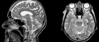
Normally, criblures are so small that they are not visible on an MRI image. However, there are cases when examination reveals expanded perivascular spaces. What does this diagnostic result mean? This suggests that criblures are visualized during an MRI examination. They look like white spots in the photo.
Differential diagnosis
Lacunar infarctions
Lacunar infarcts are small focal strokes located in the deeper parts of the brain and brain stem. They are caused by obstruction of perforating arteries that arise from the middle cerebral artery, posterior cerebral artery, basilar artery, and less commonly from the anterior cerebral artery or vertebral arteries.
Cystic periventricular leukomalacia
Perientricular leukomalacia, commonly seen in premature infants, is a leukoencephalopathy caused by prenatal or intrapartum hypoxic-ischemic brain injury.
Multiple sclerosis (MS)
MS lesions can be located anywhere in the central nervous system. Lesions in the periventricular and juccarticular white matter correspond to the location of type II VR spaces.
Cryptococcosis
Cryptococcosis is an opportunistic fungal infection caused by Cryptococcus neoformans that affects the central nervous system of patients with human immunodeficiency virus (HIV).
Mucopolysaccharidoses
Mucopolysaccharidoses are inherited metabolic disorders characterized by enzyme deficiency and failure to break down glycosaminoglycan, resulting in the accumulation of a toxic intracellular substrate. Clinical features include mental and motor retardation, macrocephaly, and musculoskeletal deformities. Urinary glycosaminoglycan levels are elevated. Brain atrophy and white matter abnormalities occur.
Cystic neoplasms
Giant dilated VR spaces may cause mass effect and suggest an eccentric location, which may be incorrectly identified as a cystic brain tumor [1]. However, cystic brain tumors often have solid components, enhance with contrast media, and exhibit perifocal edema in most cases.
Neurocysticercosis
Cysticercosis is the most common parasitic infection of the central nervous system, caused by the larval stage of Taenia solia. Fluid oval cysts with an internal scolex (cysticerci) can be located in the brain parenchyma (gray and white matter, but also in the basal ganglia, cerebellum and brainstem), subarachnoid space, ventricles or spinal cord. MR imaging findings for neurocysticercosis vary depending on the stage of infection. Lesions can be observed at different stages in the same patient.
Arachnoid cysts
Arachnoid cysts are intra-arachnoid cerebrospinal fluid-containing cysts that are not connected to the ventricular system.
Neuroepithelial cysts
Neuroepithelial cysts are rare and benign lesions that are mostly asymptomatic. Their etiology is controversial, but the underlying developmental anomalies are undeniable. The lesions are spherical, up to several centimeters in size, and may have a mass effect. They are lined with thin epithelium and have a cerebrospinal fluid signal. Neuroepithelial cysts may occur in the lateral or fourth ventricles, with which they do not communicate (intraventricular cysts). They can also be found within the cerebral hemispheres, thalamus, midbrain, pons, cerebellar vermis and medial temporal lobe [1]. Neuroepithelial cysts are not contrasted [1]. Differentiation between neuroepithelial cysts and dilated VR spaces can only be confidently made by pathological examination.
Causes
Enlarged perivascular Robin-Virchow spaces are not always a sign of pathology. This diagnostic result is also observed in completely healthy people. Most often, expansion of criblures is observed in elderly patients and is associated with age-related changes in the brain.
However, in some cases, dilated perivascular spaces may be a sign of the following diseases and conditions:
- brain atrophy;
- leukoaraiosis;
- cerebral ischemia (including cerebral infarction);
- disseminated encephalomyelitis.
In elderly people, expansion of criblures is often observed with hypertension, atherosclerosis, and dementia. These pathologies are usually accompanied by memory impairment and other cognitive impairment.
Additional diagnostic methods
What to do if the MRI results indicate that you have dilated perivascular Virchow-Robin spaces? It is necessary to show the transcript of the study to a neurologist. Only a specialist can determine whether this is a variant of the norm, an age-related feature, or a sign of pathology.
There are cases when MRI does not reveal any changes in the brain, but the image shows dilated perivascular Virchow-Robin spaces. What does this mean? As a rule, such a sign does not indicate pathology. Doctors consider an increase in criblures only in combination with other changes identified during an MRI examination.
If necessary, the doctor may prescribe additional tests:
- multislice computed tomography;
- vascular angiography;
- Dopplerography;
- cerebrospinal fluid examination.
Let's take a closer look at the most common diseases and conditions that can lead to expansion of criblures.
Perivascular zone, its structure and functions
Virchow-Robin spaces (VRO) are slit-like formations surrounding the cerebral vessel along its path from the subarachnoid region into the brain tissue.
There is still no single point of view on what perivascular spaces are. Most scientists believe that this is the area between the middle (arachnoid, arachnoid) and inner (pia) meninges. This area normally surrounds the substance of the brain and, together with the vessels, is drawn into the brain, surrounding them. The subarachnoid region, located in the cerebral cortex, is called the Virchow-Robin space.
There is no single point of view; only arteries or also veins surround the perivascular spaces. It was revealed that they continue to the level of capillaries.
It is believed that this formation is involved in the movement of cerebrospinal fluid and ensures the exchange of necessary substances between cerebrospinal fluid and neurons.
Another function is to isolate intravascular blood from brain tissue. The blood contains a number of toxic substances that normally should not enter the brain substrate due to the presence of the blood-brain barrier. Additionally, toxins are absorbed within the perivascular region.
Another challenge is immune regulation.
Brain atrophy
If the patient’s perivascular spaces are expanded and the volume of the brain is reduced, then doctors talk about organ atrophy. Most often this is a sign of the following diseases:
- senile dementia;
- atherosclerosis;
- Alzheimer's disease.
In these diseases, neurons die. This is accompanied by memory impairment, mental impairment, and mental disorders. Typically, such diseases occur in elderly patients.
In some cases, dilated perivascular Virchow-Robin spaces are detected in newborns. This may be a sign of serious genetic diseases accompanied by the death of neurons.
How to treat such pathologies? After all, it is no longer possible to restore lost neurons. You can only slow down the process of death of nerve cells. Patients are prescribed the following drugs for symptomatic therapy:
- nootropics: Piracetam, Cavinton, Nootropil;
- sedatives: “Phenazepam”, “Phenibut”;
- antidepressants: Valdoxan, Amitriptyline.
The prognosis of such pathologies is usually unfavorable, as brain atrophy and neuronal death progress.
When expansion of perivascular spaces is normal
Perivascular canals can only be seen using MRI.
Often Virchow-Robin spaces are not visualized even on MRI images due to their small area. The resolution of the tomograph matters. A size of up to 2 mm is normal and occurs in all people.
Enlarged perivascular spaces are called criblures.
Signs of dilated perivascular spaces on MRI
Their increase does not always indicate pathology. The mechanism of their expansion is still being studied. This is possible due to inflammation of the vessel wall, when the latter becomes thinner and becomes more permeable. The released liquid causes the criblure to expand. Another reason is a violation of the flow of cerebrospinal fluid, and another is the lengthening of blood vessels.
Scientists and practicing doctors have not come to a consensus on what is considered pathology and what is not. As a rule, fixation of spaces on MRI images in people of the older age group is a variant of the norm.
Criblures in the brain are often found in children in the first year of life.
They are usually localized in three areas:
- Along the course of the lenticular arteries supplying blood to the basal ganglia - the caudate nucleus, internal capsule, fence.
- Along the course of certain arteries that enter the brain from its outside , and not like most branches of the carotid and vertebral arteries from the inside.
- Along the vessels supplying the midbrain (posterior thalamoperforating and median mesencephalothalamic arteries).
Appear symmetrically. Most often, expansion of the perivascular spaces occurs in the region of the lower basal structures and very rarely in the cerebellum. As a rule, the dimensions do not exceed 5 mm.
CSF flows in the perivascular canals, so on MRI the criblures have the same plane as the latter and appear isodense.
There are 2 projections in which MRI images of the brain are usually taken: frontal and axial. In the first case, the expanded spaces appear in the form of stripes, and in the second they take on a round or oval shape corresponding to the cross-section.
The use of various MRI modes, especially T-2, helps in diagnosis. In this mode, Virchow-Robin spaces do not have a darker rim around the filled area, which indicates that this is part of the subarachnoid membrane, and not the wall of a cavity, lesion or neoplasm.
Criblurs on MRI images
Leukoaraiosis
Doctors call leukoaraiosis a thinning of the white matter of the brain. Due to structural changes in the nervous tissue, patients have enlarged perivascular spaces. This is also a sign of diseases characteristic of older people:
- hypertension;
- atherosclerosis;
- senile dementia.
Changes in the white matter of the brain cause cognitive impairment. Patients are treated symptomatically with nootropic drugs. These drugs improve the nutrition of neurons and stop their death. For atherosclerosis, statins are indicated. For high blood pressure, antihypertensive drugs are prescribed.
Expanded spaces in pathology
The localization and intensity of the MRI signal will help to distinguish pathology or age-related norms.
If criblure is visualized in an atypical place and there is an asymmetrical picture, then most likely we are talking about a disease.
Several reasons can lead to true expansion.
Psychiatric diseases
This connection is still being studied
Cryptococcosis
This is a fungal disease that occurs in people with immunodeficiency. Most often occurs in HIV-infected people. In this case, fungal spores can be localized inside the perivascular spaces, causing their expansion. Such accumulations are called gelatinous pseudocysts. Their difference from normal dilated spaces is the preservation of hyperdensity in FLAIR mode.
An MRI picture of concomitant meningitis, hydrocephalus and the presence of foci of cryptococcosis in the brain substance also helps to identify the disease.
Treatment is carried out with antifungal drugs.
Foci of cryptococcosis on MRI
Mucopolysaccharidosis
This is an inborn metabolic disease in which the decomposition of glycosaminoglycans is impaired. Excess substances accumulate, forming criblures. In the pictures they look like latticework. Hyperintense white matter is also visualized, which helps distinguish pathology from normality.
Since the disease is based on enzyme deficiency, the goal of therapy is their synthetic replacement: Aldurazyme, Elaprase.
Ischemic conditions
With ischemia, the blood supply to the brain deteriorates. This is usually a consequence of atherosclerotic changes in the vessels. The patient periodically experiences dizziness, double vision, motor coordination disorders, speech and memory disorders. Due to changes in the vessels, the spaces around their walls also expand.
Patients are prescribed nootropic drugs (Piracetam, Cerebrolysin, Actovegin), as well as drugs that normalize metabolism in brain cells (Cortexin, Ceraxon). In this case, it is very important to carry out etiotropic treatment of atherosclerosis with statins. The drugs Lovastatin, Atorvastatin, and Simvastatin are prescribed. This therapy eliminates the cause of ischemia.
Brain infarction
Often the perivascular spaces are dilated in patients who have suffered a cerebral infarction. This disease is a consequence of prolonged ischemia. In some cases, cerebral infarction is asymptomatic and goes unnoticed by the patient. Its consequences can only be seen in an MRI scan.
It is important to remember that if the patient has risk factors (high blood pressure, atherosclerosis, diabetes), then the heart attack may recur in severe form. To prevent a recurrent attack of acute ischemia, patients are prescribed antihypertensive medications, hypoglycemic agents, and blood thinners.
Interesting Facts
The perivascular spaces are named after the two scientists. However, as often happens, the area was first discovered by another person. This was done in 1843 by Durand Fardel.
Only 10 years later, the German scientist Rudolf Virchow described in detail the structure of this area. This fact is surprising, given that a conventional microscope was used for the study.
A few years later, his French colleague clarified that this is not just a gap, but a channel inside which a cerebral vessel passes.
Disseminated encephalomyelitis
Disseminated encephalomyelitis (DEM) is an acute pathology of the central nervous system. In this disease, the myelin sheath of nerve fibers is destroyed. The perivascular Virchow-Robin spaces are dilated due to damage to the white and gray matter. The MRI image shows foci of demyelination.
This pathology is of autoimmune origin. The clinical picture of the disease resembles signs of multiple sclerosis. Patients experience gait and movement disturbances, speech disorders, dizziness, and inflammation of the optic nerve.
Unlike many other demyelinating diseases, REM is treatable. Patients are prescribed corticosteroids to suppress the autoimmune reaction:
- "Prednisolone";
- "Dexamethasone";
- "Metypred."
After a course of therapy, 70% of patients experience complete recovery. In advanced cases, patients may still experience the consequences of the disease: impaired sensitivity of the limbs, gait disturbances, and visual disturbances.
Virchow-Robin spaces
Virchow-Robin spaces are slit-like spaces surrounding the vessels of the brain and spinal cord, throughout their entire course from the subarachnoid space through the brain parenchyma.
Localization
Virchow-Robin spaces are most often located along the lenticulostrial arteries, directly above the anterior perforated plate and adjacent to the anterior commissure. Less commonly, they are located along the arteries that penetrate the gray matter of the brain, sometimes penetrating deep into the white matter. Cases of localization of Virchow-Robin spaces in the projection of the subinsular region, in the dentate nucleus and hemispheres of the cerebellum, in the midbrain, corpus callosum and cingulate gyrus have been described.
Etiology
The etiology of their formation remains not fully understood. According to various authors, they can occur in arterial hypertension, in patients with migraine, episyndrome, parkinsonism and some storage diseases (mucopolysaccharidosis). In most cases, they are an incidental finding during a survey MRI of the brain.
Prevalence
It is believed that expanded Virchow-Robin spaces can occur in all age groups; in 25-30% of children they can be identified on MRI. The most typical dimensions do not exceed 5 mm. Sometimes there is a pronounced expansion of the Virchow-Robin spaces. Moreover, the latter are usually diagnosed in adult patients in the projection of the midbrain.
Diagnostics
In general, the signal intensity of the perivascular spaces matches that of the CSF on all sequences. The difference in signal intensity may be explained by partial volume effects, since the Virchow-Robin space with the vessel is smaller than the voxel volume of the MR images. Virchow-Robin spaces are not characterized by diffusion limitation on DVI, since they are communicating compartments. On T1WI with high flow sensitivity, Virchow-Robin spaces may have high signal intensity due to slice inflow effects, thereby confirming that they are Virchow-Robin spaces. Perivascular spaces do not accumulate contrast agent. In the case of moderate expansion of the Virchow-Robin spaces (2-5 mm), the surrounding brain parenchyma has normal signal intensity.
Differential diagnosis
- Lacunar infarction
- Cystic periventricular leukomalacia
- Multiple sclerosis
- Cryptococcosis
- Mucopolysaccharidosis
- Cystic neoplasms
- Neurocysticercosis
- Arachnoid cysts
- Neuroepithelial cysts
Etymology
Virchow-Robin spaces are named after the researchers who discovered them - Rudolf Virchow (German pathologist, 1821-1902) and Charles-Philippe Robin (French anatomist, 1821-1885).
Synonyms
- Robin-Virchow spaces
- His-Robin perivascular spaces
- spatia perivascularia
- surrounding vascular spaces
- intraadventitial spaces
- criblures
Prevention
How to prevent the above pathologies? It can be concluded that older patients are more susceptible to such diseases. Therefore, all people over 60 years of age need to regularly visit a neurologist and undergo an MRI examination of the brain.
It is also important to constantly monitor blood cholesterol levels and blood pressure. After all, diseases accompanied by pathological changes in the white matter most often develop against the background of atherosclerosis and hypertension.
Radiation diagnostics
The imaging characteristics of extended PVPs are fairly well described in the literature. These are, as a rule, round or oval formations with clear external and internal contours, located along the perforating arteries of the brain. PVP are identical in their radiological characteristics (according to CT and MRI) with the cerebrospinal fluid in the ventricles of the brain and SAP. There are no signs of contrast enhancement, no calcium deposits in the cyst walls, and no perifocal cerebral edema. On T2-weighted MRI and T2-Flair 25%, a slight increase in MR signal from the adjacent brain substances can be visualized around the enlarged VVP. At gigantic sizes, the mass effect on surrounding structures is determined. Since the most common location in this case is the midbrain, compression of the cerebrospinal fluid spaces and the cerebral aqueduct may be observed.
Rice. 1. Perivascular spaces of the brain. MRI in T2 (a) and T1 (b, c) modes reveals multiple small foci of changes in the MR signal in the white matter of the brain according to signal characteristics isointense to the cerebrospinal fluid. An area of increased MR signal in T2 mode is visualized around individual perivascular spaces (arrows).
Rice. 2. Perivascular spaces of the brain in the projection of the midbrain. On a series of MRI scans in T2 (a), T1 (b) and Flair (c) modes, multiple cystic formations are revealed in the projection of the right midbrain peduncle, merging into one large cyst. The MR signal from the cysts is identical to the cerebrospinal fluid, including the myelography mode (e). There is no contrast enhancement within the cystic cavities and in the surrounding brain matter (d, e).


