Such a disease as epileptic seizures has been familiar to humanity since ancient times. This disease stood alongside leprosy, since it was little studied and practically could not be treated. But today, the science of neurophysiology has developed so much that it allows epilepsy to be diagnosed and successfully treated in the early stages.
Causes of epilepsy in children
Before starting treatment, the doctor first identifies the causes of epilepsy in children. Today there are several main factors.
- Hereditary disease. A special substance, dopamine, the level of which is programmed in genes, is responsible for stopping overly excited neurons. Accordingly, if the parents of a sick child have ever suffered or are currently suffering from epilepsy, then it is likely that he received this pathology from them.
- Intrauterine brain malformation. In most cases, such causes of epilepsy in children arise due to their mother’s abuse of alcohol, cigarettes, and medications prohibited for use during pregnancy. Also, these defects can occur in primiparous, middle-aged women, the state of their health during pregnancy.
- Injuries received during childbirth. Seizures of epilepsy in children can occur due to injuries received from the midwife's forceps, the procedure for vacuum extraction of the baby, prolonged labor, or compression of the baby's neck by the umbilical cord.
- Inflammatory brain diseases. This includes meningitis, encephalitis and others.
- Increased body temperature due to a cold or viral disease. As a rule, seizures for this reason appear in children with not very healthy heredity.
In addition to the above, the following factors may also be observed:
- traumatic brain injuries. The cause of epilepsy in children who have received serious blows to the head is quite common;
- tumor neoplasms. The grown tumor puts serious pressure on the child’s fragile brain, which provokes the appearance of epileptic seizures;
- disruptions in metabolism in the body. This should include hypoglycemia, hypocalcemia and others;
- problems with cerebral blood flow;
- abuse of narcotic and serious medications. This reason is most relevant for children experiencing adolescence.
Benign myoclonic epilepsy of infancy
DMEM is diagnosed between 6 months and 3 years of age. The child's development is normal until the first myoclonic seizures appear. Approximately 20% of children present with rare seizures at birth or during the neonatal period. The patient's general condition rarely suffers; neurological disorders are not detected. The first myoclonic attacks affect the upper limbs, neck and head, and rarely the legs. They can have different intensities, including in the same child during different episodes. Severity ranges from barely noticeable twitching to visible fibrillation.
The frequency of seizures is 2-3 times a day at different time intervals. There are no long series of attacks observed. It is possible to provoke an attack with a loud sound, tactile or rhythmic light stimulation. After each episode, there may be a refractory period lasting from 20 to 120 seconds. In this time period, even intense stimulation does not cause a new attack. In this case, muscle atony is often observed. The disease is characterized by an intensification of myoclonic attacks when falling asleep (drowsiness) and their disappearance in the slow-wave sleep phase.
There are reflexive and spontaneous variants of DMEM. In the first case, myoclonic seizures develop after exposure to triggers. The spontaneous form occurs without any predictors. In the early stages of the disease and when the intensity of myoclonus is low, parents and pediatricians may mistake attacks for normal motor reactions of the child. Relatively severe myoclonic attacks can be accompanied by tilting the head forward, abductor and adduction movements, bending of the arms, and rarely by smooth rotation of the eyeballs. Often parents note a characteristic “nod” of the head lasting from 1 to 3 seconds, rarely – up to 10 seconds. (in older children). In some cases, the only clinical manifestation of DMEM is prolonged closing of the eyes.
In severe forms, generalization of convulsions is possible, accompanied by loss of balance, sudden loss of objects from hands, and rarely, disorders of consciousness. The process sometimes involves the intercostal muscles, the anterior abdominal wall and the diaphragm, which causes breathing disturbances and an expiratory noise may be heard. DMEM is characterized by an increase in the intensity of clinical manifestations up to a certain age and their subsequent complete disappearance. With a long course of the disease, a lag in psychomotor development is possible. Transformation into other forms of seizures, including absence seizures, does not occur even in the absence of specific treatment.
Symptoms of epilepsy in children
The signs of epilepsy in children are generally similar, but may differ depending on the age at which the child is at this stage.
Manifestation of epilepsy in infants
It is extremely important to detect and begin timely treatment of the first signs of epilepsy in a child of the first year of life: at this stage, while all processes are maturing, they can be adjusted as easily as possible. However, this also has its own difficulty: the signs of epilepsy in children under one year of age are very different from those in older age.
Symptoms include the following:
- sudden freezing without movement;
- suspension of swallowing;
- tilting the head back;
- eyelid tremor;
- a gaze directed into emptiness, not noticing anything or anyone;
- lack of response to the actions of parents.
In most cases, the listed symptoms begin an attack, after which the baby loses consciousness and convulsions appear. In the process, he may unknowingly perform an act of defecation or void himself.
Symptoms of epilepsy in children after one year
Epilepsy in children after one year manifests itself a little differently:
- temporary loss of consciousness;
- motor activity disorders;
- problems with sensory perception;
- a sharp change in mood, especially towards increased aggression;
- numbness of some parts of the body.
These signs of epilepsy in children are not always noticeable, but should alert parents. If you do nothing, then the following will appear after them:
- brief cessation of breathing;
- strong tone of all muscles at the same time;
- convulsions;
- uncontrolled urination, defecation;
- often biting the tongue, screaming.
Epilepsy in children is often accompanied by chronic depression and moral depression.
Forms of epilepsy in children
Depending on what triggered epilepsy attacks in children, there are three main forms:
- idiopathic: manifestations of the disease do not entail significant changes in brain function and have a genetic basis;
- symptomatic: arose as a result of brain pathology caused by a tumor, injury or malformation;
- cryptogenic: the exact cause of epilepsy in children is not clear.
Types of epilepsy
When diagnosing epilepsy in children, the location of the source of the disease is also taken into account. Depending on this, the disease is divided into several types:
- occipital;
- parietal;
- frontal (activated at night);
- temporal (loss of consciousness without characteristic convulsions);
- chronic.
There is another classification based on what caused epilepsy in children under one year of age and later:
- primary: appeared due to excessive brain activity;
- secondary: caused by injury or infectious disease;
- reflex: any stimulus can act as a provocateur, for example, a flashing lamp, any sound.
Epilepsy in childhood
Part 3. Read the beginning of the article in No. 6, 8, 2014
There are many forms of epilepsy that occur exclusively in childhood or adolescence. It is the age dependence of many types of epilepsy that is the main distinguishing feature of childhood epileptology [1–4].
Epilepsy and convulsive syndromes of the newborn period
Although the duration of the neonatal period is short, a number of epileptic syndromes are characteristic of newborns [3–5].
Benign familial seizures (convulsions) of newborns
Benign neonatal epilepsy (with an autosomal dominant type of inheritance) of three types, manifesting itself in the first 7 days of life (starting from three days). The family history necessarily includes indications of the presence of seizures in the past among members of the patient’s family (in the neonatal period). The relationship between seizures and specified inborn errors of metabolism has not been established. Benign familial neonatal seizures manifest as focal and multifocal or generalized tonic-clonic (convulsive) seizures. These seizures are characterized by short duration (1–2 minutes) and significant frequency (20–30 episodes per day). Subsequently, after 1 to 3 weeks, the attacks spontaneously stop spontaneously.
Benign nonfamilial convulsions (seizures) of newborns (“fifth day seizures”)
This epilepsy with onset in the early neonatal period also has another name (benign idiopathic neonatal seizures). The disease was first described in the late 1970s. Convulsive seizures develop in full-term newborns who previously had no signs of pathology in the central nervous system. The onset of attacks occurs by the end of the 1st week of life (in 80–90% of cases - between the 4th and 6th days), and their peak occurs on the 5th day of life (hence the name). The described seizures usually take the form of multifocal clonic seizures, which are often accompanied by apnea. In most cases, benign idiopathic neonatal seizures last no more than 24 hours (they always stop after 15 days after onset). In 80% of cases, during the convulsive period, newborns develop status epilepticus [3–5].
Early infantile epileptic encephalopathy with suppression/flare pattern on EEG (Otahara syndrome)
Early infantile epileptic encephalopathy is a rare disease related to malignant forms of childhood epilepsy. It usually debuts in the neonatal period (or at the age of 1–3 months). The disease is characterized by tonic attacks, the frequency of which varies significantly (10–300 episodes per day). Children experience rapid formation of neurological deficits and mental retardation. A specific burst/suppression pattern on electroencephalography (EEG) is present in children with Ohtahara syndrome both during sleep and while awake. Magnetic resonance imaging (MRI) of the brain reveals gross anomalies in the development of the central nervous system in patients. Among children with early infantile epileptic encephalopathy with a burst/suppression pattern on the EEG, mortality by the age of 1 year reaches 40–50%. At 4–6 months of age, Ohtahara syndrome can transform into West syndrome [3–6].
Early myoclonic (epileptic) encephalopathy
Described by J. Aicardi and F. Goutières (1978); debuts mainly in the neonatal period (sometimes before 3 months of age). In the genesis of the disease, the role of genetic factors and some “inborn errors of metabolism” (propionic aciduria, methylmalonic acidemia, urine disease with the smell of maple syrup, etc.) is assumed. Clinically manifested by frequent myoclonic seizures. The latter are usually not associated with EEG changes during an attack, but in some cases epileptiform “suppression/flare” discharges are recorded simultaneously with myoclonus. Myoclonus is often fragmentary (slight twitching of the distal limbs, eyelids, or corners of the mouth); At the same time, focal (partial) seizures, massive myoclonus and tonic spasms (isolated or serial - occur by 3-4 months) can be observed. The appearance of tonic spasms in a child suggests the presence of atypical West syndrome, but soon the main manifestations of the disease resume and persist for a long time. Focal seizures (complex partial - with eye rolling or autonomic symptoms: apnea, facial hyperemia; clonic convulsions in different parts of the body, etc.) become the main type of seizures in early myoclonic epileptic encephalopathy. During an interictal EEG study in children, a “suppression/flare” pattern is recorded, consisting of discharges lasting 1–5 seconds, alternating with almost isoelectric periods (lasting 3–10 seconds). The described EEG pattern becomes more distinct during sleep (especially in the deep sleep phase). The initial suppression/flare pattern is replaced by atypical hypsarrhythmia or multifocal paroxysms upon reaching the age of 3–5 months, but in most cases this is only a transient phenomenon. The disease is accompanied by high mortality or progressive decay of psychomotor functions (up to vegetative status), although as age increases, the frequency and severity of focal seizures and myoclonus gradually decrease [3–5, 7].
Vitamin B6-dependent epilepsy
A relatively rare hereditary disease characterized by drug-resistant seizures. Belongs to the group of metabolically caused epilepsies. Develops in newborns whose mothers received pyridoxine for a long time during pregnancy, as well as with a specific hereditary metabolic defect (with an increased need for vitamin B6). There are known cases of the onset of pyridoxine-dependent seizures in children older than 1 month and even in the second year of life. Between attacks of convulsions, children remain restless and react with muscle twitching to external irritations. The disease does not respond to conventional anticonvulsant treatment, but the administration of vitamin B6 in high doses (25 mg/kg/day) quickly leads to normalization of the condition [3–5].
Malignant migratory partial convulsions (seizures) of infancy
An extremely rare epileptic syndrome described by G. Coppola et al. (1995). To date, only approximately 50 cases of the disease have been reported from around the world. Malignant migratory partial seizures are observed in the first days of life in 50% of cases; the remaining 50% occurs between 1 and 3 months of age. At the onset, seizures are focally clonic in nature, and after several weeks they become multifocal, extremely frequent and pharmacoresistant to treatment with antiepileptic drugs. An EEG study in children reveals pronounced multifocal epileptic activity; no metabolic disorders are detected, and there are no MRI signs of pathological changes. Postmortem examination revealed signs of neuronal loss in the hippocampus [1, 3, 5, 8].
Epilepsy in children of the first year of life (1–12 months)
Upon reaching 1 month of age, the number of types of epileptic syndromes specific to the first year of a child’s life is practically not inferior to that characteristic of the newborn period.
Infantile spasms (West syndrome)
This variant of catastrophic epilepsy (generalized) can be symptomatic (the vast majority of cases) or cryptogenic (10–20%). It manifests itself in children in the first year of life (usually between 3 and 8 months). In the classic version, West syndrome is characterized at the time of an attack by a combination of flexion and extension movements, that is, pronounced myoclonic (salaam) spasms, sometimes serial short flexion movements of the head (“nodding”). Infantile spasms can develop both due to the presence of various neurological pathologies, and without any obvious previous disorders of the central nervous system. With infantile spasms, psychomotor development slows down, and in the future there is a high probability of severe developmental delay. In 80% of cases with West syndrome, microcephaly, signs of cerebral palsy, atonic-atactic disorders, etc. are found. A distinctive electrophysiological sign of West syndrome is hypsarrhythmia (according to EEG), which has the form of diffuse high-voltage peaks and slow waves located on a disorganized (slow) ) background. The prognosis of West syndrome is determined by the effectiveness of the therapy, but in general it is unfavorable [3–8].
Severe myoclonus epilepsy of infancy (Dravet syndrome)
The disease, described by C. Dravet (1978, 1992), debuts in the first year of life (between 2 and 9 months), which often follows the development of a febrile episode, soon after vaccination or infection. Dravet syndrome is characterized by the appearance of generalized or unilateral clonic seizures (usually associated with hyperthermia or fever), which occurs against the background of previous normal psychomotor development of the child during the first year of life. Gradually (over several weeks or months) the child develops afebrile myoclonic and partial (focal) seizures. A progressive increase in the frequency of myoclonus (isolated or serial) precedes the appearance of generalized seizures in patients. Children exhibit moderate cerebellar and pyramidal signs associated with gross motor deficits and gait ataxia. Psychomotor development disorders are subsequently observed in children up to approximately 4 years of age. Often, with Dravet syndrome, children develop status epilepticus (convulsive or myoclonic). EEG data during the first year of life usually remain within normal limits, although spontaneous photoinduced peak-wave discharges occur in some patients. Subsequently, ictal EEG studies in Dravet syndrome are characterized by the presence of myoclonic or clonic seizures (generalized spike-wave or polyspike-wave activity). Generalized discharges intensify in a state of relaxation; focal and multifocal peaks and sharp waves are simultaneously observed. Traditional and newer antiepileptic drugs generally do not prevent seizure recurrence in Dravet syndrome. The prognosis for intellectual development in Dravet syndrome is always unfavorable [3–5, 8].
Idiopathic benign partial epilepsies of infancy
They usually debut in children aged 3–20 months (usually between 5 and 8 months). First described by K. Watanabe et al. (1987), as a result of which they initially received the general name “Watanabe syndrome”. They are characterized by manifestations in the form of complex partial (focal) seizures and a favorable prognosis (elimination of epileptic seizures within 3 months after the onset). The average number of attacks is about 7; Some patients have exclusively complex partial seizures, others have only secondary generalized seizures, and in about half of the cases there is a combination of both. During an attack, patients are characterized by a decreased response to presented stimuli, cessation of motor activity, moderate convulsive twitching, lateral opening of the eyes and cyanosis. The main clinical signs of this group of epilepsies are the high incidence of cluster seizures, short duration of seizures, as well as initially normal interictal EEG studies (subsequently paroxysmal discharges may be detected in some children) [2, 3, 5, 6, 8].
Similar to idiopathic benign partial epilepsies of infancy, but exclusively familial paroxysmal conditions with onset in the first year of life are called “benign infantile familial seizures.” In 1997, similar cases of familial epilepsy with the subsequent formation of choreoathetosis—familial seizures with choreathetosis—were described [3–5, 8, 9].
Epilepsy in young children (1–3 years)
Young children (from 12 to 36 months) are primarily characterized by Doose syndrome, Lennox–Gastaut syndrome, benign myoclonus epilepsy of infancy, hemiconvulsions-hemiplegia syndrome, idiopathic partial epilepsy of infancy, absence epilepsy of early childhood, electrical status epilepticus. slow wave sleep, early and late childhood neuronal lipofuscinosis (types I and II).
Myoclonic astatic epilepsy of early childhood (Doose syndrome)
It is epilepsy with myoclonic-astatic seizures (of varying duration). Seizures begin between 1 and 5 years of age. More often the disease affects boys. Astatic and myoclonic seizures can be combined, with myoclonus occurring both before, during, and after an astatic seizure. The attacks come suddenly and are almost always accompanied by falls. Myoclonus is noted in the form of varying severity of symmetrical twitching in the arms and muscles of the shoulders, which is combined with a tilt of the head (“nods”). There are no signs of loss of consciousness in children at the time of the attack. Before the onset of the disease, the psychomotor development of children is usually normal. In some children, the disease is complicated by the risk of developing dementia (presumably due to the development of absence seizure status epilepticus). EEG records generalized bilaterally synchronous peak-wave complexes (3 or more per 1 second, 2–4 Hz). The prognosis for myoclonic-astatic epilepsy of early childhood is unfavorable [3–6, 8].
Lennox–Gastaut syndrome, or myokinetic epilepsy of early childhood with slow peak waves
A group of heterogeneous pathology with epileptic seizures (atonic, tonic, atypical absences), intellectual deficiency and a characteristic EEG pattern. As with West syndrome, with Lennox–Gastaut syndrome, symptomatic and cryptogenic variants of the disease are distinguished. Early forms debut at about 2 years of age. Up to 30% of cases are the result of transformation from West syndrome. Clinically, Lennox–Gastaut syndrome is characterized by myoclonic-astatic seizures, Salaam seizures (lightning-fast nodding seizures), atypical absence seizures, and tonic seizures (usually during sleep). Generalized tonic-clonic, myoclonic and focal (partial) seizures may occur. For children, a series of seizures with changes in consciousness (stupor) and a gradual transition to status epilepticus is typical. In addition to epileptic seizures, the neurological status may include cerebral paresis/paralysis, as well as atonic-astatic disorders (up to 40% of patients). In children, there is a decrease in intelligence (of varying degrees), and pronounced impairments in cognitive functions are observed. According to EEG data, changes in background activity are typical in the form of slow peak-waves < 3 Hz, night series of peaks (hypsar phenomena). Multifocal changes are often present. Neuroimaging methods can identify focal and diffuse structural disorders. Up to 75–80% of cases of the disease are resistant to therapy. The prognosis is variable, but is generally considered unfavorable [3–6, 8, 9].
Benign myoclonus epilepsy of infancy
It debuts in children under 3 years of age (usually at 1–2 years), although it can sometimes manifest itself from the age of 4–11 months. Before the onset of the disease, there are usually no previous signs of disturbances in psychomotor development. Refers to generalized/idiopathic or symptomatic epilepsies. It differs from Dravet syndrome in less severity and a more favorable prognosis. Benign myoclonus epilepsy of infancy is characterized by the appearance of short-term myoclonic seizures involving the head and upper limbs. Other types of seizures do not occur in this form of epilepsy, and the frequency and intensity of the existing paroxysms is variable. Moderate intellectual development delay is possible. The diagnosis is made based on the clinical characteristics of the attacks, as well as EEG data. An ictal EEG study reveals generalized epileptic activity with irregular peaks, peak waves, sharp waves (more often asymmetric than bilaterally synchronous); Interictally, EEG data are either not changed or moderately disturbed (sharp waves, peaks, peak-wave complexes, sharp/slow waves - with a predominance in the early stages of sleep) [3–6, 8].
Hemiconvulsion-hemiplegia syndrome (HHS)
The disease most often debuts at an early age (1–3 years), although the onset of the disease is possible between the 1st and 4th year of life (starting at approximately 5 months). The first manifestations are suddenly developed prolonged hemiconvulsions in the form of clonic convulsions (status), which are sometimes (not in all cases) associated with hyperthermia (febrile convulsions). After a convulsive episode, children experience hemiplegia. After 12–36 months, patients develop focal (partial) epilepsy. EEG in children shows peak and polypeak slow wave activity with a frequency of 10–12 Hz (mainly in the occipital leads). In some cases, hemiconvulsions-hemiplegia syndrome is considered as an atypical consequence of prolonged febrile seizures in infancy and/or early childhood [3–5].
Benign partial epilepsy of infancy and early childhood with vertex peaks and waves during sleep
Identified by Italian epileptologists G. Capovilla and F. Beccaria (2000), in contrast to benign partial epilepsy of infancy, described by K. Watanabe et al. (1987). It debuts at 13–30 months, EEG changes (characteristic peaks and waves during slow-wave sleep with localization in the vertex regions of the brain) are observed only during sleep (EEG upon awakening always does not show epileptiform changes), the clinical picture of attacks also has some differences . The prognosis for benign partial epilepsy of infancy and early childhood with vertex peaks and waves during sleep is favorable [3, 5, 8, 9].
Absence epilepsy of early childhood
This epileptic syndrome, the name of which was proposed by H. Doose et al. (1965), includes a heterogeneous group of patients. Absence epilepsy of early childhood is clinically characterized by generalized clonic-tonic seizures and/or myoclonic-astatic seizures, the presence of irregular peak-wave discharges in the EEG (2–3 Hz), and often an unfavorable prognosis. Absence epilepsy of early childhood sometimes debuts at a later age—before 5 years [3–6, 8].
Eyelid myoclonus with absence seizures (Jealons syndrome)
Age-dependent epileptic syndrome, which is a form of photosensitivity epilepsy, was described by PM Jeavons (1977). May present earlier than childhood absence epilepsy (2–5 years), although it is most common between 6–8 years of age. Slightly more common in girls. Most patients have a family history indicating the presence of idiopathic generalized epilepsy. J. Guiwer et al. (2003) emphasize that Jeavons syndrome is a myoclonic rather than an absence syndrome. Although Jeavons syndrome refers to idiopathic generalized epilepsy, it is possible that this concept includes a whole group of photosensitivity conditions. The main symptom of Jeavons syndrome is myoclonus of the eyelids, and the main trigger factor of the disease is closing the eyes in the presence of light. Absence seizures with Jeavons syndrome are not always observed; they have a short duration (3–6 seconds each), appear after closing the eyes and are accompanied by a clear rhythmic pattern of eyelid myoclonus, as well as retropulsion of the eyeballs in association with the tonic component of the muscles involved in the pathological process. Absence seizures in Jeavons syndrome do not occur without eyelid myoclonus. In most cases, children experience generalized tonic-clonic seizures, but the frequency of these seizures is relatively low. During EEG, electrophysiological paroxysms associated with eye closure and photosensitivity are recorded in patients. Impairments of consciousness in Jeavons syndrome are not as pronounced as in childhood absence epilepsy or juvenile absence epilepsy. Intellectual development in myoclonus of the eyelids with absences is practically not affected, although in some cases mild or moderate intellectual deficit is reported. The diagnosis is easily confirmed using video-EEG, since it reveals the association of generalized paroxysmal activity with eye closure. In general, the prognosis for Jeavons syndrome is favorable, although this form of epilepsy is often lifelong. There are known cases of the formation of drug resistance of the disease [3–6, 9].
Early childhood (infantile) neuronal ceroid lipofuscinosis type I (classical)
Representative of the group of progressive myoclonus epilepsies. Associated with deficiency of the enzyme α-neuraminidase (sialidase), also known as “cherry pit syndrome with myoclonus” or “Santavuori-Haltia disease”. May appear from 6 to 24 months of age. The main clinical manifestations of the disease are myoclonus and generalized tonic-clonic seizures, which are subsequently joined by ataxia with gait disorder. The disease is characterized by the presence of a “cherry pit” symptom in the fundus and a progressive decrease in vision (up to blindness). In some cases, the intellectual development of children does not suffer, but in some patients the development of dementia is noted. Using neuroimaging methods, diffuse atrophy of the hemispheres of the cerebral cortex and cerebellum can be detected. Therapy consists of the use of antimyoclonic drugs, nootropics, levocarnitine, vitamins (E, A, group B) [3, 5, 8, 9].
Late childhood (infantile) neuronal ceroid lipofuscinosis type II
Like early childhood neuronal lipofuscinosis, the late form of the disease refers to progressive myoclonus epilepsy. This type of pathology was previously also known as “Jansky-Bielschowsky disease”, or “late childhood amaurotic idiocy”. Late childhood neuronal ceroid lipofuscinosis type II usually appears later than early (type I), from the end of the 1st year of life to 2–3 years of age, although congenital forms of the disease have also been described. Myoclonic seizures and ataxia are typical for the disease. In addition to the described damage to the central nervous system, children also suffer from other internal organs and the musculoskeletal system (sensorineural hearing loss, hernias - inguinal, scrotal, umbilical; hepatosplenomegaly, pathological joint mobility, gradually giving way to limitedness, etc.). An ophthalmological examination reveals the “cherry pit” symptom, as well as clouding of the cornea. Characterized by profound mental disorders. The diagnosis is confirmed by testing neuraminidase activity in fresh fibroblasts and leukocytes. An EEG shows rapid low-voltage activity in patients, although in dementia it slows down. Generalized peak-wave bursts are absent or rare. The treatment principles used for late childhood neuronal lipofuscinosis type II correspond to those for the early form of the disease type I [3, 5, 6, 9].
Read the continuation of the article in the next issue.
V. M. Studenikin, Doctor of Medical Sciences, Professor, Academician of the Russian Academy of Economics
FSBI "NTsZD" RAMS, Moscow
Contact Information
When to see a doctor?
Only an epileptologist knows exactly how epilepsy manifests itself in children, can determine its type and prescribe adequate treatment.
Therefore, if you notice the first sign of epilepsy in a child, immediately contact a competent specialist. The earlier he diagnoses the disease, the faster and easier the therapeutic process can be.
The pediatric department of JSC “Medicine” (clinic of academician Roitberg) employs specialists who have many years of practice behind them and work with the most difficult situations.
The building, equipped with the latest specialized technology, is located in the center of Moscow, at 2nd Tverskoy-Yamskaya lane, building 10. Just a 5-minute walk away are the Mayakovskaya, Tverskaya, Chekhovskaya, and Novoslobodskaya metro stations. . You can make an appointment for your child by calling +7 (495) 995-00-33 or using the feedback form.
Diagnosis of epilepsy in children
The primary diagnosis of epilepsy in children occurs using a special device - an electroencephalograph. Then a series of functional tests is mandatory, during which the vast majority of patients demonstrate manifestations of the disease.
At the discretion of the epileptologist, neuroimaging can be performed - a technique that demonstrates the exact location and characteristics of brain damage.
Additional, but no less informative methods include:
- CT, MRI;
- general analysis of urine and blood;
- analysis to determine the level of immunoglobulins, albumin, calcium, glucose, magnesium, iron and other elements;
- Dopplerography of cerebral vessels;
- determination of the qualitative state of the cerebrospinal fluid.
Symptomatic epilepsy (Episyndrome)
Epilepsy is a chronic disease characterized by recurring episodes of uncontrolled excitation of brain neurons from time to time, which manifests itself in the form of periodic, sudden epileptic seizures of various types.
Epilepsy is one of the most common diseases of the nervous system. 5% of the population suffers at least one seizure during their lifetime. Epileptic seizures that occur in middle and older age are most often provoked by various diseases and exposure to external factors. In this case, they talk about symptomatic epilepsy or episyndrome.
The most common causes of symptomatic epileptic seizures are:
- Space-occupying formations of the nervous system;
- hereditary metabolic diseases;
- alcohol withdrawal;
- infections of the nervous system (meningoencephalitis);
- traumatic brain injury (brain contusion, traumatic or spontaneous intracranial hematoma);
- prolonged sleep deprivation and psychological stress;
- eclampsia;
- cerebral infarction, venous sinus thrombosis;
- hyperventilation, flickering light (with photosensitivity).
In most cases, the source of information about the nature of the patient’s attack is his relatives, who must answer the following questions, which are extremely important for making a complete diagnosis:
- age of onset of epileptic seizures;
- nature of the attacks (position of the head, eyes, movements in the limbs, tension or relaxation of the whole body, change in complexion, pupil size):
- onset of attack (sudden, gradual);
- duration of the attack (seconds, minutes);
- features of the patient’s behavior before and after the end of the attack (wakefulness, sleep, anxiety, irritability, excitability, etc.);
- possible provoking factors (temperature, lack of sleep, physical fatigue, psychological stress, watching TV, bright light, computer games, menstruation, etc.);
- time of occurrence of the attack (before or after waking up, during the day, before bedtime or after falling asleep, at night);
- first aid that was provided to the patient during an attack (unfastening tight clothing, ensuring airway patency, turning the head to one side, preventing bruises, injuries, administering medications, etc.).
In most patients with epilepsy, seizures occur suddenly and unexpectedly. Some patients experience nervousness, anxiety, dizziness, and headache several hours before the attack. Sometimes an aura appears before an attack. Aura (Latin “breeze”, Greek “air”) is the part of the attack that precedes the loss of consciousness, which the patient remembers after its recovery. As a rule, the aura is very short-lived and lasts only a few seconds. Depending on the nature of the sensations, auras are distinguished: somatosensory (sensations of numbness, tingling, feeling of impossibility of movement in a limb), visual (sudden short-term loss of vision, flashes of light, figures, spots of different colors, images of people, animals before the eyes, changes in the perception of the shape and size of others objects, etc.), auditory (ringing in the ears, grinding, creaking, deafness), olfactory (sudden sensations of some kind of smell, mostly unpleasant - rotting, burnt rubber, sulfur, etc.), gustatory (sensations any taste in the mouth - salty, bitter, sour, sweet), epigastric (feelings of “flying butterflies”, “whipping cream” in the upper abdomen), mental (a sudden feeling of fear, anxiety, sensations of what has already been experienced in the past or never unexperienced sensation).
An important sign of an aura is its repeatability from attack to attack. When experiencing an aura, the patient knows that it will be followed by an attack, and tries to sit or lie down at this moment in order to avoid falling or bruises during loss of consciousness. The doctor needs to know details about the nature of the sensations that the patient experiences during the aura (the period of warning signs of an attack), since they indicate the area of the brain in which the attack begins. The nature of epileptic seizures is extremely varied and for a correct diagnosis it is necessary to clearly tell the doctor what the seizures look like. In this case, such a sign as impaired consciousness at the time of the attack is of great importance. The state of consciousness of a person with epilepsy at the time of an attack is often misinterpreted. So, if he does not answer questions during an attack, relatives regard this as a violation of consciousness. At the same time, there are attacks accompanied by speech cessation with clear consciousness. On the contrary, if the patient continues the started action or movement, relatives believe that consciousness is not impaired. However, the possibility of automatic movements or actions does not exclude impairment of consciousness. Determining the nature of seizures is of fundamental importance for carrying out adequate anticonvulsant therapy.
Generalized seizures are seizures, the initial clinical and electrophysiological manifestations of which indicate the involvement of both hemispheres of the brain in the pathological process. Generalized epileptic seizures are characterized by loss of consciousness and can occur with or without convulsions.
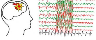
Partial seizures are seizures, the initial clinical and electrophysiological manifestations of which indicate the involvement of one or more areas of one hemisphere of the brain in the pathological process. Partial epileptic seizures are divided into simple and complex. Simple partial seizures occur with preserved consciousness, while complex partial seizures are characterized by its disturbance. To confirm the epileptic nature of the disorder of consciousness, it is necessary to conduct electroencephalography (EEG), which allows recording both normal and pathological brain activity. Electroencephalography is a safe and accessible method for recording and assessing the total electrical activity of the brain. Carrying out an EEG in a patient with seizures allows us to assess the nature of the seizures and the effectiveness of therapy. An EEG recorded during an attack clearly shows in which parts of the brain an epileptic attack occurs and how it spreads. With effective anticonvulsant therapy, as a rule, there is a significant decrease or disappearance of epileptic changes on the EEG and the disappearance of epiparoxysms.
The EEG is recorded during wakefulness, and, if necessary, during sleep, during the interictal period, and at the time of the attack. The most informative is an EEG recording during an attack. Due to its safety and painlessness, EEG can be performed repeatedly and as often as recommended by the doctor. Currently, the most informative technique for determining the nature of an attack is simultaneous video recording of the attack and EEG registration. This technique is called EEG-water monitoring.
Neuroimaging studies (computed tomography of the brain, magnetic resonance imaging, positron emission togography of the brain) are carried out to search for structural changes in the brain (malformation, tumor, injury) that may cause the development of episyndrome. Sometimes changes are found that are characteristic of certain diseases combined with epilepsy, for example, tuberous sclerosis, cytomegaly, toxoplasmosis. In some cases, the cause of seizures is hereditary pathology or metabolic diseases, for which additional research is necessary. Despite the use of the most modern research methods, it is not always possible to find the cause of the episyndrome. However, this fact should not cause relatives to doubt the diagnosis and the need for long-term treatment.
Untimely and inadequate treatment of epilepsy increases its resistance several times and can lead to social and everyday maladjustment of the patient.
Treatment of epilepsy
Treatment of epilepsy in children is primarily aimed at restoring their normal well-being and reducing the frequency of attacks. Then, with the help of well-chosen therapy, the specialist minimizes them and, if possible, completely eliminates them, while simultaneously eliminating the cause of the disease.
Therapy is carried out in a complex and may include:
- drug treatment;
- dietary nutrition (focus on fats and reduction in carbohydrates);
- neurosurgical intervention;
- psychotherapeutic support.
There is no single plan according to which therapy is carried out. Much in the scheme depends on the specific form of the disease that the child suffers from, as well as the cause that caused it.
It is extremely important that the work of the epileptologist, the young patient and his parents is carried out together; the speed and quality of treatment depends on this. It is necessary to thoroughly follow all prescribed recommendations and establish a clear daily routine for the child.
Chapter 1.
What is epilepsy, episyndrome and epi-reaction. Prognosis for attacks.
WHAT ARE EPI SEIZURES Epi seizures are repeated attacks in the form of various convulsions, motor acts, sensations or states of altered consciousness. Different people experience different types of seizures (for example, the computer program “EPI-CENTRE” that I use allows us to treat 79 types of seizures). An attack can manifest itself not only as convulsions and shudders, but also in the form of other motor phenomena, various sensations, loss or an altered state of consciousness. Some types of seizures are practically invisible to others. Therefore, epileptic seizures and epilepsy are not only those dramatic and well-known conditions when a person falls on the street, twitches, and saliva runs from his mouth. You can read a description of the different types of seizures in the recommendations: “WHAT ARE THE TYPES OF SEIZURES. HOW THEY DEVELOP AND PROCEED.” The brain consists of a dense collection of nerve cells connected to each other. These cells convert each irritation perceived by the senses into electrical impulses and transmit them further in this form. In all cases, the cause of the attack is the same - a change in the chemical state of the entire brain or its individual parts, which is accompanied by a flash of increased electrical activity (a kind of “short circuit” in the brain). In order for this “short circuit” to occur, a limited group of brain cells with increased electrical activity is needed (epileptic focus) and a decrease in the threshold of convulsive readiness on the part of the remaining brain cells (usually “healthy” cells prevent the spread of the “short circuit” from the focus to the entire brain ). A sharp increase in the electrical activity of nerve cells is accompanied by their excitation. Excitation of nerve cells leads to various changes in the psyche, including blackout. In addition, from nerve cells with increased electrical activity, along their long processes, excitation spreads to the muscles that are “controlled” by these cells. As a result of this, various involuntary movements or muscle spasms are observed during an attack. WHY THERE ARE DIFFERENT PICTURES OF SEIZURES The type of seizure is determined by which part of the brain the outbreak occurs in and how it spreads throughout the brain. If a “flare” can occur in different parts of the brain, one child may experience two or more types of seizures. Different antiepileptic drugs act differently for different attacks. Therefore, the main thing in diagnosis is the doctor’s correct diagnosis of the type of attack based on your reliable description of it. The type of attack must be specified in detail and correctly in the diagnosis made by the doctor. WHAT IS AN EPI REACTION A flash of electrical activity and a seizure can be caused in any experimental healthy animal in the laboratory by influencing its brain in special ways. And in ordinary life, the brain of any person can give a flash and attack (epi-reaction) when exposed to various environmental factors or various internal reasons. Adults sometimes unwittingly experiment on themselves to induce an epi-reaction - they may develop convulsions after severe intoxication due to fatigue or skipping meals. In children, an example of epileptic reactions is convulsions against a background of high (febrile) temperature - febrile convulsions (see special recommendations "FEBRILIC CONVVURES"). “ INFANTRY” IS NOT A DIAGNOSIS The word “infancy” is sometimes popularly used to describe seizures in infants. It is believed that these attacks occur only in young children, and will not recur at an older age. There is some truth in this, since the smaller the child, the easier his brain reacts with an epi-discharge (epi-reaction) to any harm - high temperature, transmitted disease, rickets, etc. With age, the tendency to react to these factors with seizures decreases and in many children the seizures no longer recur. However, epilepsy can also begin from the first months of life. Seizures in the first year of life can also be a manifestation of a wide variety of brain diseases. Therefore, each specific case of seizures in infancy (regardless of what people call it) requires no less serious attention (diagnosis and treatment) than seizures in older age age. EPIC SYNDROME WHAT IS IT AND HOW IS IT MANIFESTED (SYMPTOMATIC EPILEPSY) Changes in the electrical activity of brain cells can be caused by any brain disease (tumor, encephalitis, hydrocephalus, etc.), and can also be a consequence of any brain damage (head injury, oxygen starvation, brain hypoxia) . Such damage can occur before the baby is born, during childbirth, and then throughout the life of the child or adult. Therefore, when attacks occur, it is necessary to check a child or adult for all these and many other diseases, since, if they are present, these diseases must be treated first as a cause, and only then their consequence in the form of attacks. If the patient is diagnosed with such brain diseases, then an additional diagnosis is made regarding the attacks: “episyndrome”. According to the modern international classification of seizures and epilepsy, episyndrome is now called “symptomatic epilepsy” - meaning that the seizures themselves are only a symptom of some other brain disease. WHAT IS EPILEPSY However, for most people with seizures, careful examination reveals no disease or significant brain damage that can be treated to eliminate the cause of the seizures and thus the seizures themselves. In this case, repeated, regularly recurring seizures are simply a congenital or acquired feature of a given brain - idiopathic epilepsy (according to the modern international classification). The word “idiopathy” translated from Greek means “a painful disorder that occurs for no apparent reason.” This tendency of the brain to have seizures does not always appear in the first years of life - the first seizures in epilepsy (its debut) may appear for the first time after the age of majority. Modern science has accumulated a lot of data on why some people have repeated attacks for no apparent reason. This feature of the brain is a consequence of various combinations of various factors in the prenatal period of brain development, during childbirth and in the process of further brain development after childbirth. With the help of some modern, very expensive devices, it is possible to detect subtle changes in certain layers of neurons that have occurred due to the influence of these factors. However, despite the high cost of this type of research, the information obtained with their help does not change anything in the tactics of the treatment and does little to predict attacks. Some hereditary predisposition to the development of seizures is also possible, although only a few rare forms of epilepsy are purely hereditary (the issue of heredity is described in more detail in the Recommendations “PROBLEMS OF FAMILY LIFE AND MARRIAGE IN EPILEPSY.” REFLECTOR EPILEPSY Most often, seizures in epilepsy occur spontaneously by itself - from " internal" reasons. With the so-called REFLECTOR, quite rare, epilepsy, seizures can be provoked by various external factors, which, by themselves, never cause seizures in other people. Such a triggering factor can be a light (“photo” in Latin) stimulus: flickering (flashing) light, black and white contrasts (see details in the recommendations “PHOTOGENIC EPILEPSY”) Less commonly, attacks are provoked by sound stimuli (unexpected sounds), sharp touch, etc. Some other “external” causes and influences can also contribute to the occurrence specific seizure (provoke it). You should carefully look for these provoking factors and suspicious circumstances that preceded the next attack and record them in the seizure diary (for this, read and follow another Recommendations "WHAT ARE THE TYPES OF SEIZURES AND HOW THEY DEVELOP" and "LIFE STYLE DURING Epi-attacks"). However, most often, attacks can be provoked by a violation of the rules for taking medications set out in the Recommendations: “HOW AND WHAT YOU CAN HELP YOURSELF, YOUR LOVED ONE OR CHILD IN THE PRESENCE OF ATTACKS. THE MAIN METHOD FOR TREATING ATTACKS.” DO YOU OR YOUR CHILD HAVE EPILEPSY? WHAT DIAGNOSIS SHOULD BE MADE—CORRECT OR “CONVENIENT”? People who are illiterate in matters of epilepsy, including many doctors, in all cases of epilepsy consider it, firstly, to be an incurable disease for life, and secondly, necessarily leading to mental and mental degradation. This is wrong. Firstly, in the modern international classification there is, for example, a diagnosis such as “Benign infantile myoclonic epilepsy,” which goes away on its own without any treatment. Secondly, Alexander the Great, Socrates, Julius Caesar, Peter I, Dostoevsky, Napoleon, and the founder of the Nobel Prize, Alfred Nobel, had epilepsy. Therefore, the contemporaries of these greats believed that people with epilepsy, on the contrary, have special mental abilities. For example, in ancient times epilepsy was called “Hercules disease.” Epilepsy is more common than diabetes, tuberculosis or other well-known diseases. There are more than 500 thousand people with epilepsy in Ukraine. Due to the illiteracy of people, the diagnosis of “epilepsy” puts a certain stamp on a person, which can subsequently cause the wrong attitude of others towards him. Therefore, in the presence of even obvious epilepsy, when making a diagnosis, I usually limit myself to an accurate and detailed diagnosis of the type of seizure, which is necessary for proper treatment, and if it is necessary to issue any official certificates, I try to make do with the term “epi-syndrome” even in the presence of a valid diagnosis of epilepsy. PROGNOSIS OF SEIZURES AND CHANCES OF RECOVERY Both patients with seizures and their loved ones are interested in the main thing - whether the threat of repeated seizures remains for the rest of their lives and whether they can be completely cured (prognosis of seizures). According to average statistics, for one person who actually suffers from epilepsy, there are 10 those who had only one seizure during their life. Only 30 percent of those who have had one seizure will have a second one in the next 3 years. After the second seizure, the risk of their recurrence increases to 75 percent. These are statistics, and each individual case is individual. Therefore, even with one single attack, I can sometimes predict a completely different percentage of probability for a particular patient. Most patients and their loved ones associate a patient’s poor prognosis with a diagnosis of epilepsy, as opposed to a diagnosis of epi-syndrome. However, most often epilepsy is “better” than epi-syndrome, which must also be accompanied by some serious brain disease. In general, both good and not very good prognosis of seizures can occur in both epilepsy and epi-syndrome. There are benign forms of epilepsy itself, which are cured in 90-95% of cases. The prognosis depends on the type of attacks and many other features that I determine in the patient. Determining the prognosis requires special knowledge and a lot of time - and therefore cannot be carried out by a doctor at a regular appointment in the clinic. In the process of selecting treatment and monitoring its effectiveness, the accuracy of the prognosis can be significantly increased. The quality of the prescribed treatment and the implementation of these RECOMMENDATIONS significantly improves the prognosis. If the nature of the attacks is such that the tendency to recur may remain for life, this is a significant inconvenience, but not a tragedy: according to foreign scientists, the correct selection of antiepileptic drugs makes it possible to make epilepsy controllable in 70-80 percent of cases. With controlled epilepsy, despite the tendency to the resumption of attacks, they do not arise due to the constant use of antiepileptic drugs, and the development of epileptic changes in the psyche is slowed down or suspended.
To the table of contents of the article “Convulsions, epileptic seizures and epilepsy.”