Hydrocephalus (ICD code 10 G91) is a disease of the central nervous system, which is accompanied by the accumulation of excess cerebrospinal fluid in the ventricles or spaces between the membranes of the brain. The disease does not always manifest itself with symptoms of increased intracranial pressure.
All complex cases of the disease are discussed at a meeting of the expert council with the participation of candidates and doctors of medical sciences, neurologists of the highest category, who are leading experts in the field of diseases of the central nervous system. Patients requiring surgical treatment are consulted by neurosurgeons. Surgical interventions are performed in partner clinics. The staff of the neurology clinic is highly professional and attentive to the wishes of patients.
Causes of hydrocephalus
Hydrocephalus can be congenital or acquired. Congenital hydrocephalus debuts in childhood. Acquired hydrocephalus occurs under the influence of various provoking factors.
Depending on the mechanism of development of the disease, there are 3 main forms of hydrocephalus:
- occlusive hydrocephalus (ICD 10 code - G91.8);
- communicating (open, disresorptive) hydrocephalus (code G91.0);
- hypersecretory hydrocephalus (code G91.8 - other types of hydrocephalus).
Disruption of the flow of cerebrospinal fluid in occlusive (closed, non-communicating) hydrocephalus occurs due to the closure (occlusion) of the cerebrospinal fluid pathways by a blood clot, a voluminous neoplasm, or an adhesive process that develops after inflammation. If the blockage occurs at the level of the ventricular system (Aqueduct of Sylvius, foramen of Monroe, foramina of Magendie and Luschka), proximal occlusive hydrocephalus occurs.
Make an appointment
Based on the rate of progression of the disease, there are 3 forms of the disease:
- acute hydrocephalus, when no more than 3 days pass from the first symptoms of the disease to severe decompensation.
- subacute progressive hydrocephalus, developing within a month from the onset of the disease;
- chronic hydrocephalus, which develops within a period of 3 weeks to 6 months.
Depending on the level of cerebrospinal fluid pressure, hydrocephalus is divided into the following groups: hypertensive, normotensive, hypotensive. In hypertensive hydrocephalus, intracranial pressure is increased, in the case of hypotensive hydrocephalus, it is decreased. Normal pressure hydrocephalus (ICD code 10 – G91.2) is accompanied by normal values of cerebrospinal fluid pressure.
Hydrocephalus can develop after traumatic brain injury and various diseases. Hydrocephalus is formed due to the following diseases of the central nervous system:
- brain tumors localized in the brain stem or ventricles;
- acute cerebrovascular accidents;
- subarachnoid and intraventricular hemorrhages;
- encephalopathy of various origins (chronic hypoxic conditions, alcohol intoxication).
Elderly people often develop replacement hydrocephalus. Its cause is atrophy of brain tissue. When the volume of the brain decreases, the vacated space is filled with cerebrospinal fluid.
At the Neurology Clinic of the Yusupov Hospital, priority is given to the problems of diagnosis and treatment of acute and chronic hydrocephalus in non-traumatic subarachnoid hemorrhages due to disruption of arteriovenous connections and rupture of arterial vascular aneurysms, post-traumatic hydrocephalus.
Publications in the media
Hydrocephalus (dropsy of the brain) - excessive accumulation of cerebrospinal fluid in the ventricles of the brain and intrathecal spaces; manifested by symptoms of increased ICP • Occurs due to an imbalance between the formation and absorption of cerebrospinal fluid: less is absorbed than is formed. The cause may be an obstructive process in the ventricles or in the subarachnoid space, less often - true hyperproduction of cerebrospinal fluid (with villous plexus papilloma) • Characterized by excessive accumulation of cerebrospinal fluid in the ventricles of the brain or subarachnoid space with their expansion • In newborns it leads to an increase in the size of the skull and brain atrophy ( decreased metabolism in adjacent areas of the brain).
Classification.
By the presence of communication between the cavities of the ventricles of the brain and the subarachnoid space.
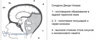
• Non-communicating (obstructive) hydrocephalus is caused by a process that causes blockage of the ventricular system •• Acquired hydrocephalus ••• Tumors of the IV ventricle (medulloblastoma, ependymoma - with them all ventricles are dilated) and the Sylvian aqueduct (glioma of the midbrain tegmentum - the lateral and III ventricles are dilated, and the size of the IV ventricle is not changed) ••• Fibrosis of the arachnoid membrane due to meningitis or intraventricular hemorrhage •• Congenital hydrocephalus ••• Stenosis of the aqueduct of Sylvius in the presence of a septum and true narrowing (#307000, Xq28, L1 cell adhesion molecule gene L1CAM, 308840, À) . Congenital stenosis is characterized by a small size of the posterior cranial fossa. ••• Dandy-Walker syndrome is congenital atresia of the foramina of Magendie and Luschka. Characterized by cystic dilatation of the IV ventricle and cerebellar hypoplasia ••• Vein of Galen aneurysms (v. cerebri magna - Galeni) and other vascular malformations ••• Benign intracranial cysts (arachnoid and ependymal) ••• X-linked hydrocephalus is rare a form of congenital hydrocephalus in men.
• Communicating (non-obstructive) hydrocephalus occurs when there is an obstruction in the cerebrospinal fluid circulation pathways distal to the exit from the fourth ventricle and at the level of the pachyonic granulations •• Acquired ••• Inflammation of the arachnoid membrane during meningitis and as a result of intraventricular hemorrhage (most often with birth trauma and in premature infants) • •• Venous thrombosis, stenosis of the dural sinuses ••• Trauma accompanied by subarachnoid hemorrhage, followed by inflammation of the arachnoid membrane ••• Normotensive hydrocephalus •• Congenital ••• Inflammation of the arachnoid membrane - intrauterine infection ••• Encephalocele ••• Agyria - developmental anomaly: absence of convolutions of the cerebral cortex; the cranial cavity is filled with cerebrospinal fluid.
Separately, functional hydrocephalus is distinguished, which develops as a result of excessive formation of cerebrospinal fluid with villous plexus papilloma. This tumor may also itself obstruct the ventricular system, causing obstructive hydrocephalus. Small hemorrhages from it cause obstruction of the subarachnoid space (after removal of the tumor, hydrocephalus may persist).
According to the predominant accumulation of cerebrospinal fluid • Internal (ventricular) hydrocephalus - hydrocephalus, characterized by the accumulation of cerebrospinal fluid mainly in the ventricles of the brain • External hydrocephalus - hydrocephalus with accumulation of cerebrospinal fluid predominantly in the subarachnoid space or (less often) in the subdural space in the presence of communication between the subarachnoid and subdural spaces • General hydrocephalus - hydrocephalus with accumulation of cerebrospinal fluid both in the ventricles and in the subarachnoid space.
Clinical picture • At the initial manifestations of the disease, the child becomes restless, then low mobility and poor appetite appear • A progressive increase in head circumference (up to 60 cm or more), thinning of the skin, dilation of the veins of the scalp • Divergence of the cranial sutures, tension, enlargement and bulging of the fontanelles , absence of their pulsation • Symptoms of intracranial hypertension due to the compliance of the skull in newborns and young children are not clearly expressed • With a significant increase in ICP - a symptom of the setting sun (the child’s eyes look down, the lower part of the iris is covered, a wide strip of sclera is visible from above; gaze paresis up) • The sound of a “cracked pot” during percussion of the skull • Swelling of the optic discs, subsequent decrease in visual acuity due to atrophy • Damage to the motor sphere: increased reflexes from the lower extremities, later - paresis • Developmental indicators lag behind normal, in advanced cases mental illness is inevitable retardation • A disease that occurs after 17–18 years of age is not accompanied by an increase in head size; signs of intracranial hypertension come to the fore.
Research methods • X-ray of the skull: dehiscence of the sutures of the skull, deepening of finger-like impressions, thinning of bones (with prolonged hypertension), intracranial calcifications (as a result of congenital infections). Small sizes of the posterior cranial fossa suggest stenosis of the Sylvian aqueduct, large ones - Dandy-Walker syndrome • Ultrasound (neurosonography) indicates the degree of ventricular dilatation, intraventricular hemorrhage • CT helps determine the size of the ventricles, the location of obstruction of the cerebrospinal fluid pathways, and allows to identify associated anomalies • MRI is the most informative , indicated for the combination of hydrocephalus with tumors, vascular malformations, and multi-chamber ventricles • Angiography helps to identify vascular anomalies • If a congenital infection is suspected, serological tests are performed for rubella, herpes, CMV, Toxoplasma gondii and Treponema pallidum viruses.
Differential diagnosis • Congenital macrocrania (normal head growth rate and family history of macrocrania) • Enlarged head with rickets (overgrowth of bone tissue of the skull and skeletal changes typical for rickets) • Volumetric intracranial formations (subdural hematomas, tumors).
Treatment is predominantly surgical.
Types of operations • Removal of the cause of obstruction of the cerebrospinal fluid pathways (brain tumor, arachnoid adhesions) • Shunting of cerebrospinal fluid into the body cavity •• Indication - progressive communicating hydrocephalus •• Methods: ventriculoperitoneal shunting (the most common); ventriculoatrial, ventriculopleural and lumboperitoneal shunting, ventriculoureteroanastomosis, ventriculocholecystostomy - less popular due to the more frequent occurrence of complications • Creation of communication between the ventricular system and the subarachnoid space •• Indication - non-communicating hydrocephalus •• Methods: Torkildsen ventriculocisternostomy, ventriculostomy III ventricle.
Conservative treatment • There is no drug for adequate treatment of hydrocephalus. As additional therapy, acetazolamide is used (reduces the formation of cerebrospinal fluid) and furosemide is used to reduce ICP • Intraventricular hemorrhages are treated with medication (symptomatic therapy), since the dilation of the ventricles is transient.
Complications often arise after shunt operations: shunt dysfunction, purulent complications (peritonitis, ventriculitis).
The prognosis for hydrocephalus in newborns is unfavorable. ICD-10 • G91 Hydrocephalus • G94* Other brain lesions in diseases classified elsewhere • O33.6 Fetal hydrocephalus leading to disproportion requiring maternal medical attention • Q03 Congenital hydrocephalus • Q05 Spina bifida [incomplete closure of the spinal canal]
Application. Normotensive hydrocephalus develops in adult patients with insufficient absorption of cerebrospinal fluid by pachion granulations, which allows the pressure to remain within physiological limits. It occurs as a result of subarachnoid hemorrhage or meningitis, sometimes appearing after several years. Clinical picture: classic triad of symptoms - dementia, gait disturbances, urinary incontinence; no signs of damage to sensory, motor or cerebellar functions. Diagnostics: radiography of the skull (increased pattern of digital impressions on the cranial vault, porosity of the “sella turcica”); CT/MRI and ventriculography: sharply enlarged cerebral ventricles in combination with relatively unexpressed atrophy of the cerebral cortex; over time (in the absence of adequate treatment), overstretching of nerve fibers leads to progressive thinning of the white matter of the brain. Surgical treatment The prognosis is relatively favorable (the operation brings a visible effect). Results are best if the method is applied within the first 6 months after the onset of symptoms. Synonyms: aresorptive hydrocephalus, hidden hydrocephalus, Hakim–Adams syndrome. ICD-10 • G91.2 Normal pressure hydrocephalus.
Normal pressure hydrocephalus
In 1965, a publication describing the classic clinical picture of normal pressure hydrocephalus first appeared. The authors of the work are neurosurgeons S. Hakim and R. Adams, who described the symptoms of chronic hydrocephalus in adults with normal cerebrospinal fluid pressure. Later, the characteristic symptoms were called the Hakim-Adams triad. Therefore, normal pressure hydrocephalus is also called Hakim-Adams syndrome.
The triad of clinical manifestations of normal pressure hydrocephalus includes:
- gait disturbance
- dementia
- urinary incontinence.
Gait disturbance is usually the first sign of disease development and occurs as a result of ataxia or damage to the frontal lobe of the brain. Gait disturbances include a magnetic gait with small strides and foot sticking to the floor, poor balance, and difficulty turning. Quite often, patients experience a shuffling gait with their legs spread wide apart.
Symptomatic normotensive hydrocephalus may result from the following situations:
- intraventricular hemorrhage
- inflammatory process in the brain
- traumatic brain injury
- perinatal brain lesions
- neoplasms in the brain
- brain surgery.
Diagnosis of idiopathic and symptomatic normotensive hydrocephalus is based on the clinical picture, medical history and results of MRI of the brain.
Normal pressure hydrocephalus is treated with surgical shunting. Surgery with the installation of a shunt allows you to obtain a long-term positive result in 50-75% of cases. The operation is most effective when diagnosing normal pressure hydrocephalus at an early stage (during the first months after the occurrence of the provoking factor).
In the postoperative period, the following complications are possible:
- subdural hematoma (requires repeat surgery);
- secondary infection;
- CSF hypotension accompanied by headaches.
After bypass surgery, the patient must undergo a course of rehabilitation to effectively restore gait and cognitive functions.
Non-occlusive hydrocephalus of the brain
The following causes of non-occlusive hydrocephalus are known:
- neoplasms of the brainstem or cerebellum;
- inflammation of the substance or membranes of the brain;
- acute and chronic cerebrovascular accidents;
- intraventricular hemorrhages;
- encephalopathy due to toxin poisoning.
Vicarious hydrocephalus of the brain in adults develops due to the replacement of the atrophied substance of the brain with cerebrospinal fluid.
The main signs of hydrocephalus in children are accelerated head growth and an enlarged, disproportionate skull. In newborns, the following symptoms of non-occlusive hydrocephalus occur:
- tense fontanel;
- frequent tilting of the head;
- downward displacement of the eyeballs, strabismus;
- pulsating rounded protrusions in places where the bones of the skull are not fused.
Adult patients with hydrocephalus complain of a feeling of heaviness in the head and headaches that get worse in the morning.
All conditions have been created for the treatment of patients with non-occlusive hydrocephalus at the Yusupov Hospital:
- experienced doctors who are leading experts in the field of neurology;
- qualified staff who are attentive to all the wishes of patients;
- comfortable conditions for patients and their relatives to stay in the neurology clinic;
- examination using equipment from leading world manufacturers;
- individual treatment regimens with drugs registered in Russia.
Patients are provided with individual personal hygiene products and dietary nutrition. Neurologists conduct dynamic monitoring of patients throughout the entire treatment period.
Neurosurgeons also perform the following operations for hydrocephalus:
- aqueductoplasty;
- ventriculocystocysternostomy;
- septostomy;
- endoscopic removal of intraventricular brain tumor;
- endoscopic installation of a shunt system.
After endoscopic surgery, the patient’s normal functioning is almost completely restored. Endoscopic surgical interventions for non-occlusive external hydrocephalus can significantly reduce the number of complications.
Diagnostics
Research methods • X-ray of the skull: dehiscence of the sutures of the skull, deepening of finger-like impressions, thinning of bones (with prolonged hypertension), intracranial calcifications (as a result of congenital infections). Small sizes of the posterior cranial fossa suggest stenosis of the Sylvian aqueduct, large ones - Dandy-Walker syndrome • Ultrasound (neurosonography) indicates the degree of ventricular dilatation, intraventricular hemorrhage • CT helps determine the size of the ventricles, the location of obstruction of the cerebrospinal fluid pathways, and allows to identify associated anomalies • MRI is the most informative , indicated for the combination of hydrocephalus with tumors, vascular malformations, and multi-chamber ventricles • Angiography helps to identify vascular anomalies • If a congenital infection is suspected, serological tests are performed for rubella, herpes, CMV, Toxoplasma gondii and Treponema pallidum viruses.
Differential diagnosis • Congenital macrocrania (normal head growth rate and family history of macrocrania) • Enlarged head with rickets (overgrowth of bone tissue of the skull and skeletal changes typical for rickets) • Volumetric intracranial formations (subdural hematomas, tumors).
Internal hydrocephalus of the brain
The disease is characterized by the accumulation of excess amounts of cerebrospinal fluid in the cavities of the brain. Depending on the rate of progression of the symptoms of the disease, 3 forms of internal hydrocephalus are distinguished:
- progressive or active – occurs with rapid accumulation of cerebrospinal fluid and severity of symptoms;
- stabilized or passive – signs of the disease do not increase;
- regressive - symptoms of the disease disappear without treatment.
Internal non-occlusive hydrocephalus of the brain develops under the influence of the following factors:
- infectious diseases of a woman during pregnancy (respiratory viral infection, mumps, herpesvirus or cytomegalovirus infections, rubella, syphilis);
- abnormalities of intrauterine development that interfere with the circulation and absorption of cerebrospinal fluid;
- brain injuries;
- inflammation of the substance and membranes of the brain (meningitis, meningoencephalitis, arachnoiditis);
- hemorrhage as a result of injuries and diseases of the cerebral vessels;
- oxygen starvation or cerebral circulatory disorders.
- intoxication with chemicals or alcohol;
- genetic defects.
Internal hydrocephalus of the brain in adults can develop in the presence of oncological tumors of the brain, which are localized in the cerebellum or brain stem, acute cerebrovascular accident (stroke), arterial hypertension, and diabetes mellitus.
The characteristic signs of internal hydrocephalus in adults are:
- shaky, unsteady gait;
- loss of urinary control;
- memory impairment;
- headaches that are not always relieved by painkillers;
- blurred vision;
- vision deteriorates, pressure is felt on the eyes;
- nausea and vomiting.
Patients experience scattered attention and decreased concentration, and loss of thinking skills. Signs of mental disorders appear:
- emotional instability;
- attacks of aggression;
- neurasthenia;
- replacement of apathy with emotional upsurge.
Impaired motor functions appear.
At the Yusupov Hospital, all conditions have been created for the treatment of patients suffering from internal hydrocephalus of the brain:
- cozy rooms of different comfort classes;
- examination using modern diagnostic devices from leading companies in the world;
- the use of effective drugs for treatment that have a minimal range of side effects;
- an individual approach to the choice of management tactics for each patient;
- attentive attitude of the staff.
The Neurology Clinic employs candidates and doctors of medical sciences, who are leading neurologists. All complex cases of internal hydrocephalus are discussed at a meeting of the expert council.
Symptoms and diagnosis of hydrocephalus
Acutely developing occlusive hydrocephalus is manifested by symptoms of increased intracranial pressure:
- headache;
- nausea and vomiting;
- drowsiness;
- congestion of the optic discs;
- symptoms of axial displacement of the brain.
Headache is most pronounced upon awakening in the morning due to an additional increase in intracranial pressure during sleep. This is facilitated by the expansion of cerebral vessels due to the accumulation of carbon dioxide, which is accompanied by blood flow, stretching of the dura mater of the brain in the area of the base of the skull and the walls of blood vessels. Nausea and vomiting worsen and sometimes lead to a decrease in headaches. The most dangerous sign of increased intracranial pressure is drowsiness. It appears on the eve of a sharp and rapid deterioration of neurological symptoms.
Walking impairment is manifested by apraxia. The patient can freely pretend to walk or ride a bicycle in a lying position, but in an upright position this ability is immediately lost. A person walks uncertainly, with his legs spread wide apart, and his gait becomes shuffling. In the later stages of hydrocephalus, paresis of the lower extremities develops. The most late and variable symptom is urinary incontinence.
Neurologists at the Yusupov Hospital diagnose occlusive hydrocephalus using computed tomography and magnetic resonance imaging. In chronic dysresorptive hydrocephalus, tomograms reveal a symmetrical expansion of the ventricular system with a balloon-like enlargement of the anterior horns, the subarachnoid fissures are not visualized, and there is a diffuse bilateral change in the white matter of the cerebral hemispheres in the form of a decrease in its density, most often around the lateral ventricles. Computed tomography also makes it possible to clarify the presence and extent of concomitant ischemic brain damage in patients with subarachnoid hemorrhages.
Patients undergo a lumbar puncture and at least 40 ml of cerebrospinal fluid are removed. She is sent to the laboratory for research. Improvement in patients' condition after the procedure is a good predictor of the patient's recovery after surgery.
Symptoms (signs)
Clinical picture • At the initial manifestations of the disease, the child becomes restless, then low mobility and poor appetite appear • A progressive increase in head circumference (up to 60 cm or more), thinning of the skin, dilation of the veins of the scalp • Divergence of the cranial sutures, tension, enlargement and bulging of the fontanelles , absence of their pulsation • Symptoms of intracranial hypertension due to the compliance of the skull in newborns and young children are not clearly expressed • With a significant increase in ICP - a symptom of the setting sun (the child’s eyes look down, the lower part of the iris is covered, a wide strip of sclera is visible from above; gaze paresis up) • The sound of a “cracked pot” during percussion of the skull • Swelling of the optic discs, subsequent decrease in visual acuity due to atrophy • Damage to the motor sphere: increased reflexes from the lower extremities, later - paresis • Developmental indicators lag behind normal, in advanced cases mental illness is inevitable retardation • A disease that occurs after 17–18 years of age is not accompanied by an increase in head size; signs of intracranial hypertension come to the fore.
Treatment of hydrocephalus
With an advanced clinical picture of the disease, conservative treatment is ineffective. Patients at the Yusupov Hospital are consulted by a neurosurgeon to decide on immediate neurosurgical intervention. In case of hemorrhage and thrombosis, the operation consists of applying external ventricular drains followed by the introduction of streptokinase into the ventricular cavity - a drug that dissolves blood clots and thereby ensures normal outflow of cerebrospinal fluid.
If the symptoms of chronic hydrocephalus do not progress in patients, they are prescribed diuretics - diacarb, mannitol, furosemide or lasix. To prevent hypokalemia, patients take asparkam. When the symptoms of occlusive hydrocephalus increase, neurosurgeons perform shunt operations. Timely surgical intervention for hydrocephalus allows for the recovery of all patients. Currently, neurosurgeons prefer to perform endoscopic operations for hydrocephalus.
If you have signs of occlusive hydrocephalus, call the Yusupov Hospital. Neurologists take an individual approach to choosing a treatment method.