Hydrocephalus of the brain (dropsy) is a local increase in the volume of cerebrospinal fluid spaces, occurring due to excessive accumulation of cerebrospinal fluid in the ventricular system against the background of its impaired secretion, circulation or absorption. According to WHO, the prevalence of the pathology around the world is as follows: about 700 thousand patients, including children, are diagnosed with hydrocephalus.
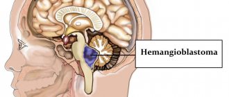
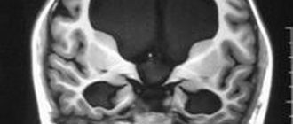
Visual model of the disease.
Causes of hydrocephalus
Such a lesion can be either acquired or congenital. According to statistics, for every 500-1000 newborns, there is 1 case of birth of a child with hydrocephalus of the brain. The clinical debut of the congenital form occurs, as a rule, in early childhood (0-6 months). A key role in the etiology of the development of a birth defect is played by:
- intrauterine infections (the main cause) and hemorrhages;
- consequences of birth trauma;
- fetal asphyxia;
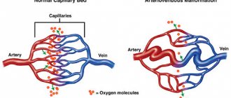
- abnormal formation of the cerebral vessels of the fetus (malformations);
- congenital neoplasms;
- genetic factor.
If we talk about the acquired form, the formation of cerebral hydrocele is most often facilitated by:
- traumatic brain injuries;
- cerebrospinal fluid infections;
- meningitis;
- tumor process in the brain;
- intracranial hemorrhages.
Symptoms of hydrocephalus of the brain
- attacks of intense headache;
- surges in blood pressure against the background of complete well-being;
- attacks of nausea, vomiting;
- episodes of urinary incontinence at night or during the day;
- changes in mental state (development of dementia);
- unsteady, shaky gait.
In children in infancy, there is a rapid increase in head size and bilateral paralysis of the extraocular muscles.
At the beginning of the disease, there are no neurological symptoms, although pathological changes in the brain are already occurring. Observed:
- cerebrovascular disorders;
- reduction of certain parts of the brain;
- organic changes in the cerebral substance.
Hydrocephalus of idiopathic origin is extremely rare.
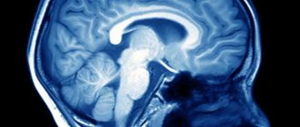
To find out the causes and nature of the disease, detailed diagnostics using hardware technologies is necessary. Magnetic resonance imaging provides the most complete information about the state of the brain. The diagnosis of hydrocephalus is most often made in children under 3 months of age. Among the adult population, pathology is registered in patients over 60 years of age.
A list of the most common causes of hydrocephalus has been determined:
- tumors of various origins;
- traumatic brain injuries;
- diseases of the vascular system;
- conditions after neurosurgical interventions;
- acute cerebrovascular accidents - strokes;
- hematomas;
- congenital pathologies of the central nervous system.
The treatment program is drawn up by the doctor. It depends on the type of disease and severity. Uncomplicated forms of cerebral hydrocele can be treated with medication. If conservative therapy has shown its failure, and the disease causes a number of severe complications, neurosurgical treatment is performed. As a rule, operations are performed on an emergency basis.
Classification of pathology by form
Hydrocephalic syndrome is classified according to localization, pathogenesis, fluid pressure level, and flow rate.
The localization of the outbreak comes in three varieties:
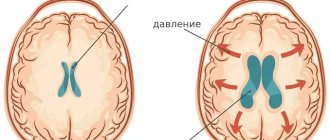
- internal - cerebrospinal fluid accumulates in excess in the lateral ventricles;
- external - overconcentration of cerebrospinal fluid is determined in the subarachnoid space;
- mixed - simultaneous accumulation of cerebrospinal fluid in the ventricles and subarachnoid space.
Based on the pathogenesis, GM hydrocephalus can be:
- occlusive (closed) – the most dangerous form, resulting from blockage (occlusion) of the cerebrospinal fluid passages by a tumor, hematoma, or post-inflammatory adhesions;
- communicating (open) - with this pathogenesis, there is a disruption of resorption processes due to damage to the structures involved in the absorption of cerebrospinal fluid into the venous system.
Based on the CBF pressure indicator, they are distinguished:
- hypertensive hydrocephalus – the level of intracranial pressure is increased;
- normotensive – ICP remains within normal limits;
- hypotensive – the pressure inside the skull is reduced.
Hydrocephalus is diagnosed based on the rate of progression:
- acute - from the appearance of the first signs to the phase of severe clinical decompensation it takes no more than 72 hours;
- subacute – develops within 30 days;
- chronic - formation occurs at a slow pace, over months and even years (more common in open forms).
In ICD-10, hydrocephalus is assigned a general code of G91. Each form has its own alphanumeric symbol: communicating - G91.0; occlusal – G91.1; normotensive – G91.2; post-traumatic unspecified – G91.3; another type – G91.8; hydrocephalus of unspecified origin – G91.9.
Clinical symptoms of increased liquor spaces
Signs of dilation of the ventricles and subarachnoid cavities in newborns:
- Constant irritability;
- Negative reaction to strong noises, flashes of light;
- Restless sleep;
- Frequent regurgitation;
- Strabismus;
- Different pupil sizes;
- Moodiness when weather changes;
- Slow overgrowth of the fontanel;
- Twitching of the chin.
Deviations must be detected immediately after their occurrence. Correction of changes should be carried out immediately after detection.
Clinical symptoms of subarachnoid enlargement in adults:
- Dyspeptic disorders (vomiting and nausea);
- Headache;
- Drowsiness;
- Constant dizziness;
- Increased intracranial pressure;
- Dementia;
- Memory problems;
- Disorders of spatial orientation;
- Unsteady gait.
Determining clinical signs in a child and an adult requires contacting a neurologist. The specialist will detect the pathology and give a referral for an MRI of the convexital cerebrospinal fluid cavities.
At the initial stages there are no manifestations of pathology. Symptoms depend mainly on the severity of the deformity and the causes of the pathology. In small children, the disease is provoked by arachnoiditis, birth trauma, and meningitis. Tumors are a common etiological factor in the occurrence of pathology in adults. Compression of the cerebral parenchyma causes defects in the destruction of white and gray matter. The formed cavities are filled with liquor.
Analysis of brain tomograms in people with enlarged cerebrospinal fluid cavities: atrophic changes in the cortex of the frontal, occipital, and temporal regions. The number of convolutions is smoothed out, and blood accumulations are visible.
The presence of concomitant complications in a child is dangerous, so invasive diagnostic measures are postponed to the second year of life. A reliable diagnosis can be made after taking a biopsy of the enlarged cavities. Microscopic assessment of the properties of cerebrospinal fluid makes it possible to differentiate inflammatory, tumor, and ischemic changes.
Magnetic resonance imaging reveals accompanying pathological changes:
- Leukomalacia is a pathology of impulse signal transmission due to softening of the meninges;
- Inflammatory foci;
- Collections of blood.
An MRI picture of a moderate expansion of the external cerebrospinal fluid spaces shows morphological changes, but a whole range of diagnostic methods is required to make a correct diagnosis.
Tomograms of hypertensive hydrocephalus
Signs of the disease
The symptoms of the disease are dictated mainly by reduced perfusion of brain tissue, overstretching of groups of nerve fibers (conducting pathways) due to increased ICP.
- In acute pathogenesis, weak microcirculation (hypoperfusion) leads mainly only to functional disorders of intracranial metabolism. This is
a change in energy metabolism, a reduction in the level of creatine phosphate and ATP, an increase in the concentration of lactic acid and inorganic salts of phosphoric acids. The acute clinical picture is reversible. - The long-term existence of hypoperfusion causes irreversible transformations at the structural level. These are
defects in the vascular endothelium and disruption of the BBB, axonal damage (destruction of axons, up to their complete disappearance). Dropsy of prolonged duration ultimately causes brain atrophy. - The morphology of signs in hydrocephalus in combination with high intracranial pressure is characterized, first of all, by atrophy of the substance of the brain and periventricular edema. There is also damage to the vascular mesenchyme, disruption of brain homeostasis, axonal lesions, and in rare cases, neuronal death. These signs are combined with the clinical picture of the primary pathology that provoked hydrocephalic syndrome.
The symptomatic complex characteristic of hydrocephalus in early childhood includes such distinctive features as:
- increased head size;
- frequent regurgitation;
- restless behavior of the child;
- bulging of a large fontanel;
- divergence of cranial sutures;
- the severity of the venous pattern on the scalp;
- delayed psychomotor development, less often physical;
- throwing the head back;
- “setting sun” syndrome (Graefe);
- congestive optic disc;
- paraplegia of the lower extremities (in severe, advanced conditions).
In adults and older children, the clinical picture depends on the rate of progression of hydrocephalus. In the acute form of the disease, combined with high ICP, the following are observed:
- a bursting and pressing headache that spreads to the orbits of the eyes (one of the features is the peak of pain in the morning after a night's sleep, and then, during the day, the severity of the pain syndrome decreases);
- nausea, which usually accompanies morning headaches (vomiting often occurs in the morning, after which a person notices an improvement in the condition);
- visual disturbances usually include blurred vision, blurred vision, double vision and a burning sensation in the eyes;
- fatigue, drowsiness, lethargy;
- convulsive phenomena (like an epileptic seizure);
- when the brain stem is compressed due to dislocation of brain structures - oculomotor disorders, forced head position syndrome, clouding of consciousness (up to a coma), respiratory failure.
Hydrocele of the brain in the chronic stage manifests itself:
- signs of dementia (dementia), emotional instability;
- apraxia of walking, more often it manifests itself with a shaky and uncertain gait, disproportionately large steps (being in a lying position, patients often do not experience difficulties in imitating walking and twisting a “bicycle” with their legs);
- decreased muscle strength, sometimes patients complain of pain in the neck;
- severe imbalance (in the final stages), which is expressed by a person’s inability to move and sit independently;
- partial or complete loss of sensitivity (not always!):
- urinary and/or fecal incontinence (with a massive lesion).
The pathology is dangerous due to its life-threatening complications! In no case should you ignore an urgent visit to the doctor if one or more symptoms from the lists provided are identified. Timely visit to the hospital for the purpose of diagnosis and receiving adequate medical care increases the chances of a favorable prognosis, including complete recovery.
On average, out of 10 patients who do not receive treatment at the right time, 6-7 people die soon (this also applies to children). Those who did not undergo therapy but survived are doomed to disability with neurological disorders, mental and physical disabilities with a tendency to progress.
Stages of expansion of subarachnoid spaces
The location of the liquor-containing spaces determines the possibility of free flow from one tank to another. An increase in intracranial pressure is promoted by compression of blood vessels and ventricles of the brain by external formations, accumulation of blood (hematoma), edema, and inflammatory foci.
The main stages of expansion of the subarachnoid spaces:
- Moderate – exceeding the size by up to two millimeters;
- Average - up to four millimeters;
- Heavy – over 4 mm.
In an adult, the expansion of liquor-containing spaces is proportional to the growth of the head and swelling of the fontanelles. Detection of external changes requires timely treatment to prevent irreversible complications. Correct therapy allows you to restore dilated ventricles and liquor spaces to normal.
Diagnosis of dropsy of the brain
Clinical manifestations are so specific that they allow a neurology specialist to suspect hydrocephalus already during the initial examination of the patient. Despite this, the diagnosis of pathology always involves differentiating hydrocephalic syndrome from other possible diseases that have similar symptoms.
To differentiate, as well as establish the localization, degree and form of hydrocephalus, the etiological factor of its development, according to the doctor’s decision, the leading visual diagnostic tools are prescribed in a certain combination:
- magnetic resonance imaging (the most informative);
- conventional or multislice CT;
- echoencephalography (shows the level of ICP);
- neurosonography (done on infants through an open large fontanel to determine ICP);
- radiography (more of a backup method, sometimes recommended for assessing the condition of the skull bones).
If cerebral vascular pathology is suspected, the patient is examined using MR angiography. Dropsy of infectious origin additionally involves performing a PCR analysis to identify the type of infection. All patients are prescribed ophthalmological examinations, including examination of the fundus with an ophthalmoscope, eye perimetry, and visometry.
Principles for diagnosing convexital dilatation
Let's consider the main diagnostic methods:
- Neurosonography is an ultrasound examination of the brain structure through open slits of the fontanelles. It is carried out for each child after birth;
- Computed tomography (CT) measures the size of the cerebral ventricles, subarachnoid space;
- Comprehensive MRI of the head – monitors soft tissue changes. Layer-by-layer mapping of brain areas followed by three-dimensional remodeling shows the spatial structure of objects;
- Cisternography – determines the direction of movement of the cerebrospinal fluid, the type of dropsy (hydrocephalus).
Additional diagnostic methods are contrast angiography, PET/CT. Positron emission tomography is a radiation examination, therefore it is performed according to strict indications to limit the level of radiation exposure to tissues.
Treatment of hydrocephalus in children and adults
Treatment tactics are determined by a specialist based on the severity of hydrocephalus and the disease that gave rise to excessive accumulation of CBF. In the most isolated cases, for example, with a mild form, a conservative approach can be used (based on the use of diuretics to reduce ICP), but it does not lead to a complete cure. Conservative therapy can also be used as a preparatory step for surgery.
For patients of all ages with this diagnosis, surgical intervention is recommended, and on an emergency basis. The danger of the pathology is that even from a mild form it can quickly reach a critical stage at any time with a disappointing prognosis for the patient.
Today, depending on the indications, various neurosurgery techniques are successfully used to eliminate hydrocephalus. They are united by a common goal - creating the necessary conditions to ensure the removal of excess cerebrospinal fluid and maintaining normal cerebrospinal fluid pressure. Thus, brain functionality is restored, ICP is stabilized, neurological and cognitive symptoms disappear or are noticeably reduced. Let's consider what operations can achieve this goal.
Liquor shunt interventions
CSF shunting is the installation of elastic silicone implant systems to remove cerebrospinal fluid outside the central nervous system. The systems are equipped with catheters in the form of flexible hollow tubes, as well as valves with an anti-siphon (reverse) mechanism and with fixed or adjustable opening pressure.
The operation can be performed in various ways. But surgeons consider ventriculoperitoneal (the most commonly used method) or ventriculoatrial shunting to be the most successful in terms of safety. Interventions are performed under endotracheal anesthesia, manipulations are controlled by intraoperative fluoroscopy, CT, and ECG.
- Ventriculo-peritoneal shunt. The principle of the procedure is based on the implantation of silicone catheters, through which an excess of CBF goes into the intra-abdominal cavity, where it is resorbed between the intestinal loops.
- The procedure begins by making an incision on the scalp, after which a small burr hole is created in the skull. The dura mater is opened sparingly.
- A ventricular catheter is introduced through the created access, its end is placed in the lateral ventricle of the brain.
- The valve element is implanted in the area of the auricle (at the back or slightly above). The ventricular (ventricular) and distal catheter (DC) are fixed to it.
- Next, the neurosurgeon places a distal catheter into the abdominal cavity through a specially formed subcutaneous channel.
- Upon reaching the desired abdominal area, the specialist makes a small incision (no more than 10 mm) and inserts the end of the DC into the abdominal cavity.
- The procedure ends with thorough disinfection of the surgical field, followed by closing the wound areas with antiseptic dressings (sutures are applied if necessary).
- Ventriculoatrial shunting. The essence of this operation is to divert cerebrospinal fluid through installed shunts from the ventricle of the brain into the right atrium.
- Dissection of tissue in the neck along the anterior sternocleidomastoid muscle is performed to open the common facial or internal jugular vein.
- The atrial catheter is removed into one of the indicated veins, fixing it with specially designed ligatures.
- The shunt is directed through the catheterized vein to the right atrium. The end of the atrial shunt is generally located in the superior vena cava.
- For the installation area of the distal end of the vascular catheter, yes, the superior vena cava is often preferred. Here the blood flow is turbulent, and this reduces the likelihood of thrombosis of the drainage system by blood clots.
- The cranial part of the intervention, when the ventricular element of the system is implanted, a valve and two catheters are connected to it, is identical to VP shunting.
For adults, shunts are permanently implanted. In childhood, they are periodically replaced with elongated models. We emphasize that patients after surgery with shunt implantation are shunt-dependent people.
Endoscopic operations
Endoscopic surgical techniques are used in the treatment of occlusive hydrocephalus, among them:
- ventriculocisternostomy;
- ventricular cystocisternostomy;
- ventriculoplasty of the aqueduct of Sylvius;
- septostomy;
- etiotropic endoscopy (getting rid of the causative factor - removal of a tumor, cyst, hematoma, etc.).
In 90% of cases, the method of endoscopic ventriculocisternostomy is used. The purpose of this operation is perforation of the bottom of the third ventricle of the brain under endoscopic control through a miniature trepanation window. The anastomosis created during the endoscopic procedure allows you to restore the natural path of outflow of cerebrospinal fluid between the third ventricle and the basal cisterns of the brain.
Endoscopy of any kind is a more gentle tactic of neurosurgery; it does not require the implantation of foreign bodies into the body and is less likely to cause postoperative consequences. Despite the promising characteristics of endoscopic methods, in some cases it is impossible to do without shunting or the use of open microsurgery.
Modern neurosurgery technologies have been honed to perfection in the Czech Republic; brain surgery in this country is a leading field of medicine. Neurosurgical care in clinics in the Czech Republic is no worse than in Germany and Israel, but the price is much lower (about 2 times). People with this diagnosis are operated on here at the most exemplary level, and upon completion of the full course of rehabilitation they are discharged with excellent and good results.
Hypertensive-hydrocephalic syndrome in children of the first year of life. What do you need to know about him?
Firstly, it should be noted that a true increase in intracranial pressure occurs as a result of a fairly serious number of reasons: a volumetric formation of the brain (vessel aneurysm, tumor, etc.), inflammation of brain structures, increased production of cerebrospinal fluid, blockage of outflow cerebrospinal fluid (disturbance in the structure of cerebrospinal fluid containing spaces, hemorrhage into the ventricles of the brain, etc.).
Secondly, any of these conditions is characterized by a number of symptoms, accompanied by an increase in intracranial pressure, which is manifested by persistent expressed anxiety of the child, sleep disturbances, appetite, vomiting, bulging fontanelles and gradual separation of the sutures between the bones of the skull, a progressive increase in the size of the ventricles of the brain according to ultrasound data brain (NSG - neurosonography).
If you notice similar symptoms in a child, you must strictly contact a neurologist to rule out serious diseases manifested by these symptoms. At the same time, there are a number of symptoms that often serve as the basis for a diagnosis: intracranial hypertension. However, the presence of only one of them in a child casts doubt on the diagnosis of “increased intracranial pressure.”
Greffe's sign (the appearance of a strip of white (sclera) between the upper eyelid and the iris of the eye) is a fairly common phenomenon in children in the first months of life. This is due to some discrepancy between the sizes of the orbit and the eyeball. During growth, the volume of the orbit increases, the eyeball takes a deeper position, and the strip of sclera above the iris disappears.
An increase in head circumference serves as an indicative sign for a neurologist by which intracranial hypertension can be suspected, provided that other symptoms are present.
Normally, head circumference (CH) increases most actively in the first months of life: 1 month. – up to 3.5 cm; 2 months - 2 cm and 3 months. – 2 cm. Then the rate decreases and the increase averages 1-1.5 cm per month for up to 6 months. In premature babies, these rates are even higher. The idea that the increase is 1-1.5 cm per month is wrong!
There are no absolute standards for head circumference. It is correct to start from the indicators at birth, because if at birth the head circumference was 33 or 35 cm, and the increase in 1 month was 3 cm, then in the end the indicator is 1 month. will be 36 and 38 cm, respectively, and both values are normal.
Increase in the size of liquor-containing spaces according to NSG - neurosonography
Exceeding the size by 1-2 mm, without any other neurological symptoms and dynamics, should be considered as a possible individual norm for a particular child. After all, we do not treat data from brain ultrasound (NSG - neurosonography), it is necessary to look at the whole situation in its entirety.
Also, a slight increase in size does not at all indicate an increase in intracranial pressure. Intracranial pressure is reliably measured only during puncture, and the expansion of cerebrospinal fluid-containing spaces on neurosonography (NSG) only indicates their expansion, and not an increase in intracranial pressure. Liquor fills the expanded spaces compensatory, which ensures constant intracranial pressure and a shock-absorbing effect.
Nosko Anastasia Sergeevna, PhD, neurologist.