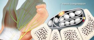
Cubital tunnel syndrome is a condition that occurs due to excessive pressure on the ulnar nerve, which runs through the elbow joint. In this place it is located in the bone groove on the surface of the internal epicondyle on the posterior side.
When a person hits the inside of the elbow, he may feel a short electric shock that radiates to the outside of the hand - this is a manifestation of irritation of the ulnar nerve. When this happens systematically due to increased levels of pressure or, for example, due to injury, it often leads to cubital tunnel syndrome.
Surgery options for cubital tunnel syndrome
The syndrome belongs to the group of compression-ischemic neuropathies. The very first symptoms are pain and problems with movement, as nerve function is impaired. First, disorders arise, after which the problem is aggravated by a decrease in muscle strength. Based on the reasons, motor and sensory disorders can occur simultaneously.
There are two types of syndromes:
Cubital tunnel syndrome
The main symptoms of this syndrome are:
- pain in the area of the ulnar fossa, which radiates to the forearm;
- burning, tingling, twitching;
- pain worsens at night and when moving the elbow joint;
- over time, the pain intensifies; decreased sensitivity in the elbow joint;
- movement disorders and muscle weakness;
- the hand begins to lose weight due to muscle atrophy;
- deformation of the hand occurs.
Guyon's canal syndrome (ulnar carpal syndrome)
The symptoms are largely similar to the previous syndrome, but there are some differences in presentation:
- pain in the area of the wrist joint;
- pain intensifies when moving the hand;
- decreased sensitivity in the area of the ring and little fingers;
- motor disorders of the ring and little fingers;
- the brush may take on the appearance of a “bird’s foot”;
- muscular atrophy of the hand.
If the primary symptoms of the disease are ignored, the symptoms can be combined and confused due to the fact that the nerve will be increasingly subject to compression. The presence of neuropathy in the elbow joint can be determined by tapping a neurological hammer in the place where, in the opinion of a specialist, it is compressed.
If the main cause is gradual compression of the nerve, and the symptoms have appeared recently, then first of all they begin conservative treatment methods (medicines, traditional medicine) and only if there is no result, they begin surgical intervention due to the ineffectiveness of conservative treatment.
Diagnostic measures
The diagnosis is made based on the patient’s complaints, external examination, and medical history. Indirect confirmation of a pinched nerve is the presence of previous injuries, endocrine, neurogenic, and articular pathologies. The Froman tests are the most informative in diagnosing compression of nerve fibers. The doctor asks the patient to squeeze the table top so that it is between his thumbs and forefingers. Even with minor pinching, the thumbs will move sideways or bend at the interphalangeal joints.
The next test is to grip a paper sheet with the side surfaces of straightened fingers. A patient with a pinched ulnar nerve will not be able to cope with the task. He will be able to hold a sheet of paper, but at the same time he will bend his fingers (“claw-shaped bird’s hand”).
Froman test.
Then the doctor identifies Tinnel’s symptom and determines the degree of its severity. It presses on the skin located above the cubital canal of the elbow joint. This movement causes a tingling sensation in the thumb, index and middle fingers. The severity of the symptom is detected by changing the intensity of pressure.
| Instrumental and biochemical studies to identify pinched ulnar nerve | The essence of the diagnostic measure |
| Radiography | It is carried out to assess the consequences of injuries and the condition of the elbow joint. The resulting images visualize bone structures and the contours of joint spaces. The study allows you to detect bone growths (osteophytes) that compress nerve fibers |
| MRI or CT | The method is most informative for inflammation of tendons, muscles, ligaments, and soft tissues. Helps to dynamically assess the blood supply to all connective tissue structures of the elbow |
| Electromyography | The study is carried out to identify muscle damage, localization and spread of pathology. Allows you to detect one of the leading signs of pinching - muscle atrophy |
| Electroneurography | This method of studying the speed of electrical impulses along nerves helps to detect innervation disorders. A functional test reveals damage to peripheral nerves, the level and nature of the damage |
| Biopsy | A biochemical study is carried out if there is a suspicion of compression of the nerve by a neoplasm. It helps differentiate malignant and benign tumors |
Prices for treatment of ulnar nerve tunnel syndrome
Specialist consultations
*Surgeons N.S. Romanyutina And Alieva T.V. Free consultation until January 30, 2022.
| Primary appointment (examination, consultation) with a surgeon | 1 500₽ |
| Repeated appointment (examination, consultation) with a surgeon | 1 500₽ |
Ulnar nerve tunnel syndrome
| Ligamentotomy for cubital tunnel syndrome | 150 000₽ |
| Neurolysis, decompression of nerves in carpal tunnel syndrome | 110 000₽ |
| Relocation of the ulnar nerve in cubital tunnel syndrome with increased mobility of the ulnar nerve | 180 000₽ |
Anesthesia
| Local anesthesia | 5 000₽ |
| Intravenous anesthesia (IVVA) | 20 000₽ |
| General anesthesia (GENA). The cost depends on the duration of the operation (from 2 to 8 hours) | from 30,000₽ to 40,000₽ |
Hospital stay
| Stay of patients in the ward after general anesthesia (1 day) | 10 000₽ |
| Patients' stay in the ward after intravenous anesthesia (day) | 5 000₽ |
View full price list
How to treat elbow pain
Adequate treatment of elbow pain syndrome is possible only after a preliminary examination. It is impossible to carry it out at home. Non-severe patients are examined on an outpatient basis, but for acute illnesses and injuries, patients with elbow pain often require hospitalization.
Diagnostics
First, the doctor conducts a clinical examination of the patient, then he is sent for additional examination, including:
- Laboratory diagnostics - clinical, biochemical and immunological blood tests reveal inflammatory, metabolic and autoimmune processes.
- Instrumental studies:
- X-ray of the elbow joint - reveals dislocations, fractures and bone changes in inflammatory and degenerative diseases;
- MRI and CT are more accurate studies needed in doubtful cases when clarification of the diagnosis is required;
- Ultrasound – changes in soft articular and periarticular tissues;
- Diagnostic arthroscopy - performed according to indications when a purulent inflammatory process and the presence of blood in the joint cavity are suspected.
Methods for treating elbow pain
Specialists at the Moscow Paramita clinic treat diseases of the elbow joint only based on the results of the examination. Patients who require surgical care are hospitalized with a recommendation for rehabilitation in our clinic after discharge from the hospital.
If the patient does not require hospitalization, he is provided with qualified medical care. An individual treatment plan is drawn up for each patient, taking into account his underlying and concomitant diseases. Complex therapy includes:
- modern Western methods of treating diseases and injuries of the elbow joint, including drug therapy, effective complexes of therapeutic exercises, physiotherapy;
- traditional oriental treatment methods that help restore energy potential and eliminate pathological foci (courses of acupuncture, moxibustion, acupressure;
- various types of joint immobilization to eliminate additional injury to the elbow area, for example, taping; fixation with adhesive elastic tape, etc.
The Paramita Clinic has extensive experience in treating and restoring the function of the elbow joint. Carrying out preventive courses of conservative therapy allows our patients to forget about pain in the elbow and lead a normal life.
More about the procedure
Causes of Carpal Tunnel Syndrome
Ulnar neuropathy occurs due to pressure on the ulnar nerve. Common causes of this disease are the following factors:
- systematic support on the elbow on a hard surface;
- the elbow joint is in a bent position for a long time;
- abnormal bone growth;
- heavy or frequent physical activity;
- a profession in which the joint is under constant load (working on a computer, telephone, etc.);
- injuries.
Diagnostics
Typically, a history and a thorough physical examination are sufficient to suspect ulnar tunnel syndrome. Treatment largely depends on the stage of development of the disease and the cause of its occurrence, which can be determined during the diagnostic process. For this purpose, the following methods are used:
- Electroneuromyography is a study that gives the doctor the opportunity to evaluate the work of the muscles of the forearm and hand, determine the speed of impulse transmission along the nerve - the degree of conduction disturbance (nerve function).
- X-ray, ultrasound, MRI are also used in the diagnostic process, especially if there is a suspicion of injury, fracture, arthritis, gout and other diseases.
Preparing for surgery
There are no special recommendations for preparing for surgery. On the morning of surgery, do not eat or drink anything. If you take medication regularly, take it with one sip of water.
Contraindications
- Diseases that prevent anesthesia (discussed during consultation with a doctor)
- Purulent-inflammatory diseases in the area of surgery
Features of the operation
The operation consists of freeing the nerve from compressing tissue. If there is excessive mobility of the nerve, it is moved, covered with muscles so that it does not stretch when the arm is bent at the elbow joint.
Postoperative period
- After surgery, it is imperative to continue treatment with medications: painkillers; vitamins; decongestants; improving conductivity and nerve trophism.
- It is imperative to use medications in conjunction with physical therapy and physiotherapeutic methods. Only in this case will it be possible to achieve the effect in 3-6 months. In rare cases, with an advanced stage and severe muscle atrophy, complete restoration of sensory and motor functions is not possible, so it is very important to consult a specialist as soon as the first symptoms of the disease appear.
Cubital tunnel syndrome (ulnar nerve compression syndrome)
In this article we will talk about cubital tunnel syndrome, which is sometimes called cubital tunnel ulnar nerve compression syndrome. Nerves carry impulses from the brain and spinal cord to all organs of the body, and these impulses also give signals to muscles to move and are responsible for the sensitivity of the skin. When a nerve is compressed, then, accordingly, it cannot work normally and the conduction of nerve impulses is disrupted. Naturally, the disturbance in the conduction of nerve impulses is stronger when the pressure on the nerve is greater.
We are all familiar with the situation when, having hit the back-inner part of the elbow on something hard, we feel a sharp pain, a shooting along the forearm, which can reach the fingers. In such situations, we just hit the ulnar nerve, and the lumbago from the elbow down to the little finger goes exactly through the area that the ulnar nerve provides impulses (innervates). Yes, this is a strong and sharp pain that occurs with a single blow to the ulnar nerve, but imagine what will happen if you constantly press on the ulnar nerve? Not as hard as when hit, but a little and constantly? It is this situation, when the ulnar nerve is compressed in the canal, that is called compression syndrome, and the resulting disturbance in the conduction of impulses along the nerve in such a situation is called ulnar nerve neuropathy. Ulnar nerve neuropathy itself can occur due to other reasons, for example, due to compression in other places (lower or higher), but if the ulnar nerve is compressed precisely in the cubital canal, then, accordingly, in this case the diagnosis can be formulated as follows: neuropathy of the ulnar nerve due to compression syndrome in the ulnar (cubital) canal.
Cubital tunnel syndrome is the second most common cause of compression neuropathies (ie, pain due to nerve compression) after carpal tunnel syndrome.
Anatomy
The ulnar nerve emerges from the cervical plexus of nerves, travels down the arm and into the fingers. The ulnar nerve is responsible not only for the sensitivity of the skin of the little finger and the adjacent half of the ring finger, but also gives commands to some muscles that move the hand (the interosseous muscles that extend and spread the fingers to the sides, and the muscles that adduct the thumb). If the ulnar nerve is damaged, gripping with the hand will be difficult.
On its way, the nerve bends around the elbow from behind and slightly inwards - this place is called the cubital (ulnar) canal. This canal is formed by muscle tendons (flexor carpi ulnaris, flexors and pronators), ligaments and bone (internal epicondyle of the humerus and olecranon).
Ulnar nerve and cubital tunnel
Causes of compression of the ulnar nerve in the cubital canal
There is no clear and single important cause of compression of the ulnar nerve in the cubital canal. There are several reasons for the development of this syndrome, the importance of each of which varies from person to person.
It is believed that one of the most common causes of compression of the ulnar nerve is repeated trauma. For example, during repeated monotonous movements or when playing sports. Tension of the flexor carpi ulnaris (the beginning of this muscle in the form of an arched tendon forms one of the walls of the cubital tunnel) leads to compression of the ulnar nerve. Of course, normally the tension of this muscle will not lead to compression of the nerve, but if there is a lot of movement, then inflammation will develop - the edges of the inflamed tendon arch become thicker, the canal space, accordingly, becomes smaller, and the nerve will begin to hurt. Often this mechanism for the development of neuropathy occurs during the bench press, when the hand holding the barbell is tense for a long time.
The ulnar nerve can also be compressed by external reasons - if, for example, you like to put your elbow on the side of the car door next to the glass.
Another common cause is a change in the anatomy of the cubital canal. After fractures of the condyles of the humerus, fractures of the olecranon, the formation of cysts, bone spurs, the space of the cubital canal may become narrower, which will lead to compression of the ulnar nerve.
Cubital tunnel syndrome can also appear after a severe injury to the elbow.
Symptoms
Cubital tunnel syndrome most often manifests itself (not necessarily all of these symptoms are present at the same time):
- A feeling of stiffness, pain, numbness in the little finger and in the half of the ring finger adjacent to it.
- Weakness and pain when gripping with the hand and extending the fingers.
- Pain in the cubital canal itself, a feeling of tightness in the elbow.
These problems may begin gradually and progress. Often complaints are stronger in the morning, after sleep, when the elbow has been in a stationary, often bent position for a long time - many people like to place their bent arm under their head or under a pillow. In this case, problems may arise when moving the hand - it is difficult to pick up a kettle, type on the keyboard, play the guitar, etc. After a few minutes to hours, the severity of these symptoms usually decreases. If cubital tunnel syndrome is not treated promptly, the problems may worsen and the complaints become permanent.
If the initial symptoms of cubital tunnel syndrome do not go away within a few weeks, you should consult a doctor.
Diagnosis
The doctor examines the hand, forearm, elbow and shoulder, and neck, trying to determine the level at which compression of the ulnar nerve occurs. The sensitivity of the fingers, certain movements of the hand, Tinnel's symptom (increased symptoms when tapping the fingers along the cubital canal) are checked.
A doctor's examination for cubital tunnel syndrome can be unpleasant or even painful, but this is necessary to clearly determine the cause of the pain.
In most cases, the diagnosis can be established only by examination, but sometimes, in doubtful cases, the doctor may prescribe an additional examination that will determine the presence of non-standard causes of compression of the ulnar nerve (bone spurs, fractures and their consequences, arthritis, etc.) . The most commonly prescribed are x-rays, computed tomography and MRI.
X-rays show bones, CT scans also show bones, but in more detail, and MRI (magnetic resonance imaging) shows soft tissues (muscles, ligaments, cartilage, etc.). Sometimes the doctor uses ultrasound to make a diagnosis.
You don’t need to prescribe these studies yourself before an examination or consultation with a doctor - in most cases, this simply leads to a pointless waste of your time and money, but it can also lead you down the wrong path, because the conclusions of these studies should only be evaluated together with the examination!
In some cases, your doctor may order electromyoneurography (EMNG), which can evaluate the degree of nerve conduction disturbance and sometimes help determine the level at which the nerve is being compressed (neck, shoulder, elbow, forearm, or hand). Electromyoneurography is also useful in those rare cases when nerve compression occurs at two levels simultaneously.
Treatment
In most cases, in the early stages, when symptoms are inconsistent, conservative treatment is effective, i.e. non-surgical treatment .
Conservative treatment includes limiting exercise, stopping training, and anti-inflammatory drugs.
It is necessary to eliminate repeated repetitive monotonous movements, give up the habit of placing your elbow on the side of the car door next to the window. If your job does not allow you to temporarily abandon monotonous elbow movements, then give yourself breaks more often.
If you work at a desk, place something soft under your elbow. Do not let your elbow hang over or rest on the edge of the table when using the mouse or keyboard.
In other words, you need to eliminate as much as possible all those movements that cause pain or discomfort.
If you sleep with your bent arm under your head or under a pillow, try tying a roll of a terry towel to the front (flexor) surface of your elbow to limit flexion. This type of nighttime bandaging can be effective not only when you sleep with your arm bent, but also in other cases of cubital tunnel syndrome.
It is important to use non-steroidal anti-inflammatory drugs, for example, Voltaren gel 3-4 times a day for 2-3 weeks. You need to smear your elbow with anti-inflammatory drugs constantly, regardless of whether there is pain or not. The fact is that these drugs not only relieve pain, but fight the inflammation itself, which is accompanied by swelling, thickening of the soft tissue component of the walls of the cubital canal, and, accordingly, compression of the nerve itself.
In some cases, vitamin B-6 tablets can help treat cubital tunnel syndrome.
Conservative treatment usually gives effect within 3-4 weeks, but sometimes, in persistent cases, it is continued for up to 12 weeks. If sufficient improvement does not occur within 12 weeks, surgery is performed.
Surgery
Surgery may be necessary not only in cases where, after 12 weeks, conservative treatment has not brought sufficient results, but also when:
- despite treatment, complaints due to nerve conduction disorders progress
- initially the complaints were constant and quite pronounced
- there are gross anatomical disorders of the cubital canal (consequences of fractures, the presence of a plate fixing the fracture, etc.).
The operation belongs to the category of “simple”, if, of course, this can be said about operations in general.
The operation is performed under anesthesia - under general anesthesia, intravenous sedation, or regional anesthesia is used, which numbs only one arm.
There are several options for operations that are performed from an incision 5-8 cm long.
Simple decompression. During this operation, part of the thickened canal walls is removed and the tendon arch is dissected. This operation is technically simple, but, unfortunately, it does not always give a lasting effect and after a certain period of light the complaints return again. Sometimes decompression is supplemented by resection (partial removal) of part of the epicondyle of the humerus.
Nerve transposition. The nerve is completely removed from the cubital canal and moved 3-4 centimeters anteriorly. If the nerve is transplanted into the space between the muscle and the subcutaneous fat, then this operation is called anterior subcutaneous transposition. And if the nerve is moved deeper, under the muscle, then, accordingly, the operation is called anterior axillary transposition. The choice of transposition option depends on many features.
According to modern statistical studies, simple decompression and transposition of the nerve are equally effective, but, nevertheless, this does not mean that it is necessary to abandon the relatively complex transposition in favor of simpler decompression.
Scheme of anterior axillary transposition of the ulnar nerve when it is compressed in the cubital canal
After surgery, the elbow is usually immobilized for 10-14 days after subcutaneous transposition and 20-25 days after axillary transposition. After this, movements begin, but after axillary transposition, restrictions on the intensity of physical activity remain for up to 4-6 weeks.
Full recovery from surgery may take several months. It is important to note that in general, a correctly performed operation is very effective, and the long period of gradual recovery is due to the long restoration of nerve cells. Yes, contrary to the well-known proverb, nerve cells are still restored, but very slowly.
The growth rate of axons (processes) of nerve cells is one millimeter per day, and if the distance from the elbow to the tip of the little finger is 30-40 cm, i.e. 300-400 mm, then complete recovery may take 300-400 days. Fortunately, the condition of the nerve is improving gradually, and there is no situation where on the 299th day everything is still bad, but on the 300th everything is fine. In severe cases, when there is neurogenic muscle contracture, i.e. the muscles have already died, deprived of nerve impulses, a complete solution to the problem is impossible, and it is possible to try to restore movements only with the help of other, more complex operations.
| The author of the article is Candidate of Medical Sciences Sereda Andrey Petrovich |
Features of the anatomical structure of the elbow
The elbow is a complex articular joint formed by the radius, ulna and humerus. Each bone involved in creating an articulation plays an important role and has certain characteristics:
- humerus - a tubular bone, the ends of which have strong differences (the upper one is predominantly rounded, and the lower one is triangular), which provides the opportunity for ideal articulation;
- ulnar - in combination with the radial one, forms the forearm, is thicker and resistant to external influences;
- radial - is a structural element of the forearm, is smaller in thickness, but is surrounded by an impressive muscle frame (a set of flexor/extensor and rotatory muscles).
The joint is nourished by a large number of veins and arteries. Innervation (supply of tissues with nerves) is realized thanks to large nerve fibers, which explains the increased sensitivity of the upper extremities.
Why does pain appear in the elbow joints of the hands?
Pain in the left/right elbow joint can occur due to various triggering factors, which can be found in our table below.
| Initiating factor | How and with what is it manifested? |
| Traumatic injuries | These can be strong blows to the joint, bruises, unsuccessful flexion, extension of the upper limb and heavy load (for example, when carrying heavy bags). Even a minor blow with a bent elbow causes sharp pain due to the proximity of the ulnar nerve. As for serious injuries, they require long-term treatment and, as a rule, make the upper limb incapacitated for the period of treatment. |
| Epicondylitis | Tissue damage of a degenerative-inflammatory nature, developing at the points of fixation of the tendons of the forearm to the epicondyles of the tubular bone of the shoulder. The disease manifests itself as pain in the right/left elbow joint, which intensifies with extension or grasping, or prolonged similar rotations. |
| Arthrosis | It is a chronic disease characterized by progression in the form of destruction of cartilage tissue and an increase in pathological changes not only in the capsule, but also in neighboring bones and ligaments. It manifests itself as painful symptoms, a feeling of morning stiffness, and restrictions of movement. As the pathology develops, the clinical manifestations intensify, and in the later stages severe dysfunction of the upper limb is possible. |
| Arthritis | It is represented by a whole group of inflammatory lesions, covering cartilaginous tissue, synovial membranes, capsule and joint as a whole. Clinical manifestations, in addition to pain, include swelling, redness, increased temperature in the area of inflammation and joint deformities. |
| Osteochondrosis of the cervical spine | A chronic pathological condition characterized by degenerative processes of the vertebrae and discs. It manifests itself as pain symptoms that cover the entire left or right upper limb. It goes numb and hurts even more during hypothermia. |
| Bursitis | Inflammation of the bursa of a joint in acute or chronic form due to prolonged compression or infection of the joint. It is characterized by the formation and accumulation of exudate, pain symptoms localized in the back of the elbow, along with swelling, redness, local increase in body temperature, and limitation of movements. |
| Tendinitis | Inflammatory processes involving the tendon, with its subsequent degeneration. They can be acute and chronic. The pain is initially short-term, appearing exclusively during stress on the affected area. They are absent at rest. As the disease progresses, they become more pronounced and become paroxysmal in nature. Other clinical manifestations include swelling, a local increase in temperature, and crunching when moving. |
| Damages to the cardiovascular or nervous system |
|
More about arthritis

