Compression neuropathy of the median nerve (carpal tunnel syndrome - CTS) develops most often at the level of the carpal tunnel due to compression of the nerve under the transverse carpal ligament. CTS is the most common tunnel neuropathy of the upper extremities [1], occurring in approximately 3.8% of the population [2]. Every 5th patient who complains of pain, paresthesia and numbness in the upper limb suffers from CTS [3]. In women, compressive neuropathy of the median nerve develops 5-6 times more often than in men, with a peak incidence between the ages of 40 and 60 years [4, 5].
Diagnosis of compression neuropathy of the median nerve is based on a typical clinical picture, consisting of impaired sensitivity in the area of the palmar surface of the I-III fingers, numbness and pain in the wrist and I-III fingers, mainly at night, which leads to frequent awakenings of patients, as well as clumsiness or weakness of the hand [6]. Specific provocative tests make it possible to establish a diagnosis of CTS with a high probability, even in the absence of instrumental diagnostic methods [7]. Due to the high prevalence of the disease, in many countries it is customary to use specialized questionnaires to establish a diagnosis and determine the severity of symptoms in CTS [6]. Instrumental research methods, such as electroneuromyography (ENMG) and ultrasound examination of the median nerves, help confirm the diagnosis [8]. According to a number of authors [9, 10], patients with compressive neuropathy of the median nerve are unreasonably prescribed MRI of the cervical spine, which often reveals its degenerative changes, which often causes an erroneous diagnosis.
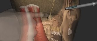
Treatment of CTS is conservative and surgical. At the initial stage of the disease (usually within the first 6 months from the onset of symptoms), conservative treatment is indicated - wearing an orthosis at night in a neutral position of the hand and administering glucocorticosteroids (GCS) inside the carpal tunnel [11-13]. According to randomized clinical trials [7, 11, 14], there is a significant decrease in the severity of symptoms with this therapy compared to placebo.
In patients with severe disease, symptoms lasting more than 6 months, and when conservative treatment is ineffective, surgical decompression of the median nerve is advisable, which has been used since 1950 [15]. According to randomized clinical trials [16], when the clinical picture of the disease is severe, surgical treatment is more effective than wearing an orthosis and intracanal administration of corticosteroids. Successful results of surgical treatment are observed in more than 75% of cases [17].
The purpose of the study is to analyze typical medical practice, diagnostic errors and treatment tactics for compressive neuropathy of the median nerve.
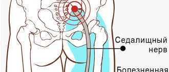
Causes of median nerve neuropathy
The causes of median nerve neuropathy include:
- trauma - bruises and wounds with partial rupture of fibers, fractures of the shoulder or forearm (if the nerve was damaged by bone fragments), fractures inside the elbow, wrist (the place where the hand and forearm connect) joints - compression of the median nerve of the arm;
- dislocations, inflammation of these joints;
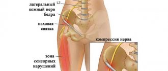
- the nerve is pinched by a tumor or hematoma, rarely in the presence of a process on the humerus (compressive ischemic neuropathy of the median nerve);
- endocrine dysfunction;
- gout and rheumatism lead to changes in tissues, which can cause neuropathy.
But most often, median nerve neuropathy affects people with specific professional activities: builders, packers, musicians. Breastfeeding women are also susceptible to the disease because they hold the baby for a long time so that his head rests on her forearm.
Characteristics of peroneal nerve neuropathy according to ICD-10
In medical terminology, “ICD-10” refers to the international classification of diseases, which was revised in 2010 (for the tenth time). This classification contains codes that are intended to designate all diseases currently known to medicine. Peroneal nerve neuropathy according to ICD-10 is a non-inflammatory damage to nerve fibers, classified in class 6 diseases of the nervous system. According to ICD-10, the code for neuropathy of the peroneal nerve is G57.8.
Diagnostics
A neurologist can diagnose median nerve neuropathy by performing an examination and electroneuromyography. Also, to prescribe the correct treatment and make a diagnosis, the doctor must examine the structures of the bones and muscles by conducting x-rays, ultrasound and tomography.
During the examination, the neurologist asks the patient to undergo several tests, based on the results of which a diagnosis is made:
- make a fist; with neuropathy, the first two fingers cannot be assembled;
- cross your second (index) finger with your nail;
- stretch the paper sheet, grasping it only with the thumb and index (first and second) fingers;
- bring the tip of your thumb and little finger together.
To diagnose the affected area, a neurologist examines pain along the nerve by tapping at the site of injury. This diagnostic method is called Tinel's symptom.
To exclude diseases with similar symptoms (polyneuropathy, brachial plexitis, vertebrogenic syndromes), electroneuromyography is necessary. Joints and bones are also examined using ultrasound, MRI, and CT. The patient also needs to take a blood test for RF, proteins, sugar and to study hormones.
Depending on the root cause of the disease, together with a neurologist, the diagnosis should be carried out by an endocrinologist, orthopedist or traumatologist.
The occurrence of MN neuropathy in children
The development of peroneal nerve neuropathy in children follows the same principle as in adults. Factors that provoke this condition may be vascular disorders, viral and infectious lesions, and exposure to toxic substances. But most often, pathology in children develops against the background of various injuries associated with increased activity of children.
The main manifestations of the disease are impaired motor abilities of the affected limb. If characteristic symptoms occur in a child, he should be immediately taken to the nearest medical facility. Late diagnosis and treatment of such a disease in a child can lead to consequences that can leave an imprint on the child’s entire future life.
Treatment of median nerve neuropathy
Depending on the onset and development of the disease, median nerve syndrome is treated not only by a neurologist, but also by orthopedic traumatologists, surgeons and endocrinologists.
The first step is to eliminate the cause of median nerve neuropathy - hematoma, tumor, injury, and treat the diseases that caused the neuropathy. Simultaneously with the elimination, pain is relieved and inflammation is relieved. During treatment, it is important to improve the nutrition of the nerve with neurometabolites and vascular agents.
If conservative therapy for median nerve neuralgia was not effective, especially with trauma, then surgical treatment is prescribed.
During the recovery period, it is necessary to engage in physical therapy, hand massage, manual therapy, electrical myostimulation, treatment with mud and ozokerite is indicated. In special neurological cases, surgery to restore the median nerve of the arm is indicated.
What could be the consequences?
Diagnosed neuropathy of the peroneal nerve is a serious disease that requires timely treatment. Otherwise, the patient may lose his ability to work and become disabled. Complications of this disease are paresis, which reduces the strength capabilities of the limb. Possible complications and consequences can be prevented if, when the first signs of the disease occur, you undergo a professional examination in a hospital setting and a prescribed course of treatment followed by rehabilitation.
Pathogenesis of the disease
The abducens nerve of the eye is included in the category of fibers of the central nervous system. Its main function is to ensure the mobility of the visual organs. The nerve is responsible for the movements of the eyeballs, eyelids, dilation and constriction of the pupil. With neuropathy of the abducens nerve, a malfunction of the extraocular muscles occurs, and communication with the central nervous system of the body is disrupted. Thanks to the work of the abducens nerve, a person can look around without turning his head. Impaired functioning leads to ophthalmological pathologies, the most common of which is strabismus - strabismus.
Lesions of the abducens nerve cause the eyeball to not move evenly in different directions. Problems arise when the pupil moves in the opposite direction from the bridge of the nose. The mobility of the rectus lateral muscle of the eye is impaired. This leads to the development of a converging form of heterotropia, as the intrinsic muscle “pulls” the eyeball towards itself. People who are faced with this problem suffer from dizziness, diplopia - double image, and lose orientation in space.
Strabismus is not the only problem that a person with abducens neuropathy may face. This pathology is especially dangerous in childhood. It often causes the development of amblyopia. This condition is often called "lazy eye." The disease occurs due to the fact that the organs of vision do not participate in the visual process at the same time. Against the background of neuropathy, paresis often develops - peripheral paralysis. This disease is included in the neurological group. It is characterized by damage to the facial nerve, impaired facial expressions, and disruptions in the production of tear fluid.
Clinical picture
The symptoms of neuritis of the lower extremities certainly depend on the location of the nerve damage.
The main symptoms are as follows:
- problems with sensitivity;
- severe pain in the area of the affected nerve.
In addition, the disease is accompanied by:
- swelling of the legs;
- periodic sensation of pins and needles and numbness on the surface of the legs;
- convulsions and involuntary spasms may occur;
- patients have difficulty walking, mainly due to pain.
If the tibial nerve is damaged, flexion of the toes is impossible, the movement of the foot is limited - it is abducted outward.
Patients may complain of:
- feeling of coldness in the foot;
- burning;
- pain around the ankle that moves down to the toes;
- difficulty while walking.
Signs of peroneal nerve neuritis:
- loss of temperature, pain and tactile sensitivity of the spot on the back, side-front and in the area of the toes,
- pain in the lateral surface of the foot and lower leg, which intensifies when the limb is flexed,
- difficulty in straightening the toes,
- weakness, to the point of complete impossibility, in the concept of the outer edge of the foot,
- “cock gait” - the leg is bent at the knee and hip joint,
- sagging and turning of the foot inward,
- amyotrophy,
- change in skin color in the affected area - from blanching to a brown or bluish color.
Patients complain that while walking they are forced to step first on their toes and then on their heels.
In addition, it is not possible to straighten the toes and rotate the foot into an anatomically correct position. The patient cannot stand on his heels and walk on them .
G50—G59 Lesions of individual nerves, nerve roots and plexuses
Add a comment Cancel reply
List of classes
- Class I. A00-B99. Some infectious and parasitic diseases
Excluded: autoimmune disease (systemic) NOS (M35.9)
disease caused by the human immunodeficiency virus HIV (B20 - B24) congenital anomalies (malformations), deformations and chromosomal disorders (Q00 - Q99) neoplasms (C00 - D48) complications of pregnancy, childbirth and the postpartum period (O00 - O99) certain conditions arising in the perinatal period (P00 - P96) symptoms, signs and abnormalities identified during clinical and laboratory tests, not classified elsewhere (R00 - R99) trauma, poisoning and some other consequences of external causes (S00 - T98) endocrine diseases, nutritional disorders and metabolic disorders (E00 - E90).
Note. All neoplasms (both functionally active and inactive) are included in class II. The corresponding codes in this class (for example, E05.8, E07.0, E16-E31, E34.-) can, if necessary, be used as additional codes to identify functionally active neoplasms and ectopic endocrine tissue, as well as hyperfunction and hypofunction of the endocrine glands, associated with neoplasms and other disorders classified elsewhere. Excluded: certain conditions arising in the perinatal period (P00 - P96), some infectious and parasitic diseases (A00 - B99), complications of pregnancy, childbirth and the puerperium (O00 - O99), congenital anomalies, deformities and chromosomal disorders (Q00 - Q99 ), diseases of the endocrine system, nutritional disorders and metabolic disorders (E00 - E90), injuries, poisoning and some other consequences of external causes (S00 - T98), neoplasms (C00 - D48), symptoms, signs and deviations from the norm identified in clinical and laboratory studies, not classified elsewhere (R00 - R99).Chapter IX Diseases of the circulatory system (I00-I99)
Excluded
: diseases of the endocrine system, nutritional disorders and metabolic disorders (E00-E90) congenital anomalies, deformations and chromosomal disorders (Q00-Q99) some infectious and parasitic diseases (A00-B99) neoplasms (C00-D48) complications of pregnancy, childbirth and postpartum period (O00-O99) certain conditions arising in the perinatal period (P00-P96) symptoms, signs and abnormalities identified during clinical and laboratory tests, not classified elsewhere (R00-R99) systemic connective tissue disorders (M30- M36) trauma, poisoning and some other consequences of external causes (S00-T98) transient cerebral ischemic attacks and related syndromes (G45.-)