Classification of subaxial injuries of the cervical spine
Clinical classification of subaxial ( C3 - C7 vertebrae) injuries of the cervical spine includes the following types of injuries:
- compression fracture,
- burst fracture,
- flexion-distraction injury of the neck,
- dislocation of the articular processes (unilateral or bilateral),
- fracture of the articular processes.
There is also a classification by Allen and Ferguson for injuries of the cervical spine, which is used in specialized literature and in scientific research. This classification of subaxial ( C3 - C7 vertebrae) injuries of the cervical spine is based on radiographic data and the mechanism of injury:
- flexion-compression,
- vertical compression,
- flexion-distraction: subluxation of the articular process,
- unilateral dislocation of the articular processes,
- bilateral dislocation of the articular processes with 50% displacement,
- complete dislocation (100% displacement),
Diagnosis of neck injury and cervicocranial syndrome
To diagnose injury and bruise of the neck (cervical spine) and cervicocranial syndrome, you should consult a doctor for a neurological examination, during which the biomechanics of the cervical spine should be assessed (range of motion, muscle tone and strength, the presence of fibromyalgia in the neck muscles, etc.). d.).
A neurological examination of a patient with a neck injury may reveal:
- symptoms of monoradiculopathy,
- symptoms of spinal cord compression.
Monoradiculopathy occurs in patients with unilateral dislocation. Unilateral dislocation of the articular processes at the level of the C5 - C6 vertebrae usually manifests itself in the form of radiculopathy C6 root. In this case, the patient complains of muscle weakness when extending the hand, numbness and tingling in the fingers.
The pattern of segmental innervation on the surface of the human body helps to clarify the localization of the level of radiculopathy and spinal cord compression.
Unilateral dislocation of the articular processes at the level of the C6 - C7 vertebrae usually manifests as radiculopathy C7 root. In this case, the patient complains of muscle weakness when extending the arm at the elbow (triceps), when bending the hand, as well as numbness and tingling on the index and middle fingers.
Neurological symptoms of spinal cord compression occur with bilateral dislocations of the cervical vertebrae, which may worsen as the subluxation increases.
Based on the results of the examination, a clinical diagnosis can be made and treatment can be suggested. In case of an unspecified diagnosis, additional diagnostic appointments may be given:
- REG, USDG of vessels of the neck and brain
- X-ray of the cervical spine with functional tests
- cervical spine
- MRI of the cervical spine
Magnetic resonance imaging (MRI) of the cervical spine (lateral view) helps diagnose disc damage and determine the position of the cervical vertebrae when they are displaced.
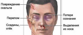
Brain contusion (concussion) - symptoms and treatment
A patient with suspected traumatic brain injury should receive medical attention as soon as possible. Since it is impossible to reverse brain damage resulting from trauma, treatment measures should be aimed at stabilizing the patient's condition and preventing secondary damage.
Conservative treatment of brain contusions
In the setting of traumatic illness, decreased or increased intracranial pressure, decreased blood oxygen saturation, high temperature, and increased blood glucose levels can have a detrimental effect on the brain. In this regard, the main directions of therapy for people with severe bruises are identified [4]:
- breathing support (ventilators, oxygen mask);
- correction of blood circulation and infusion therapy (medication);
- treatment of intracranial hypertension;
- neuroprotection - protection of neurons from damage, carried out with medications.
Basic therapy. For victims with severe TBI, an open airway is created by removing saliva, blood and vomit. Sedatives and muscle relaxants are used as needed (designed to relax muscles, block nerve impulses and reduce pain.). Correction of elevated body temperature is necessary (using antipyretic drugs and/or physical conditioning methods) [4].
The development of convulsive syndrome in persons with a brain contusion is considered dangerous and requires immediate response. Seizures are always accompanied by rapid intracranial hypertension, an increase in the volume of intracranial hematomas, increased oxygen consumption in the brain, its blood supply and increased cerebral edema [13]. Prophylactic use of anticonvulsants (according to multicenter studies) in people with TBI reduces the likelihood of seizures in the acute period, but does not reduce the risk of their occurrence in the long-term period.
All persons with a cerebral contusion require prevention of thromboembolic complications (deep vein rhombosis of the lower extremities and pulmonary embolism), which involves the use of compression stockings, increased physical activity and anticoagulant therapy whenever possible. Mortality in thrombosis of the veins of the lower extremities reaches 9-50% [7].
It is also necessary to control blood glucose levels and prevent complications from the gastrointestinal tract (stress ulcers, gastrointestinal bleeding). The main reason for the development of stress ulcers is the release of catecholamine hormones during injury, which are produced in response to stress, insufficient blood supply to the upper gastrointestinal tract and disruption of its self-regulation.
Respiratory support. Indications for respiratory support [3][10]:
- depression of the level of wakefulness to stupor or coma;
- lack of own breathing;
- acutely developed breathing rhythm disturbances, pathological type of breathing (Cheyne-Stokes, Kussmaul);
- tachypnea (rapid shallow breathing) more than 30 respiratory movements per minute;
- clinical signs of hypoxemia (lack of oxygen in the blood) and/or hypercapnia (excess carbon dioxide): partial pressure of oxygen in arterial blood (PaO2) less than 60 mm Hg. Art.; hemoglobin saturation with arterial blood oxygen (SaO2) is less than 90%; partial pressure of carbon dioxide in arterial blood (PaCO2) more than 55 mm Hg. Art.;
- status epilepticus (epileptic seizures do not stop for more than 30 minutes);
- maxillofacial trauma combined with trauma to the facial skeleton, cranio-orbital region (near the orbit and adjacent areas) and/or chest.
The main task of respiratory support is to maintain normal carbon dioxide tension (PaCO2 - 30-35 mm Hg) and the necessary oxygenation of blood in the arterial bed (PaO2 more than 100 mm Hg) [6][7].
If the duration of mechanical ventilation is more than 48 hours after the start of breathing support, a tracheostomy should be performed (an operation to create an external opening in the wall of the trachea to establish breathing). When a brain contusion is combined with trauma to the facial skeleton and chest, it is preferable to perform a tracheostomy immediately upon admission of the victim to the hospital [10].
Correction of blood circulation and infusion therapy. More than half of patients with a decrease in wakefulness to stupor and coma are in a state of dehydration upon admission to the hospital. Causes include bleeding, insufficient fluid intake, overheating, vomiting and/or diabetes insipidus. Performing adequate infusion therapy (intravenous administration of medicinal solutions and drugs using a dropper) makes it possible to achieve a normal total blood volume, normalize cardiac output and the delivery of blood and oxygen to the brain.
The injured brain is extremely sensitive to low blood pressure (below 90 mm Hg), which a healthy brain tolerates normally. Therefore, the management of patients with severe TBI involves maintaining blood pressure (BP should be at least 90 mm Hg), which is necessary for adequate brain perfusion (providing it with oxygen and glucose) in conditions of edema [4][5][6] [7][9]. However, aggressive attempts to maintain blood pressure using vasopressors (vasoconstrictors) should be avoided due to the risk of respiratory distress syndrome (pulmonary edema with difficulty breathing) and cerebral edema [4][7].
Treatment of intracranial hypertension. To select adequate treatment tactics, it is necessary to distinguish between intracranial and extracranial causes of high ICP. The former include intracranial hematomas, bruises, edema and/or cerebral ischemia, epileptic seizures, and meningitis. The second is a lack of oxygen supply, inadequate sedation or ventilation, impaired venous outflow from the cranial cavity, increased intrathoracic and intra-abdominal pressure [1][3]. All these reasons can accompany brain contusion.
Sedation and analgesia are first-line interventions for the treatment of intracranial hypertension [4][7]. The head of the bed must be raised by 30-40° to improve venous outflow from the cranial cavity. To treat increased intracranial pressure and protect the brain from secondary damage, craniocerebral hypothermia (lowering brain temperature) is used. It is sufficient to carry out moderate hypothermia (T = 30-31 °C) [1][4]. The use of hyperbaric oxygenation (saturation of the patient with oxygen under high pressure) is pathogenetically justified in persons with brain contusion, since this method increases the oxygen tension in arterial blood and improves oxygen saturation of the brain.
To reduce intra-abdominal pressure, drugs are used that improve intestinal motility and normalize the function of the gastrointestinal tract [4][6]. If intracranial hypertension is not responsive to drug therapy, decompressive craniotomy is indicated.
Neuroprotection. Currently, the use of neuroprotective drugs is a promising direction in the treatment of brain contusions [9]. Based on their mechanism of action, neuroprotectors are divided into several types:
- Calcium channel blockers (Nimodipine, Breinal, Dilceren, Nimopin, Nimotop). They reduce the entry of calcium ions into the cell, reduce the level of damage and death of nerve cells under the influence of neurotransmitters and prevent apoptosis (programmed cell death).
- Antioxidants and antihypoxants (Actovegin, polyethylene glycol superoxide dismutase, Solcoseryl, Neurox, Mexidol, Armadin, Cytoflavin, Neurocard, Astrox, Meksifin, tocopherol, Methostabil, ascorbic acid, Ascovit, Nootropil, Piracetam, Noocetam, Melatonin, Cavinton, Vinpocetine, Cohen winter Q10). These drugs are nitrogen synthase antagonists, they prevent the formation of free radicals, restore the activity of antioxidant enzymes, accelerate glycolysis (the process of glucose oxidation), increase resistance to hypoxia and improve cerebral blood flow.
- NMDA receptor antagonists (Memantine, Memantal, Memorel, Noodzheron, Akatinol, Memikar, Mirvedol, Memaneurin). Reduce the damaging effects of glutamate.
- Blockers of the inflammatory and immune response (Cycloferon, COX-2 inhibitors, CD11 and CD18 antibodies). Reduce the severity of the inflammatory reaction.
- Stabilizers of cell membranes: intermediate products of phosphatidylcholine synthesis (Recognan, citicoline, Ceraxon, Proneuro, Quinelle, Neuropol, Neurocholin, Noocil, Ceresil Canon), magnesium and potassium preparations (magnesium sulfate, Aspangin, Pamaton, Asparkam, Panaspar, Panangin).
- Drugs that improve synaptic transmission (precursors for acetylcholine synthesis - Cereton, Cerepro, Gleatser, Holitylin, Delecit, Gliatilin).
- Apoptosis blockers (caspase-3 inhibitors, calpain inhibitors).
- Drugs with a neuron-specific neurotrophic effect (Cortexin, Cerebrolysate, Cerebrolysin).
- Immunosuppressants (cyclosporine A). Suppress the immune response.
Nutrition for severe brain contusion . With serious damage, patients, as a rule, cannot eat on their own, so early nutritional support (i.e., the introduction of nutrients into the body) is necessary. Such support should match the patient's protein and energy needs. Basic energy metabolism in patients with severe brain contusion corresponds to 20-25 kcal/kg per day [4][6].
Surgical treatment of brain contusions
Some patients with severe traumatic brain injury are transported to the operating room after a head CT scan. The purpose of surgery for severe brain contusion is to remove massive intracranial hematomas or correct elevated ICP.
Not all patients with TBI require emergency intervention. Since the size of a brain contusion and the volume of an intracranial hematoma may increase during the first hours/days after injury, dynamic monitoring is recommended. Treatment of such patients is carried out in an intensive care unit. If neurological disorders increase or intracranial pressure increases (if an ICP monitoring sensor was installed), a control CT scan is performed. If a significant increase in the volume of the hematoma or displacement of the brain is diagnosed, the safest option will be surgical intervention to prevent brain death.
Before surgery, the hair over the affected area of the brain is shaved. After cutting the scalp and removing the skin flap, the bone is cut out. The bone is removed and the underlying dura mater is opened with the utmost care. The doctor removes the hematoma or brain contusion. In the presence of severe cerebral edema, plastic surgery of the dura mater is performed using a patch from one’s own tissue or artificial replacement material. This is necessary to prevent further displacement of the brain. In such cases, the bone is not put back in its original place.
Before “closing” the wound, it is possible to install an ICP sensor if it has not been installed previously. Upon completion of the operation, the patient is transferred to the intensive care unit for intensive treatment measures aimed at combating cerebral edema and preventing infectious and thromboembolic complications.
Decompressive craniotomy is the most aggressive treatment for intracranial hypertension [3][9]. It is used when conservative therapy is ineffective. The main goal of decompressive trephination is to increase intracranial volume. As a result, the pressure in the cranial cavity is reduced and the blood supply to the brain is normalized.
Decompressive trepanation includes infratemporal and bifrontal decompression, temporal lobectomy and hemicraniectomy. They are carried out in cases of pronounced displacement of brain structures and persistent intracranial hypertension in patients with clinical and CT signs of brain compression.
Possible postoperative complications. In the long-term postoperative period of TBI, epilepsy is registered in 13% of patients, and post-traumatic hydrocephalus in 11% [3]. Post-traumatic epilepsy occurs due to the formation of a pathological focus in the brain. Post-traumatic hydrocephalus is caused by impaired circulation (formation of adhesions) and absorption of cerebrospinal fluid.