An axon in human anatomy is a connecting neural structure. It connects nerve cells to all organs and tissues, thereby ensuring the exchange of impulses throughout the body.
Axon (from Greek - axis) is a brain fiber, a long, elongated fragment of a brain cell (neuron), a process or neurite, an area that transmits electrical signals at a distance from the brain cell (soma) itself.
Many nerve cells have only one process; cells in small numbers without any neutrites.
An axon looks like an elongated cone-shaped process, the duration and circumference of which varies and depends on the size of the brain cell.
Despite the fact that the axons of individual nerve cells are short, as a rule, they are characterized by a very significant length. For example, the processes of motor spinal neurons that transmit the muscles of the foot can reach a length of up to 100 cm. The base of all axons is a small triangular fragment - a neutrite mound - branching from the neuron body itself. The outer protective layer of the axon is called axolemma (from the Greek axon - axis + eilema - sheath), and its internal structure is axoplasm.
Cell membrane
This element provides a barrier function, separating the internal environment from the neuroglia located outside. The thinnest film consists of two layers of protein molecules and phospholipids located between them. The structure of the neuron membrane suggests the presence in its structure of specific receptors responsible for recognizing stimuli. They have selective sensitivity and, if necessary, “turn on” in the presence of a counterparty. The connection between the internal and external environments occurs through tubules that allow calcium or potassium ions to pass through. At the same time, they open or close under the influence of protein receptors.
Thanks to the membrane, the cell has its potential. When it is transmitted along the chain, excitable tissue is innervated. Contact between the membranes of neighboring neurons occurs at synapses. Maintaining a constant internal environment is an important component of the life of any cell. And the membrane subtly regulates the concentration of molecules and charged ions in the cytoplasm. At the same time, they are transported in the required quantities for metabolic reactions to occur at an optimal level.
Classification
Structural classification
Based on the number and arrangement of dendrites and axons, neurons are divided into axonless neurons, unipolar neurons, pseudounipolar neurons, bipolar neurons, and multipolar (many dendritic arbors, usually efferent) neurons.
Axonless neurons
- small cells, grouped near the spinal cord in the intervertebral ganglia, which do not have anatomical signs of division of processes into dendrites and axons. All processes of the cell are very similar. The functional purpose of axonless neurons is poorly understood.
Unipolar neurons
- neurons with a single process, present, for example, in the sensory nucleus of the trigeminal nerve in the midbrain. Many morphologists believe that unipolar neurons do not occur in the body of humans and higher vertebrates.
Bipolar neurons
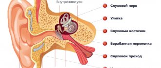
- neurons with one axon and one dendrite, located in specialized sensory organs - the retina, olfactory epithelium and bulb, auditory and vestibular ganglia.
Multipolar neurons
- neurons with one axon and several dendrites. This type of nerve cells predominates in the central nervous system.
Pseudounipolar neurons
- are unique in their kind. One process extends from the body, which immediately divides in a T-shape. This entire single tract is covered with a myelin sheath and is structurally an axon, although along one of the branches the excitation goes not from, but to the body of the neuron. Structurally, dendrites are branches at the end of this (peripheral) process. The trigger zone is the beginning of this branching (that is, it is located outside the cell body). Such neurons are found in the spinal ganglia.
Functional classification
Based on their position in the reflex arc, afferent neurons (sensitive neurons), efferent neurons (some of them are called motor neurons, sometimes this not very accurate name applies to the entire group of efferents) and interneurons (interneurons) are distinguished.
Afferent neurons
(sensitive, sensory, receptor or centripetal). Neurons of this type include primary cells of the sensory organs and pseudounipolar cells, whose dendrites have free endings.
Efferent neurons
(effector, motor, motor or centrifugal). Neurons of this type include the final neurons - ultimatum and penultimate - non-ultimatum.
Association neurons
(interneurons or interneurons) - a group of neurons communicates between efferent and afferent ones.
Secretory neurons
- neurons that secrete highly active substances (neurohormones). They have a well-developed Golgi complex, the axon ends at axovasal synapses.
Morphological classification
The morphological structure of neurons is diverse. Several principles are used to classify neurons:
- take into account the size and shape of the neuron body;
- number and nature of branching of processes;
- axon length and the presence of specialized sheaths.
According to the shape of the cell, neurons can be spherical, granular, stellate, pyramidal, pear-shaped, fusiform, irregular, etc. The size of the neuron body varies from 5 μm in small granular cells to 120-150 μm in giant pyramidal neurons.
Based on the number of processes, the following morphological types of neurons are distinguished:
- unipolar (with one process) neurocytes, present, for example, in the sensory nucleus of the trigeminal nerve in the midbrain;
- pseudounipolar cells grouped near the spinal cord in the intervertebral ganglia;
- bipolar neurons (have one axon and one dendrite), located in specialized sensory organs - the retina, olfactory epithelium and bulb, auditory and vestibular ganglia;
- multipolar neurons (have one axon and several dendrites), predominant in the central nervous system.
Structure of neurons
Neuron diagram
Cell body
The body of a nerve cell consists of protoplasm (cytoplasm and nucleus), bounded on the outside by a membrane of lipid bilayer. Lipids consist of hydrophilic heads and hydrophobic tails. The lipids are arranged with hydrophobic tails facing each other, forming a hydrophobic layer. This layer allows only fat-soluble substances (eg oxygen and carbon dioxide) to pass through. There are proteins on the membrane: in the form of globules on the surface, on which growths of polysaccharides (glycocalyx) can be observed, thanks to which the cell perceives external irritation, and integral proteins that penetrate the membrane through, in which ion channels are located.
A neuron consists of a body with a diameter ranging from 3 to 130 microns. The body contains a nucleus (with a large number of nuclear pores) and organelles (including a highly developed rough ER with active ribosomes, the Golgi apparatus), as well as processes. There are two types of processes: dendrites and axons. The neuron has a developed cytoskeleton that penetrates its processes. The cytoskeleton maintains the shape of the cell; its threads serve as “rails” for the transport of organelles and substances packaged in membrane vesicles (for example, neurotransmitters). The cytoskeleton of a neuron consists of fibrils of different diameters: Microtubules (D = 20-30 nm) - consist of the protein tubulin and stretch from the neuron along the axon, right up to the nerve endings. Neurofilaments (D = 10 nm) - together with microtubules, provide intracellular transport of substances. Microfilaments (D = 5 nm) - consist of actin and myosin proteins, especially pronounced in growing nerve processes and in neuroglia. ( Neuroglia
, or simply glia (from ancient Greek νεῦρον - fiber, nerve + γλία - glue), is a collection of auxiliary cells of the nervous tissue. Makes up about 40% of the volume of the central nervous system. The number of glial cells in the brain is approximately equal to the number of neurons).
A developed synthetic apparatus is revealed in the body of the neuron; the granular endoplasmic reticulum of the neuron is stained basophilically and is known as the “tigroid”. The tigroid penetrates the initial sections of the dendrites, but is located at a noticeable distance from the beginning of the axon, which serves as a histological sign of the axon. Neurons vary in shape, number of processes, and functions. Depending on the function, sensitive, effector (motor, secretory) and intercalary are distinguished. Sensory neurons perceive stimuli, convert them into nerve impulses and transmit them to the brain. Effector (from Latin effectus - action) - generate and send commands to the working organs. Intercalary neurons - communicate between sensory and motor neurons, participate in information processing and the generation of commands.
There is a distinction between anterograde (away from the body) and retrograde (toward the body) axon transport.
Dendrites and axon
Main articles: Dendrite
and
Axon
Scheme of neuron structure
An axon is a long extension of a neuron. Adapted for carrying excitation and information from the body of a neuron to a neuron or from a neuron to an executive organ. Dendrites are short and highly branched processes of a neuron, which serve as the main site for the formation of excitatory and inhibitory synapses affecting the neuron (different neurons have different ratios of axon and dendrite lengths), and which transmit excitation to the body of the neuron. A neuron may have several dendrites and usually only one axon. One neuron can have connections with many (up to 20 thousand) other neurons.
Dendrites divide dichotomously, while axons give off collaterals. Mitochondria are usually concentrated at branching nodes.
Dendrites do not have a myelin sheath, but axons may have one. The place of generation of excitation in most neurons is the axon hillock - a formation at the point where the axon departs from the body. In all neurons, this zone is called the trigger zone.
Synapse
Main article: Synapse
Synapse
(Greek σύναψις, from συνάπτειν - hug, clasp, shake hands) - the place of contact between two neurons or between a neuron and the effector cell receiving the signal. It serves to transmit a nerve impulse between two cells, and during synaptic transmission the amplitude and frequency of the signal can be adjusted. Some synapses cause depolarization of the neuron and are excitatory, while others cause hyperpolarization and are inhibitory. Typically, stimulation from several excitatory synapses is necessary to excite a neuron.
The term was introduced by the English physiologist Charles Sherrington in 1897.
Damaged wrapper
Figure 5. Sensory impairment of the polyneuritic type. The name "sock-glove" is due to the fact that the anatomical areas corresponding to nerve damage are similar to the areas covered by these items of clothing.
website dic.academic.ru
It seems to me that the following rule is quite suitable for the human body: if there is an organ, then there must be a disease associated with it. In principle, this rule can be extended to molecular processes: if there is a process, there are also diseases associated with disruption of this process. In the case of myelin, these are demyelinating diseases. There are quite a few of them, but I will talk in more detail about two - Guillain-Barré syndrome and multiple sclerosis. In these disorders, damage to myelin leads to disruption of adequate signal transmission along the nerves, which causes the symptoms of the disease.
Guillain-Barré syndrome (GBS) is a disease of the peripheral nervous system in which the myelin sheath formed by Schwann cells is destroyed. GBS is a classic autoimmune disease. As a rule, it is preceded by an infection (often caused by the microbe Campylobacter jejuni). The presence of various pathogens in the human body triggers autoimmune damage to the myelin of nerve fibers by T- and B-lymphocytes. Clinically, this is manifested by muscle weakness, sensory impairment of the “socks-gloves” type (polyneuritic type) (Fig. 5). In the future, muscle weakness can increase until complete paralysis of the limbs and damage to the trunk muscles. Damages to the sensitive nervous system can also be varied: from a decrease in the ability to distinguish one’s own movements (impaired deep sensitivity) to severe pain. In severe forms of GBS, the main danger is the loss of the ability to breathe independently, requiring connection to a mechanical ventilation device (ALV). To treat GBS, plasmapheresis (purification of plasma from harmful antibodies) and intravenous infusions of human immunoglobulin preparations are currently used to normalize the immune response. In most cases, treatment leads to lasting recovery.
Multiple sclerosis (MS) is markedly different from GBS. First, this demyelinating disease leads to damage to the central nervous system, that is, it affects the myelin synthesized by oligodendrocytes. Secondly, there is still a lot of uncertainty about the causes of MS: too many genetic and environmental factors are involved in the pathogenesis of the disease. The fundamental point in the initiation of MS is a violation of the impermeability of the blood-brain barrier (BBB) for immune cells. Normally, brain tissue is fenced off from the rest of the body by this reliable filter, which prevents many substances and cells, including immune cells, from entering it. The BBB appears already in the embryonic period of development, isolating brain tissue from the developing immune system. At this time, the human immune system “gets acquainted” with all existing tissues, so that in the future, during adulthood, it does not attack them. The brain and a number of other organs remain “not presented” to the immune system. When the integrity of the BBB is disrupted, immune cells have the opportunity to attack unfamiliar brain tissue. Third, MS is characterized by more severe symptoms that require different therapeutic approaches. Symptoms depend on where the damage to the nervous system is located (Fig. 6 and 7). This may include unsteadiness of gait, sensory disturbances, and various cognitive symptoms. To treat MS, high doses of glucocorticoids and cytostatics are used, as well as interferon drugs and specific antibodies (natalizumab). Apparently, new methods of treating MS will be developed in the future, based directly on the restoration of the myelin sheath in damaged areas of the brain. Scientists point to the possibility of transplanting oligodendrocyte precursor cells or enhancing their growth by introducing insulin-like growth factor or thyroid hormones [11]. However, this is still ahead, and for now, more “molecular” treatment methods are not available to neurologists.
Figure 6. Lesions of the central nervous system in multiple sclerosis appear as white plaques on MRI.
website neurology.org
Figure 7. Depending on the location of damage to the nervous system in multiple sclerosis, there may be different symptoms: from tremor and ataxia with damage to the cerebellum to emotional disorders with localization of lesions in the frontal lobes.
www.diagnosisms.com
Literature
- Polyakov G.I., On the principles of neural organization of the brain, M: Moscow State University, 1965
- Kositsyn N. S. Microstructure of dendrites and axodendritic connections in the central nervous system. M.: Nauka, 1976, 197 p.
- Nemechek S. et al. Introduction to neurobiology, Avicennum: Prague, 1978, 400 p.
- Brain (collection of articles: D. Hubel, C. Stevens, E. Kandel, etc. - issue of Scientific American (September 1979)). M.:Mir, 1980
- Savelyeva-Novoselova N. A., Savelyev A. V. Device for modeling a neuron. A. s. No. 1436720, 1988
- Savelyev A.V.
Sources of variations in the dynamic properties of the nervous system at the synaptic level // Journal “Artificial Intelligence”, NAS of Ukraine. - Donetsk, Ukraine, 2006. - No. 4. - P. 323-338.
Important wrapper
Myelination (the gradual insulation of nerve fibers by myelin) begins in humans already in the embryonic period of development. The first to pass this path are the subcortical structures. During the first year of life, myelination occurs in the parts of the peripheral and central nervous system responsible for motor activity. Myelination of areas of the brain that regulate higher nervous activity ends by the age of 12–13 years. From this it is clear that myelination is closely related to the ability of parts of the nervous system to perform functions specific to them. It is probably the active work of the fibers before birth that triggers their myelination.
The differentiation of oligodendrocyte precursor cells depends on a number of factors related to the functioning of neurons. In particular, working neuronal processes can secrete the protein neuroligin 3, which promotes the proliferation and differentiation of progenitor cells [4]. Subsequent maturation of oligodendrocytes occurs due to a number of other factors. In an article with the characteristic title “How Big is the Myelinating Orchestra?” describes the origin of oligodendrocytes in different parts of the brain [5]. First, in different parts of the brain, oligodendrocytes begin to mature at different times. Secondly, different cellular factors are responsible for their maturation, which also depends on the region of the nervous system (Fig. 3). We may have a question: are the oligodendrocytes that appeared with such a discrepancy in the starting data similar to each other? And how similar is their myelin? In general, the authors of the article believe that there are indeed differences between populations of oligodendrocytes from different parts of the brain, and they are largely due to the location of the cells and the influence of surrounding neurons on them. Yet the types of myelin synthesized by different pools of oligodendrocytes do not differ so much that they are not interchangeable.
Figure 3. Differences in the timing of oligodendrocyte formation in different parts of the brain and in the cellular factors influencing their development.
[5]
The process of myelination of nerve fibers in the central nervous system occurs as follows (Fig. 4). Oligodendrocytes send out several processes to the axons of different neurons. Coming into contact with them, the processes of oligodendrocytes begin to wrap around them and spread along the length of the axon. The number of turns gradually increases: in some areas of the central nervous system their number reaches 50. The membranes of oligodendrocytes become increasingly thin, spreading along the surface of the axon and “squeezing out” the cytoplasm. The earlier the layer of myelin was wrapped around the nerve ending, the thinner it will be. The innermost layer of the membrane remains quite thick - for metabolic function. New layers of myelin are wound on top of the old ones, overlapping them as shown in Figure 4 - not only on top, but also increasing the area of the axon covered by myelin.
Figure 4. Myelination of nerve fiber. The oligodendrocyte membrane wraps around the axon, gradually becoming denser with each turn. The inner layer of membrane adjacent to the axon remains relatively thick, which is necessary for metabolic function. Different parts of the figure ( a-c ) from different angles show the gradual winding of new layers of myelin onto the axon. The thicker, metabolically active layer is highlighted in red, and the new thickening layers are highlighted in blue. The inner layer of myelin (inner tongue in part b ) is covered by more and more layers of membrane, not only on top, but also on the sides ( c ), along the axon.
[6]
Myelination of nerve fibers by oligodendrocytes also significantly depends on the protein neuregulin 1. If it does not affect oligodendrocytes, then a myelination program is launched in them, which does not take into account the activity of the nerve cell. If oligodendrocytes received a signal from neuregulin 1, then they will then begin to focus on the work of the axon, and myelination will depend on the intensity of glutamate production and its activation of specific NMDA receptors on the surface of oligodendrocytes [6]. Neuregulin 1 is a key factor for triggering myelination processes in the case of Schwann cells [7].
Neuron structure
The figure shows the structure of a neuron. It consists of a main body and a core. Numerous fibers branch off from the cell body, which are called dendrites.
Powerful and long dendrites are called axons, which in reality are much longer than in the picture. Their length varies from a few millimeters to more than a meter.
Axons play a leading role in transmitting information between neurons and ensure the functioning of the entire nervous system.
The junction of a dendrite (axon) with another neuron is called a synapse. Dendrites, in the presence of stimuli, can grow so much that they begin to pick up impulses from other cells, which leads to the formation of new synaptic connections.
Synaptic connections play a significant role in the formation of human personality. Thus, a person with an established positive experience will look at life with love and hope, a person who has neural connections with a negative charge will become a pessimist over time.
Functions
The main task of neurons is data processing. With their help, information is received, processed, and transmitted to parts of the nervous and other systems of the body.
If dendrites conduct signals towards the body of the nerve cell (perikaryon), then the axonal process transmits impulses from the perikaryon to other cells.
The main function of axons is to conduct impulses within the neural network and to the executive organs. Axonal branches are among the primary pathways in the nervous system. An auxiliary function is the transport of substances. With the help of axonal transport, the movement of proteins synthesized in the body, neurotransmitters, and organelles occurs. Many substances can move in both directions.
In the peripheral segments of the axon, viruses and toxic substances can penetrate into it, moving to the body of the nerve cell and damaging it. Axonal transport depends on the amount of ATP energy. If the energy level of ATP decreases by more than 2 times, axonal transport is blocked.
The functions of the axon are to transmit impulses. When an axon interacts with the body of another neuron, an axosomatic contact is formed. If an axon interacts with the dendrites of other cells, axodendritic contact occurs. Interaction with the axon of another cell leads to the formation of axo-axonal contact, which rarely occurs in the nervous system, and supports inhibitory reflex reactions.
Fibers
Glial sheaths are located independently around the nerve processes. Together they form nerve fibers. The branches in them are called axial cylinders. There are unmyelinated and myelinated fibers. They differ in the structure of the glial membrane. Unmyelinated fibers have a fairly simple structure. The axial cylinder approaching the glial cell bends its cytolemma. The cytoplasm closes over it and forms a mesaxon - a double fold. One glial cell can contain several axial cylinders. These are “cable” fibers. Their branches can pass into neighboring glial cells. The pulse travels at a speed of 1-5 m/s. Fibers of this type are found during embryogenesis and in the postganglionic areas of the autonomic system. The myelin segments are thick. They are located in the somatic system, innervating the skeletal muscles. Lemmocytes (glial cells) pass sequentially, in a chain. They form a strand. An axial cylinder runs through the center. The glial membrane contains:
- The inner layer of nerve cells (myelin). It is considered the main one. In some areas between the layers of the cytolemma there are extensions that form myelin notches.
- Peripheral layer. It contains organelles and a nucleus - the neurilemma.
- Thick basement membrane.
Complex wrapper
Myelin surrounds the processes of nerve cells, insulating them from external influences. This is necessary for more reliable and faster signal transmission through the nervous system. Thanks to the insulation of the nerve fiber, the electrical signal does not dissipate and reaches its destination without interference. The speed of signal transmission along myelinated and unmyelinated fibers can differ by three orders of magnitude: from 70 to 140 m/s and from 0.3 to 0.5 m/s, respectively.
Essentially, myelin is the cell membrane of glial cells wrapped repeatedly around an axon. The membrane itself consists of 70–75% lipids and 25–30% proteins. In the peripheral nervous system, Schwann cells become membrane donors, and in the central nervous system, oligodendrocytes. These cells carefully wrap their membranes around valuable communication channels to ensure reliable communication between the nervous system and peripheral organs. Myelin does not cover the entire nerve fiber: there are gaps between the layers of myelin, called nodes of Ranvier (Fig. 1). There is a direct relationship between the distance from one gap to another and the speed of propagation of the nerve impulse along the fiber: the greater the distance between the nodes of Ranvier, the higher the speed of signal transmission in the nerve [1].
Figure 1. Nerve fiber wrapped in myelin. The nucleus of Schwann cells and nodes of Ranvier are visible - areas of the axon that are not covered by the myelin sheath.
theclickercenterblog.com
If we talk about the proteins that make up myelin, we must clarify that these are not only simple proteins. Myelin contains glycoproteins - proteins to which short carbohydrate sequences are attached. An important component of myelin is myelin basic protein (MBP), first isolated about 50 years ago. MBP is a transmembrane protein that can repeatedly “stitch” the lipid layer of a cell. Its various isoforms (Fig. 2) are encoded by a gene called Golli (gene in the oligodendrocyte lineage). The structural basis of myelin is an isoform with a mass of 18.5 kilodaltons [2].
Figure 2. Different isoforms of myelin basic protein (MBP) are created from the same gene. For example, to synthesize the 18.5 kDa isoform, all exons except exon II are used.
[2]
Myelin contains complex lipids called cerebrosides. They are the amino alcohol sphingosine combined with a fatty acid and a carbohydrate residue. Peroxisomes of oligodendrocytes take part in the synthesis of myelin lipids. Peroxisomes are lipid vesicles with various enzymes (in total, about 50 types of peroxisomal enzymes are known). These organelles are involved, in particular, in the β-oxidation of fatty acids: very long chain fatty acids (VLCFAs), some eicosanoids and polyunsaturated fatty acids (PUFAs). Since myelin can contain up to 70% lipids, peroxisomes are essential for the normal metabolism of this substance. They use N-acetylaspartate, produced by the nerve cell, to constantly synthesize new myelin lipids and maintain its existence. In addition, peroxisomes take part in maintaining the energy metabolism of axons [3].
Internal structure of neurons
Core
the neuron is usually large, round, with finely dispersed chromatin, 1-3 large nucleoli. This reflects the high intensity of transcription processes in the neuron nucleus.
Cell membrane
A neuron is capable of generating and conducting electrical impulses. This is achieved by changing the local permeability of its ion channels for Na+ and K+, changing the electrical potential and its rapid movement along the cytolemma (depolarization wave, nerve impulse).
All general purpose organelles are well developed in the cytoplasm of neurons. Mitochondria
are numerous and provide the high energy needs of the neuron, associated with significant activity of synthetic processes, the conduction of nerve impulses, and the operation of ion pumps.
They are characterized by rapid wear and renewal (Figure 8-3). The Golgi complex
is very well developed.
It is no coincidence that this organelle was first described and demonstrated in a cytology course in neurons. With light microscopy, it is detected in the form of rings, threads, and grains located around the nucleus (dictyosomes). Numerous lysosomes
ensure constant intensive destruction of worn-out components of the neuron cytoplasm (autophagy).
R
is. 8-3. Ultrastructural organization of the neuron body.
D. Dendrites. A. Axon.
1. Nucleus (nucleolus shown by arrow).
2. Mitochondria.
3. Golgi complex.
4. Chromatophilic substance (sections of the granular cytoplasmic reticulum).
5. Lysosomes.
6. Axon hillock.
7. Neurotubules, neurofilaments.
(According to V.L. Bykov).
For normal functioning and renewal of neuron structures, the protein synthesizing apparatus must be well developed (Fig. 8-3). Granular cytoplasmic reticulum
in the cytoplasm of neurons it forms clusters that are well stained with basic dyes and are visible under light microscopy in the form of clumps
of chromatophilic substance (
basophilic, or tiger substance, Nissl's substance).
The term “Nissl substance” was preserved in honor of the scientist Franz Nissl, who first described it. Lumps of chromatophilic substance are located in the perikarya of neurons and dendrites, but are never found in axons, where the protein synthesizing apparatus is poorly developed (Fig. 8-3). With prolonged irritation or damage to a neuron, these accumulations of granular cytoplasmic reticulum disintegrate into individual elements, which at the light-optical level is manifested by the disappearance of Nissl's substance ( chromatolysis
, tigrolysis).
Cytoskeleton
neurons are well developed, forming a three-dimensional network represented by neurofilaments (6-10 nm thick) and neurotubules (20-30 nm in diameter).
Neurofilaments and neurotubules are connected to each other by cross bridges; when fixed, they are glued into bundles 0.5-0.3 microns thick, which are stained with silver salts. At the light-optical level, they are described as neurofibrils
They form a network in the perikarya of neurocytes, and in the processes they lie parallel (Fig. 8-2). The cytoskeleton maintains the shape of cells and also provides a transport function - it is involved in the transport of substances from the perikaryon to the processes (axonal transport).
Inclusions
in the cytoplasm of the neuron are represented by lipid droplets, granules
of lipofuscin
- the “aging pigment” - yellow-brown in color of lipoprotein nature.
They are residual bodies (telolysosomes) containing products of undigested neuron structures. Apparently, lipofuscin can accumulate at a young age, with intense functioning and damage to neurons. In addition, in the cytoplasm of neurons of the substantia nigra and locus coeruleus of the brain stem there are pigment inclusions of melanin
.
Glycogen
inclusions are found in many neurons of the brain .
Neurons are not capable of division, and with age their number gradually decreases due to natural death. In degenerative diseases (Alzheimer's disease, Huntington's disease, parkinsonism), the intensity of apoptosis increases and the number of neurons in certain areas of the nervous system sharply decreases.
Sensory function[edit | edit code]
Different sensory receptors are stimulated by different types of nerve fibers.
Proprioceptors are excited by type Ia, Ib and II sensory fibers, mechanoreceptors - by type II and III sensory fibers and type nociceptors and thermoreceptors. Sensory fiber types
| Types | Classification | Diameter | Myelin | Conduction speed | Associated sensory receptors |
| Aα | 13-20 microns | Yes | 80-120 m/s | Primary receptors of muscle spindle | |
| Ib | Aα | 13-20 microns | Yes | 80-120 m/s | Golgi tendon organ |
| Aβ | 6-12 microns | Yes | 33-75 m/s | Secondary receptors of muscle spindle All cutaneous mechanoreceptors | |
| III | 1-5 microns | Thin | 3-30 m/s | Free nerve endings of touch and pressure Nociceptors of neospinothalamic tract Cold thermoreceptors | |
| IV | 0.2-1.5 microns | No | 0.5-2.0 m/s | Nociceptors of paleospinothalamic tract Warmth receptors |
Nerve cells
To provide multiple connections, the neuron has a special structure. In addition to the body, in which the main organelles are concentrated, there are processes. Some of them are short (dendrites), usually there are several of them, the other (axon) is one, and its length in individual structures can reach 1 meter.
The structure of the nerve cell of the neuron is designed in such a way as to ensure the best interchange of information. Dendrites are highly branched (like the crown of a tree). With their endings they interact with the processes of other cells. The place where they meet is called a synapse. This is where the impulse is received and transmitted. Its direction: receptor – dendrite – cell body (soma) – axon – reacting organ or tissue.
The internal structure of a neuron is similar in composition to organelles to other structural units of tissue. It contains a nucleus and cytoplasm bounded by a membrane. Inside there are mitochondria and ribosomes, microtubules, the endoplasmic reticulum, and the Golgi apparatus.
How an axon is formed
The elongation and development of these neuron processes is ensured by the location of their location.
Elongation of axons becomes possible due to the presence of filopodia at their upper end, between which are located, like corrugations, membrane formations - lamelopodium. Filopodia actively interact with nearby structures, penetrating deeper into the tissue, resulting in directed elongation of axons. The filopodia itself sets the direction for the axon to increase in length, establishing the certainty of the organization of fibers. The participation of filopodia in the directional elongation of neutrites was confirmed in a practical experiment by introducing cytochalasin B into embryos, which destroys filopodia. At the same time, the axons of the neurons did not grow to the brain centers.
The production of immunoglobulin, which is often found at the junction of areas of axonal growth with glial cells and, according to the hypotheses of a number of scientists, this fact determines the direction of axonal elongation in the decussation zone. If this factor promotes axon elongation, then chondroitin sulfate, on the contrary, slows down the growth of neutrites.
Synapses
With their help, the cells of the nervous system connect with each other. There are different synapses: axo-somatic, -dendritic, -axonal (mainly inhibitory). Electrical and chemical ones are also released (the former are detected quite rarely in the body). Synapses are divided into post- and presynaptic parts. The first contains a membrane in which highly specific protein (protein) receptors are present. They respond only to certain mediators. There is a gap between the pre- and postsynaptic parts. The nerve impulse reaches the first and activates special vesicles. They move to the presynaptic membrane and enter the cleft. From there they influence the postsynaptic film receptor. This provokes its depolarization, which is transmitted, in turn, through the central process of the next nerve cell. In a chemical synapse, information is transmitted in only one direction.
Development
The formation of nervous tissue occurs in the third week of the embryonic period. At this time, a plate is formed. From it develop:
- Oligodendrocytes.
- Astrocytes.
- Ependymocytes.
- Macroglia.
During further embryogenesis, the neural plate turns into a tube. In the inner layer of its wall there are stem ventricular elements. They proliferate and move outward. In this area, some cells continue to divide. As a result, they are divided into spongioblasts (components of microglia), glioblasts and neuroblasts. From the latter, nerve cells are formed. There are 3 layers in the tube wall:
- Internal (ependymal).
- Medium (raincoat).
- External (marginal) - represented by the white medulla.
At 20-24 weeks, the formation of bubbles begins in the cranial segment of the tube, which are the source of the formation of the brain. The remaining sections serve for the development of the spinal cord. Cells involved in the formation of the crest extend from the edges of the neural groove. It is located between the ectoderm and the tube. From these same cells, ganglion plates are formed, which serve as the basis for myelocytes (pigmented skin elements), peripheral nerve ganglia, melanocytes of the integument, and components of the APUD system.
Variable wrapper
Myelin is constantly formed and destroyed in the human body. The synthesis and breakdown of myelin can be influenced by environmental factors. For example, education. From 1965 to 1989, Romania was led by Nicolae Ceausescu. He established strict control over reproductive health and the institution of marriage in his country: he complicated the divorce procedure, banned abortion and introduced a number of incentives and benefits for women who had more than five children. The result of these measures was the expected increase in the birth rate. Along with the birth rate, the number of criminal abortions, which did not add to the health of Romanians, increased, and the number of abandoned children increased. The latter were brought up in orphanages, where the staff did not actively communicate with them. Romanian children have fully experienced what is called social deprivation - deprivation of the opportunity to fully communicate with other people. If we are talking about a small child, then the consequences of social deprivation will be a violation of the formation of emotional attachments and attention disorders. When the Ceausescu regime fell, Western scholars had to fully evaluate the results of the social policies of this dictator. Romanian children with severe problems with attention and establishing social contacts subsequently began to be called Ceausescu's children.
In addition to differences in the performance of neuropsychological tests, even the structure of the brain was different in Ceausescu’s children compared to children who were not in such conditions [8]. When assessing the state of the white matter of the brain, scientists use the fractal anisotropy indicator. It allows you to evaluate the density of nerve fibers, the diameter of axons and their myelination. The greater the fractal anisotropy, the more diverse the fibers that are found in that area of the brain. In Ceausescu's children, there was a decrease in fractal anisotropy in the white matter bundle connecting the temporal and frontal lobes in the left hemisphere, that is, the connections in this region were not sufficiently complex and diverse, with disturbances in myelination. This state of connections interferes with the normal transmission of signals between the temporal and frontal lobes. The temporal lobe contains emotional response centers (amygdala, hippocampus), and the orbitofrontal cortex of the frontal lobe is also associated with emotions and decision making. Disruption of the formation of connections between these parts of the brain and problems in their work ultimately led to the fact that children raised in orphanages experienced difficulties in establishing normal relationships with other people.
Myelination can also be affected by the composition of the food given to the child. With protein-energy malnutrition, there is a decrease in myelin formation. The lack of fatty acids also negatively affects the synthesis of this valuable substance, since more than 2/3 of it consists of lipids. Deficiency of iron, iodine and B vitamins leads to decreased myelin formation [9]. Basically, these data were obtained from the study of laboratory animals, but history, unfortunately, has given people the opportunity to assess the impact of lack of food on the developing brain of a child [10]. Hungry winter (Dutch hongerwinter) 1944–1945 in the Netherlands resulted in many children being born whose mothers were malnourished. It turned out that under conditions of starvation, the brains of these children were formed with disturbances. In particular, a large number of disorders were observed in the white matter, that is, problems arose with the formation of myelin. As a result, this led to a variety of mental disorders.
Classification
Neurons are divided into types depending on the type of mediator (mediator of the conductive impulse) released at the axon terminals. This can be choline, adrenaline, etc. Depending on their location in the parts of the central nervous system, they can relate to somatic neurons or autonomic ones. There are receptive cells (afferent) and transmitting feedback signals (efferent) in response to irritation. Between them there may be interneurons responsible for the exchange of information within the central nervous system. Depending on the type of response, cells can inhibit excitation or, conversely, increase it.
According to their state of readiness, they are distinguished: “silent”, which begin to act (transmit an impulse) only in the presence of a certain type of irritation, and background, which constantly monitor (continuous generation of signals). Depending on the type of information perceived from the sensors, the structure of the neuron also changes. In this regard, they are classified into bimodal, with a relatively simple response to irritation (two interrelated types of sensation: a prick and, as a result, pain, and polymodal. This is a more complex structure - polymodal neurons (specific and ambiguous reaction).
What is a neuron? Neural connections
Translated from Greek, neuron, or as it is also called neuron, means “fiber”, “nerve”. Neuron
- this is a specific structure in our body, which is responsible for the transmission of any information within it, in everyday life called a nerve cell.
Neurons operate using electrical signals and help the brain process incoming information to further coordinate the body's actions.
These cells are an integral part of the human nervous system, the purpose of which is to collect all signals coming from outside or from one’s own body and make a decision about the need for one or another action. It is the neurons that help cope with this task.
Each of the neurons has a connection with a huge number of the same cells, a kind of “web” is created, which is called a neural network. Through this connection, electrical and chemical impulses are transmitted in the body, bringing the entire nervous system to a state of rest or, conversely, excitation.
For example, a person encountered a certain significant event. An electrochemical impulse (impulse) of neurons occurs, leading to the excitation of the uneven system. A person’s heart begins to beat faster, their hands sweat, or other physiological reactions occur.
We are born with a given number of neurons, but the connections between them have not yet been formed. The neural network is built gradually as a result of impulses coming from outside. New impulses form new neural pathways; it is along them that similar information will flow throughout life. The brain perceives each person's individual experience and reacts to it. For example, a child grabbed a hot iron and pulled his hand away. This is how he developed a new neural connection.
A child develops a stable neural network by the age of two. Surprisingly, from this age those cells that are not used begin to weaken. But this does not in any way interfere with the development of intelligence. On the contrary, the child learns about the world through already established neural connections, and does not aimlessly analyze everything around him.
Even such a child has practical experience that allows him to cut off unnecessary actions and strive for useful ones. This is why, for example, it is so difficult to wean a child from the breast - he has formed a strong neural connection between the attachment to mother's milk and pleasure, safety, and calmness.
Learning new experiences throughout life leads to the death of unnecessary neural connections and the formation of new and useful ones. This process optimizes the brain in the most efficient way for us. For example, people living in hot countries learn to live in a certain climate, but northerners need a completely different experience to survive.
Components
There are 5-10 times more gliocytes in the system than nerve cells. They perform different functions: supporting, protective, trophic, stromal, excretory, suction. In addition, gliocytes have the ability to proliferate. Ependymocytes are distinguished by their prismatic shape. They make up the first layer, lining the brain cavities and the central spinal cord. The cells are involved in the production of cerebrospinal fluid and have the ability to absorb it. The basal part of ependymocytes has a conical truncated shape. It passes into a long thin process that penetrates the medulla. On its surface it forms a glial delimiting membrane. Astrocytes are represented by multi-processed cells. They are:
- Protoplasmic. They are located in the gray matter of the brain. These elements are distinguished by the presence of numerous short branches and wide endings. Some of the latter surround blood capillary vessels and participate in the formation of the blood-brain barrier. Other processes are directed to the neural bodies and carry nutrients from the blood through them. They also provide protection and insulate synapses.
- Fibrous (fibrous). These cells are found in the white matter. Their endings are weakly branched, long and thin. They have branches at their ends and form demarcation membranes.
Oliodendrocytes are small elements with extending short tails located around neurons and their endings. They form the glial membrane. Through it, impulses are transmitted. At the periphery, these cells are called mantle cells (lemmocytes). Microglia are part of the macrophage system. It is presented in the form of small mobile cells with poorly branched short processes. The elements contain a light core. They can be formed from blood monocytes. Microglia restore the structure of a nerve cell that has been damaged.
Neuroglia
Neurons are not able to divide, which is why the statement appeared that nerve cells do not regenerate. That is why they should be protected with special care. Neuroglia cope with the main function of the “nanny”. It is located between the nerve fibers.
These small cells separate neurons from each other and hold them in place. They have a long list of features. Thanks to neuroglia, a constant system of established connections is maintained, the location, nutrition and restoration of neurons is ensured, individual mediators are released, and genetically foreign substances are phagocytosed.
Thus, neuroglia perform a number of functions:
- supporting;
- delimiting;
- regenerative;
- trophic;
- secretory;
- protective, etc.
In the central nervous system, neurons make up the gray matter, and outside the brain they accumulate in special connections, nodes - ganglia. Dendrites and axons create white matter. At the periphery, it is thanks to these processes that the fibers that make up the nerves are built.
Neuron structure
Plasma membrane
surrounds the nerve cell.
It consists of protein and lipid components that are in a liquid crystalline state (mosaic membrane model)
: the bilayer of the membrane is created by lipids forming a matrix in which protein complexes are partially or completely immersed. The plasma membrane regulates the exchange of substances between the cell and its environment, and also serves as the structural basis for electrical activity.
Core
separated from the cytoplasm by two membranes, one of which is adjacent to the nucleus and the other to the cytoplasm. Both of them converge in places, forming pores in the nuclear envelope, which serve for the transport of substances between the nucleus and the cytoplasm. The nucleus controls the differentiation of the neuron into its final form, which can be very complex and determines the nature of intercellular connections. The nucleus of a neuron usually contains a nucleolus.
Rice. 1. Structure of a neuron (with modifications according to):
1 - body (soma), 2 - dendrite, 3 - axon, 4 - axon terminal, 5 - nucleus,
6 - nucleolus, 7 - plasma membrane, 8 - synapse, 9 - ribosomes,
10 - rough (granular) endoplasmic reticulum,
11 - Nissl substance, 12 - mitochondria, 13 - agranular endoplasmic reticulum, 14 - microtubules and neurofilaments,
15 - myelin sheath formed by Schwann cell
Ribosomes
produce elements of the molecular apparatus for most cellular functions: enzymes, carrier proteins, receptors, transducers, contractile and supporting elements, membrane proteins.
Part of the ribosomes is in a free state in the cytoplasm, the other part is attached to an extensive intracellular membrane system, which is a continuation of the nuclear membrane and diverges throughout the soma in the form of membranes, channels, cisterns and vesicles (rough endoplasmic reticulum).
In neurons, a characteristic accumulation of rough endoplasmic reticulum (
Nissl's substance
) is formed near the nucleus, serving as a site of intense protein synthesis.
Golgi apparatus -
a system of flattened sacs, or cisterns, has an internal, forming side and an external, secreting side. From the latter vesicles bud, forming secretory granules. The function of the Golgi apparatus in cells is to store, concentrate and package secretory proteins. In neurons it is represented by smaller clusters of cisterns and its function is less clear.
Lysosomes -
structures enclosed in a membrane that do not have a permanent shape form the internal digestive system.
In adults, lipofuscin granules
from lysosomes form and accumulate in neurons. They are associated with the aging process, as well as some diseases.
Mitochondria
have a smooth outer and folded inner membrane and are the site of synthesis
of adenosine triphosphoric acid (ATP)
- the main source of energy for cellular processes - in the glucose oxidation cycle (in vertebrates). Most nerve cells lack the ability to store glycogen (a polymer of glucose), which increases their dependence for energy on the oxygen and glucose levels in the blood.
Fibrillar structures: microtubules
(diameter 20-30 nm),
neurofilaments
(10 nm) and
microfilaments
(5 nm). Microtubules and neurofilaments are involved in the intracellular transport of various substances between the cell body and outgoing processes. Microfilaments are abundant in growing nerve processes and appear to control membrane movements and fluidity of the underlying cytoplasm.
Synapse -
the functional connection of neurons through which electrical signals are transmitted between cells.
The gap junction
provides an electrical mechanism for communication between neurons
(electrical synapse).
Rice. 2. Structure of synaptic contacts:
a - gap junction, b - chemical synapse (modified according to):
1 - connecton, consisting of 6 subunits, 2 - extracellular space,
3 - synaptic vesicle, 4 - presynaptic membrane, 5 - synaptic
cleft, 6 - postsynaptic membrane, 7 - mitochondria, 8 - microtubule,
9 - neurofilaments
Chemical synapse
differs in the orientation of membranes in the direction from neuron to neuron, which is manifested in the unequal degree of compaction of two adjacent membranes and the presence of a group of small
vesicles
near the synaptic cleft.
This structure ensures signal transmission by exocytosis of the mediator
from the vesicles.
Synapses are also classified depending on what they are formed by: axo-somatic, axo-dendritic, axo-axonal and dendro-dendritic.
Dendrites
Dendrites are tree-like extensions at the beginning of neurons that serve to increase the surface area of the cell. Many neurons have a large number of them (however, there are also those that have only one dendrite). These tiny projections receive information from other neurons and transmit it as impulses to the neuron's body (soma). The point of contact between nerve cells through which impulses are transmitted - chemically or electrically - is called a synapse.
Characteristics of dendrites:
- Most neurons have many dendrites
- However, some neurons may only have one dendrite
- Short and highly branched
- Participates in the transmission of information to the cell body
Soma
The soma, or body of a neuron, is the place where signals from dendrites are accumulated and transmitted further. The soma and nucleus do not play an active role in the transmission of nerve signals. These two formations serve rather to maintain the vital activity of the nerve cell and preserve its functionality. The same purpose is served by mitochondria, which provide cells with energy, and the Golgi apparatus, which removes cell waste products beyond the cell membrane.
Axon hillock
The axon hillock, the portion of the soma from which the axon arises, controls the transmission of impulses by the neuron. It is when the overall signal level exceeds the threshold value of the colliculus that it sends an impulse (known as an action potential) further along the axon to another nerve cell.
Axon
An axon is an elongated extension of a neuron that is responsible for transmitting a signal from one cell to another. The larger the axon, the faster it transmits information. Some axons are covered with a special substance (myelin) that acts as an insulator. Axons covered with a myelin sheath are able to transmit information much faster.
Axon Characteristics:
- Most neurons have only one axon
- Participates in the transmission of information from the cell body
- May or may not have a myelin sheath
Terminal branches
At the end of the Axon there are terminal branches - formations that are responsible for transmitting signals to other neurons. At the end of the terminal branches there are synapses. They use special biologically active chemicals - neurotransmitters - to transmit signals to other nerve cells.
Tags: brain, neuron, nervous system, structure
Have something to say? Leave a comment!:
Axon growth and development[edit | edit code]
Neuron
Axon growth occurs through its environment, in the form of a growth cone, which is located at the axon tip. The growth cone has a broad leaf-like extension called lamellipodia, which contains protrusions called filopodia. Filopodia is a mechanism representing the process of holding surfaces. It analyzes the immediate environment. Actin plays a major role in the motility of this system. Environments with high levels of cell adhesion molecules or "CAM" create an ideal environment for axonal growth. This appears to provide a "sticky" surface for axons to grow forward. Examples of CAM specific to nervous systems include: N-CAM, neuroglial CAM or NgCAM, TAG 1, MEG, and DCC, all of which are part of the immunoglobulin superfamily. Another set of molecules called extracellular matrix adhesion molecules also provide a sticky base for axons to grow forward. Examples of these molecules include laminin, fibronectin, tenascin, and perlecan. Some of them are surface bound to cells and thus act as short range attractants or repellents. Others are difusible ligands and thus can long-last range effects.
Bell cells and pointer cells help in guiding neuronal axon growth. These cells are typically other, sometimes immature, neurons.
Conclusion
Human physiology is striking in its coherence. The brain has become the greatest creation of evolution. If we imagine the body in the form of a coherent system, then neurons are wires through which signals pass from the brain and back. Their number is huge, they create a unique network in our body. Thousands of signals pass through it every second. This is an amazing system that allows not only the body to function, but also contact with the outside world.
Without neurons, the body simply cannot exist, so you should constantly take care of the state of your nervous system
It is important to eat right, avoid overwork, stress, and treat diseases in a timely manner.
Consequences of dendrite destruction
Although they, after eliminating the conditions that caused disturbances in their construction, are able to recover, completely normalizing metabolism, but only if these factors have a short, insignificant effect on the neuron, otherwise, parts of the dendrites die, and since they do not have the opportunity to leave the body , accumulate in their cytoplasm, provoking negative consequences.
In animals this leads to disturbances in forms of behavior, with the exception of the simplest conditioned reflexes, and in humans it can cause disorders of the nervous system.
In addition, a number of scientists have proven that in cases of dementia in old age and Alzheimer's disease, the processes in neurons are not tracked. The dendrite trunks externally become charred (charred).
No less important is the change in the quantitative equivalent of spines due to pathogenic conditions. Since they are recognized as structural components of interneuronal contacts, disturbances that occur in them can provoke quite serious dysfunctions of brain activity.