Physiology
The red nuclei resulted from a large accumulation of neurons along the entire length of the midbrain. They are red in color because neurons have a large number of capillaries and iron-containing substances. The kernels consist of two parts:
- Parvocellular. In this part lies the beginning of the red nuclear olivary tract. This part began to develop in the brain due to the fact that a person began active movement on two limbs. Over the millennia it has developed more and more.
- Large cell. In this part lies the beginning of the rubrospinal tract. This part was always with the ancient man. In essence, it is a motor center.
Thanks to connections between the red nuclei and the cerebellum, the extrapyramidal system influences all skeletal muscles. In addition, they have projections to the nuclei of the spinal cord.
How to develop efficiency
Regular training can negate the age-related characteristics of the midbrain. The main thing is that they are daily. The midbrain will help you maintain your sanity at any age. You definitely need to lead a healthy lifestyle, walk a lot in the fresh air, and play sports. Intellectual development in the form of reading books, solving puzzles or learning foreign languages has a positive effect on all parts of the brain. Monitor your blood pressure, because it is responsible for the integrity of blood vessels. Do not forget to visit doctors and undergo all necessary examinations.
Functions of red kernels
Their main function is to ensure communication and transfer of information coming from the cerebellum and brain, more precisely its cortex, to all underlying structures. In a sense, this can be called the regulation of unconscious automatic movements. In addition to the main function, the red cores perform other, equally important tasks:
- Providing an open pathway between the extrapyramidal system and the spinal cord.
- Supports the active work of all skeletal muscles of the body.
- Coordination of movements with the cerebellum.
- Control of automatic movements, for example, changing body position during sleep.
The role of red nuclei
Their role is to ensure the transition of efferent signals from the nucleus itself to other neurons along a special path. After successful passage of the signal, the motor muscles of the limbs receive all the necessary information. Through a special tract, the red nuclei help facilitate the onset of active motor neuron activity, and the neurons also help regulate the motor abilities of the spinal cord.
But what happens if this path is damaged? After disruption of connections with the red nucleus of the midbrain, the syndromes presented below begin to develop, which in most cases are fraught with death.
Brain training
Every year our body ages, and this cannot be avoided - these are age-related features. The midbrain can develop simultaneously with the other hemispheres.
The following exercises are used for training and prevention:
- Aerial drawings. Use your fingers to make imaginary shapes in the air. You can draw geometric shapes, numbers, write names or more complex elements. During the process it is necessary to change hands.
- It is considered a good exercise to write texts with both hands at the same time. Take two sheets of paper, two pens. Choose a simple text. This will use all parts of the human brain.
- Computing. They teach you to count in your head at school. But over time, without proper training, this becomes more difficult to do. To keep your mind sharp, practice a few examples of addition, subtraction, division, or multiplication each day.
- List of words. This exercise develops memory. We write down any 10 words on paper and read the list several times. We remove the leaf. Then we remember all the written words. This will not only help train the brain, but also after reading the text, the necessary information will be stored in memory on its own.
- It is useful to do gymnastics for visual endings. There are many exercises for the eyes. Just 5 minutes every day can preserve your vision for many years.
- You can develop your sense of smell using essential oils. The main thing is not to smell many aromas at the same time, this can negatively affect the sensations.
Pathologies in violation
It all started when science received a description of severe muscle tension in animals. Tension was created by breaking the connections of the red core. This break is called decerebrate rigidity. Based on this observation, a conclusion was made, the essence of which is that when the connection between the red and vestibular nuclei is lost, severe tension occurs in the skeletal muscles, muscles of the limbs, as well as the muscles of the neck and back.
The above muscles are distinguished by their ability to counteract gravity, so it was concluded that this development of events is associated with the vestibular system. As it turned out later, the vestibular nucleus of Deiters is capable of triggering the work of extensor motor neurons. The activity of these neurons is significantly slowed down under the influence of the red nuclei and Deiters nucleus.
It turns out that active muscle work is the result of the joint work of the entire complex. In humans, decerebrate rigidity occurs as a result of traumatic brain injury. You can also encounter this phenomenon after a stroke. It should be understood that such a condition is a bad sign. You can find out about its presence by the following signs:
- arms straight, spread in different directions;
- hands lie palms up;
- all fingers are clenched except the thumbs;
- legs extended and folded together;
- feet extended;
- toes curled;
- the jaws press tightly against each other.
In case of injuries, severe infectious diseases, all kinds of internal damage to organs, including the brain, as well as tumor processes and aggression of the immune system - all this leads to disruption of the brain. Thus, if connections with the red nuclei are disrupted, decerebrate rigidity may occur, as well as disruption of the eyeball and eyelid muscles, the latter being an easier reaction of the body to the rupture of connections.
Claude syndrome
In 1912, when the famous transatlantic liner Titanic sank and the first metro line was opened in Hamburg, Henri Claude first described the syndrome, which was named after its discoverer. The essence of Claude's syndrome is that when the lower part of the red nuclei is damaged, the fibers from the cerebellum to the thalamus, as well as the oculomotor nerve, are damaged.
After the lesion, the patient’s eyelid muscles stop working, causing them to droop or one eyelid to droop on the side where the disorder occurred. Pupil dilation is also observed, and divergent strabismus appears. Body weakness and hand tremors are observed.
Claude's syndrome is caused by damage to the lower part of the red nucleus, through which the third nerve root passes. In addition, there are dento-rubral connections passing through the superior cerebellar peduncle. When these important connections are disrupted, a person begins to experience intentional tremors, hemiataxia, and muscle hypotonia.
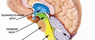
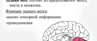
Midbrain: structure and functions
1.3. Midbrain nuclei: general information
The midbrain centers are represented by a number of nuclear groups located at this level of the central nervous system, but this section discusses only the most important of them.
Superior colliculus nuclei
. These nuclei are represented by sensory, intercalary and motor neurons. The axons of retinal ganglion cells converge on their sensory neurons, which in the form of collaterals branch from the axons of the optic nerve and follow to the neurons of the superior colliculus. Sensitive neurons in the superior colliculus receive afferent auditory signals from the inferior colliculus and temporal auditory cortex, as well as signals from cortical areas that control eye movements (eye fields of the occipito-parietal, frontal cortical areas). The neurons of the superior colliculus receive signals from the substantia nigra, thalamus, basal ganglia, cerebellum and other areas of the central nervous system. Through the nuclei of the superior colliculi, reflex movements of the eyes and head are triggered in response to the action of light or sounds, while the movements are given a certain direction towards the target - the source of light or sound (sentinel reflexes).
However, the superior colliculi cannot independently ensure sufficient accuracy of the movements performed. To achieve this, the neurons of the superior colliculus nuclei send a copy of the motor commands to the cortex, thalamus and cerebellum. The latter is an obligatory part of the brain necessary for organizing precise movements of the eyes and head towards the source of irritation.
The nuclei of the superior colliculus and lateral geniculate body are considered to be the primary centers of vision, in which undifferentiated perception of light signals and their simplest analysis occur. The results of this analysis are used to implement sentinel reflexes to the action of light.
Nuclei of the inferior colliculi.
The neurons of these nuclei are part of complex auditory pathways for transmitting and analyzing sound signals.
They receive auditory signals from the axons of the neurons of the underlying auditory nuclei - the inferior olive, the opposite inferior colliculus, the primary auditory (temporal) cortex and the cerebellar cortex. Nuclear neurons are signal switches in the auditory pathways. In this case, the signals of high-frequency sounds are switched in the ventral part of the nucleus, and low-frequency sounds in the dorsal part (as in the cochlea). The nucleus directly serves the function of auditory attention. Processed and analyzed auditory signals are transmitted by neurons of the inferior colliculus to the medial geniculate body and further to the primary auditory cortex, the opposite inferior colliculus, superior colliculus, and cerebellum. Thus, the inferior colliculus is the nucleus that switches auditory signals to the cerebral cortex and cerebellum and localizes the sound source in space.
The nuclei of the inferior colliculus and medial geniculate body are considered to be the primary centers of hearing. They perceive auditory signals, activate auditory attention, and form an undifferentiated auditory sensation. The results of the analysis are used to implement acoustic, including guard reflexes in the form of turns of the head and eyes towards an unexpected sound stimulus.
Pretectal nuclei.
They are represented by sensory neurons located in the roof of the pretectal region.
Receiving signals about retinal illumination through the axons of ganglion cells, these nuclei play a primary role in the implementation of pupillary reflexes, regulation of pupil lumen and maintaining optimal illumination of the retina.
The neurons of the nuclei send processed signals about the illumination of the retina to the motor preganglionic neurons of the parasympathetic nervous system of the Edinger-Westphal nucleus, located in the complex of subnuclei of the oculomotor nucleus of the midbrain. Nuclei of the oculomotor nerve (III pair of cranial nerves)
.
The oculomotor nucleus is located at the level of the superior colliculus. It is represented by somatic and visceral motor neurons. Somatic motor neurons innervate with their axons the muscle that lifts the eyelid and all the external muscles of the eyeball, with the exception of the lateral rectus, which is innervated by the axons of neurons of the abducens nerve nucleus, and the superior oblique, innervated by fibers of the trochlear nerve. The somatic nucleus is represented by subnuclei that innervate individual eye muscles. The neurons of the parasympathetic division of the ANS (autonomic nervous system) contained in the nucleus of the oculomotor nerve are included in the concept of the Yakubovich-Edinger-Westphal nucleus.
Neurons of the somatic part of the nucleus of the oculomotor nerve receive signals from the cerebral cortex via cortico-reticulo-bulbar fibers, from the diencephalon (nucleus of Cajal, rostral interstitial nucleus of the medial longitudinal fasciculus), the pons and medulla oblongata (vestibular nuclei, abducens nucleus), and cerebellum.
Neurons of the visceral part of the nucleus receive signals from neurons of the pretectal nuclei. The axons of the neurons of the Edinger-Westphal nucleus go together with the axons of somatic neurons up to the orbit. In the orbit they separate and travel to the ganglion neurons of the ciliary ganglion. Postganglionic fibers of ciliary ganglion neurons innervate the constrictor pupillary muscle and the ciliary muscles. Damage to the visceral component of the oculomotor nerve leads to dilation of the pupil, which becomes insensitive to light or impaired accommodation.
Damage to the nucleus of the oculomotor nerve or damage to the oculomotor nerve after it leaves the brain stem leads to the development of paralysis of the muscles innervated by its fibers. This is manifested by ptosis, disruption of eye alignment, development of double vision (diplopia), paresis of the sphincter of the pupil and ciliary muscles, which leads to dilation of the pupil of the ipsilateral eye ( on the same side)
, its insensitivity to light and impaired accommodation.
Trochlear nerve nuclei (IV pair of cranial nerves).
The nucleus is located in the ventral part of the central gray matter of the midbrain. The trochlear nerve nucleus consists of motor neurons that innervate the superior oblique muscle of the eye with axons. The neurons of the nucleus receive signals from neurons of the cerebral cortex via corticobulbar fibers and from the superior and medial vestibular nuclei via fibers of the medial longitudinal fasciculus.
When the nuclei of the trochlear nerve are damaged, paresis of the contralateral superior oblique muscle occurs, and when the nerve is damaged after it leaves the brainstem, paresis or paralysis of the ipsilateral superior oblique muscle develops. This muscle rotates the eye inwards, downwards and abducts. When the trochlear nerve is damaged, patients complain of vertical double vision (especially when looking down while going down stairs).
Mesencephalic nucleus of the trigeminal nerve.
The neurons of the nucleus receive signals of proprioceptive sensitivity from the masticatory muscles and periodontal membranes via the fibers of the mesencephalic tract. The results of the analysis of these signals are used for reflex regulation of chewing movements.
Pigment nucleus (locus ceruleus)
localized in the rostral pons and caudal part of the midbrain.
Contains 30–50 thousand pigmented cells that contain melanin granules. Nuclear pigmentation decreases in Parkinson's disease. Macula neurons provide noradrenergic innervation to most areas of the central nervous system.
The axons of macular neurons branch widely and are scattered throughout the brain, including the thalamus, hypothalamus, cerebellum, sensory nuclei of the brainstem, and spinal cord. It is believed that the neurons of this nucleus are involved in the regulation of sleep-wake cycles, breathing and rapid eye movements during the paradoxical phase of sleep.
Black substance
is a collection of non-pigmented neurons and neurons containing the pigment melanin and iron compounds. The substantia nigra is located between the cerebral peduncle and the tegmentum. The nature of the neural connections of the substantia nigra suggests that it plays an important role in the regulation of movement. Synaptic signal transmission by neurons of the substantia nigra is carried out using dopamine (pigmented neurons), acetylcholine and GABA (non-pigmented neurons). There is a certain pattern of neuronal loss in the substantia nigra in some brain diseases and especially dopaminergic neurons in Parkinson's disease. Diseases in which the substantia nigra is involved in the pathological process are almost always manifested by the development of parkinsonism and disorders such as tremor, rigidity, and decreased motor activity.
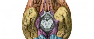
Red core
located in the tegmentum of the midbrain. It is richly vascularized and has a pinkish tint on fresh sections. This circumstance explains the name of the kernel. The neurons of the red nucleus receive signals from the premotor and primary motor areas of the cerebral cortex (along the corticorubral pathway) and from the deep cerebellar nuclei.
Neurons of the red nucleus send efferent signals along the rubrospinal pathway to neurons of the ventral horns, which innervate the distal muscles of the limbs. Like the neurons of the motor cortex that form the corticospinal tract, the neurons of the red nucleus through the rubrospinal tract facilitate the activation of flexor motor neurons and inhibit extensor motor neurons. Neurons of the red nucleus through the rubrospinal tract are directly involved in the coordination of motor functions of the spinal cord. When the nucleus or fibers of the rubrospinal tract are damaged, contralateral limb tremor occurs.
Interstitial nucleus of Cajal
located in the rostral midbrain.
The neurons of the nucleus have extensive connections with the rostral and caudal structures of the brain. They receive signals from the frontal eye field, the deep cerebellar nuclei and, through the medial longitudinal fasciculus, from the vestibular nuclei. The axons of the neurons of the Cajal nucleus follow to the neurons of the nuclei of the oculomotor and trochlear cranial nerves, as well as to the nuclei of the brain stem and spinal cord. Neurons of the interstitial nucleus control the implementation of rotational and vertical eye movements and their pursuit movements
.
Rostral interstitial nucleus of the medial longitudinal fasciculus
.
This nucleus is located rostral to the nucleus of Cajal and the nucleus of the third pair of cranial nerves, almost at the border of the junction of the midbrain and diencephalon. The neurons of the nucleus receive signals from the vestibular nucleus through the medial longitudinal fasciculus and from the horizontal gaze nucleus of the pons. The axons of the neurons of the rostral nucleus follow to the neurons of the subnucleus of the inferior rectus muscle of the oculomotor nucleus and control the implementation of eye movement downwards. Neurons of the interstitial nucleus of Cajal and the rostral interstitial nucleus of the medial longitudinal fasciculus form a neural network that functions as a center for vertical eye movements (vertical gaze)
. If it is damaged, restriction or impossibility of vertical eye movements may develop.
Central periaqueductal gray matter.
The periaqueductal gray matter of the midbrain is located around the aqueduct of Sylvius and is represented by scattered neurons.
Signals to gray matter neurons come from the hypothalamus, amygdala, reticular formation of the brain stem, locus coeruleus, and spinal cord. When gray matter is activated, its neurons release enkephalin, substance P, neurotensin, serotonin, dynorphin, and somatostatin. The central gray matter is involved in the formation of pain. The neurotransmitters of its neurons act on the serotonergic neurons of the medulla oblongata, which send axons to afferent neurons that conduct pain signals in the dorsal horn of the spinal cord and, depending on the activation of neurons in various parts of the central gray matter, cause a decrease in pain sensitivity (analgesia) or its increase. In addition, the central gray matter is involved in vocalization, control of reproductive behavior, modulation of the activity of the respiratory centers of the brain stem, and the formation of aggressive behavior.
Benedict's syndrome
The Austrian doctor Moritz Benedict in 1889 described the human condition and his behavior when the red nuclei are affected. In his works, he wrote that after such a violation, the connection between the structure of the oculomotor nerve and the cerebellum ceased.
The doctor's observation was aimed at the fact that on the damaged side the pupil was dilating, and on the opposite side the patient began to have a strong tremor. The patient also began to make erratic, chaotic, wriggling movements of his limbs.
It was these observations that formed the basis of Benedict's syndrome. Benedict's syndrome occurs when the midbrain is damaged at the level of the red nucleus and the cerebellar-rednuclear tract. It combines oculomotor nerve palsy and facial tremors on the opposite side.