March 1, 2019
141923
0
3.5 out of 5
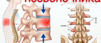
The human spine is the basis of the musculoskeletal system. At the same time, it not only performs a supporting function and provides the ability to walk upright, but also represents a fairly flexible axis of the body, which is achieved due to the mobility of the vast majority of its individual parts. In this case, the anterior part of the spine participates in the formation of the walls of the thoracic and abdominal cavities. But one of its most important functions is to ensure the safety of the spinal cord that runs inside it.
general information
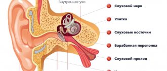
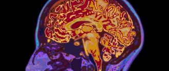
The anatomy of the spinal cord differs from the brain in its oblong structure. In Latin the organ is called medulla spinalis. It is a thickened tube with a small channel inside, slightly flattened in front and behind. It is this structure that ensures the normal transportation of nerve impulses from the main organ located in the cranium to the peripheral structures of the nervous system.
Locally, the organ is located in the spinal canal, where soft and bone tissues and nerve endings responsible for many functions of the human body are concentrated. Without a normally functioning spinal cord, natural breathing, digestion, heartbeat, reproductive activity, and any motor activity are not possible.
In humans, it begins to form at about 4 weeks of development inside the mother’s womb. But in what form it is observed in an adult, it appears much later; at first it is a neural tube, gradually developing into a full-fledged organ. It completes its formation within 2 years after birth.
Age and gender characteristics of the spine
The length of the spinal column in newborns does not exceed 40% of the total height. But during the first 2 years of life, its length almost doubles. All this time, all parts of the spine are growing at a high speed, but mainly in width. From 1.5 to 3 years, the growth rate decreases, especially in the cervical and upper thoracic regions. At about 3 years of age, active growth of the lumbar and lower thoracic spine begins. From 5 to 10 years, a phase of smooth, uniform growth in all parameters begins, followed by a phase of active growth, lasting from 10 to 17 years. After this, the growth of the cervical and thoracic regions slows down, but the growth of the lumbar region accelerates. The entire process of development of the spinal column is completed at 23-25 years of age.
Thus, in an adult man, the length of the spine is on average 60-75 cm, and in a woman - 60-65 cm. Over the years, degenerative changes occur in the intervertebral discs, they flatten and cease to fully cope with their functions, and physiological bends increase. As a result, not only various diseases arise, but also the length of the spinal column decreases in old age by about 5 cm or more.
Thoracic kyphosis and lumbar lordosis are more pronounced in women than in men.
Thus, the human spine has a complex structure, a dense network of nerves and blood vessels. This largely explains the difficulty of performing surgical interventions on it and the possible risks. Therefore, today all efforts are aimed at finding the least invasive methods of performing operations that involve minimal tissue trauma, which sharply reduces the likelihood of developing complications of varying severity.
Structure
The local location of the spinal cord along the entire back has its own characteristics. This physiology ensures that the organ performs its basic functions. The organ begins at the level of the 1st cervical vertebra, where it is gently rebuilt into the brain, but there is no clear division in them. At the junction there is a crossover of the pyramidal tracts responsible for the motor activity of the limbs. The spinal cord ends in the region of the 2nd lumbar vertebra, so it is shorter in length than the entire spine as a whole. This feature allows lumbar puncture to be performed at the level of the 3-4 lumbar vertebrae, without the risk of damaging the spinal cord.
What is special about the structure? The oblong tube has two grooves at the front and back. The brain is covered by three membranes:
- Solid. It is the tissue of the periosteum of the spinal canal, followed by the epidural space and the outer layer of the dura mater.
- Cobweb. A thin, colorless plate that fuses with the hard shell in the area of the intervertebral foramen. In the place where there is no fusion, the subdural space is located.
- Vascular. A soft membrane separated from the previous one by a subarachnoid space with cerebrospinal fluid. The membrane is adjacent to the spinal cord and consists mainly of choroid plexuses.
The space between them is filled with cerebrospinal fluid - cerebrospinal fluid. The gray matter is located in the center of the organ. It consists of intercalary and motor neurons. It also contains two types of horns: the anterior ones, which contain motor neurons, and the posterior ones, where interneurons are located.
External characteristics
The external structure of the spinal cord largely follows the contours of the spine, since the structures adapt to its physiological curves. Two thickenings are observed in the area of the neck and lower thoracic, the beginning of the lumbar calving. These places are characterized as exits of the roots of the spinal nerves responsible for the innervation of the arms and legs.
The external structure can be briefly described by the following characteristics:
- The shape is cylindrical, flattened on the front and back sides.
- Visually, the spinal cord looks like an elongated “cord” with processes.
- On average, the length of the organ is 42-44 cm, but directly depends on the person’s height.
- The mass is 34-38 g, which is 50 times less than the organ of the brain.
- There are two grooves in front and behind, which visually divide the organ into two symmetrical parts.
- In the middle there is a canal, which in the upper part communicates with one of the ventricles of the brain. Inferiorly, the central canal expands, forming the terminal ventricle.
The thickness of the spinal cord is uneven and depends on in which part the measurement is taken. The organ also has four surfaces: two rounded lateral, a convex posterior and a flattened anterior. The external structure is in many ways reminiscent of the internal part of the ridge, since the organ fills the entire canal. The organ is reliably protected by bone tissue.
Internal structure
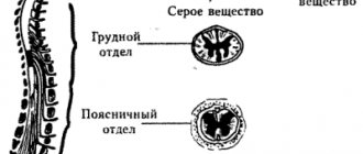
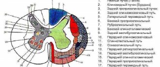
The spinal cord is made up of nerve tissue cells called neurons. They are concentrated closer to the center and form gray matter. According to rough estimates by scientists, the entire organ contains about 13 million cells, which is many times less than in the head section. The gray matter is located inside the white matter, and if you make a cross section, it will be shaped like a butterfly. This is especially clearly visible in the diagram.
Schematic cross-section of the spinal cord
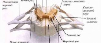
This unique anatomy allows the spinal cord to be divided into several structures. It is arranged as follows:
- Front horns. They are distinguished by their rounded, wide shape and consist of neurons responsible for transmitting nerve impulses to the muscles. It is precisely because they perform such a task that they are called motor. The anterior roots of the spinal nerves begin in the anterior horns.
- Hind horns. They are distinguished by a long, narrow shape and consist of interneurons. They bear this name due to their ability to receive incoming signals from the sensory roots of the spinal nerves, otherwise they are called dorsal roots.
- Side horns. They are present only in the lower segments of the organ, and contain vegetative nuclei responsible for the dilation of the pupils, or the functioning of the sweat glands.
The physiological function of nerve endings is to transmit signals from the brain to the spinal cord, as well as deliver received impulses in the opposite direction. This ensures interconnection at all levels and areas of the nervous system. Nerve fibers are combined into bundles and are present along the entire length of the spinal cord.
Metamer and segmental structure
Each part of the spinal cord is a constituent element of a specific metamere of the body. Moreover, if there is a “piece” of the spinal cord that includes a section of gray matter with a pair of roots, then the metamer includes the spinal segment itself, muscle fiber (myotome), a section of the epidermis (dermatome), a bone component (scletorome), an internal organ (splanchiotome), controlled by this segment. In humans and higher representatives of the animal world, radicular metamerism is observed - the spinal cord is confined to certain parts of the body.
The skin areas of the body consisting of sensory fibers that approach the corresponding segment of the spinal cord are called dermatomes. They are strips of epidermis controlled by sensitive nerve endings. They are located throughout the body and sometimes overlap each other.
Visual representation of the connection between the skin and the spinal cord
Myotomes are muscle groups that receive motor fibers from certain areas of the brain. Thanks to the study and knowledge of their location, the process of damage and diagnosis of spinal cord lesions is greatly simplified. Damage to a certain segment of the spinal cord provokes sensory and motor disorders.
Connection between the spinal cord and muscle fiber
Segmental structure
The spinal cord is conventionally divided into five sections, although it is a single whole. The name of each directly depends on its location in the body. In total, a person may have 31-33 segments, which consist of:
- Cervical area - includes 8 segments.
- Thoracic region - 12 segments.
- Lumbar region – 5 segments.
- Sacral – 5 segments.
- Coccygeal – 1-3 segments.
This division allows us to examine the organ in more detail and simplify the process of diagnosing various pathologies.
White and gray matter
In cross-section, the symmetrical halves can be seen in detail and the anterior medial fissure and connective tissue septum can be seen. The part located inside is darker and is called gray matter (GM), it is located in a lighter substance - white matter (WM). Most of the SV is located in the lumbar region, the least is observed in the thoracic region. What are the main functions of gray matter:
- Transmission of pain impulses.
- Response to temperature changes.
- Closure of reflex arcs.
- Obtaining information from muscle tissue, tendons, ligaments.
- Formation of pathways.
What is the structure of white matter? It consists of myelinated, unmyelinated nerve fibers, blood vessels and a small amount of connective tissue. Its main task is to trigger simple reflexes and provide connections with skeletal muscles.
Blood vessels
The blood supply to the spine is realized through fairly large arteries that pass either in close proximity to the vertebral bodies or along them. The arteries of the cervical vertebral bodies originate from the subclavian artery, the thoracic vertebrae are supplied by the intercostal arteries, and the lumbar vertebrae are supplied by the lumbar arteries. As a result, the spine is actively supplied with blood at all levels, and the pressure in the vessels is at fairly high levels. But if the bone structures have a direct blood supply, then the intervertebral discs are deprived of this. Their nutrition is carried out through the diffusion of substances during compression/straightening of the disc during physical activity.
The lumbar and intercostal arteries are located along the anterolateral surfaces of the vertebral bodies. In the area of the intervertebral natural foramina, posterior branches branch off from them, which are responsible for feeding the soft tissues of the back and dorsal parts of the vertebrae. In turn, spinal branches depart from them, which deepen into the spinal canal, where the blood vessels are again divided into 2 branches: anterior and posterior. The anterior branch is larger in size and is located transversely to the anterior part of the vertebral body, and on the posterior surface it unites with a similar vessel on the opposite side of the body. The posterior branch extends along the posterolateral surface of the spinal canal and connects with a similar artery on the opposite side.
Thus, the spinal arteries form an anastomotic network that covers the entire spinal canal and has transverse and longitudinal branches. Numerous vessels responsible for feeding the vertebral bodies and spinal cord are diverted from it. The arteries penetrate into the vertebral bodies near the midline, but they do not pass into the intervertebral discs.
The spinal cord has 3 blood supply basins:
- Cervicothoracic, where the first 4 segments are fed from the anterior spinal artery, formed by the fusion of 2 vertebral arteries, the next 5 segments have absolutely independent nutrition, and the blood supply is provided by 2-4 large radicular-spinal arteries, branching from the vertebral arteries, the ascending and deep cervical arteries.
- The intermediate (middle) thoracic basin, including segments T3-T8, is supplied exclusively by one single artery located at level 5 or 6 of the thoracic root. Due to such anatomical features, there is a high risk of developing severe ischemic lesions in this part of the spinal cord.
- Lower thoracic and lumbosacral basin - blood supply is provided by one large anterior radicular artery.
As for the venous system, the spine has 4 venous plexuses: 2 external, localized on the anterior surface of the vertebral bodies behind the arches, and 2 internal. The largest venous plexus is the anterior intravertebral plexus. Its large vertical trunks are interconnected by transverse branches. It is firmly fixed to the periosteum along the posterior surface of the vertebrae by a large number of jumpers. The posterior venous intravertebral plexus can easily move because it does not have strong connections with the vertebral bodies. But at the same time, all 4 venous plexuses of the spine are closely interconnected by numerous vessels that penetrate the vertebral bodies, as well as the yellow ligaments. In general, they form a single whole and extend from the base of the skull to the tailbone.
Venous blood is drained through the system of the superior and inferior vena cava, into which it enters from the vertebral, intercostal, lumbar and sacral veins. All intervertebral veins exit through the corresponding openings of the spine. At the same time, they are firmly attached to the periosteum of the bony edges of the foramina.
The spinal cord itself has 2 venous blood outflow systems: anterior and posterior. In this case, the veins of the surface of the organ are united by a large anastomotic network. Therefore, if it is necessary to ligate one or more veins, the likelihood of developing spinal disorders is close to zero.
Functions
Functional anatomy implies that, being part of the central nervous system, the spinal cord performs a reflex and conductive function. In the first case, the body controls the implementation of the simplest actions at the level of reactions contained in the subconscious. A striking example is the initiation of a motor function by withdrawing the hand if the surface is too hot. The limb does this before the person himself understands what happened. The second task of the organ is to transmit nerve impulses to the head section of the central nervous system, along the ascending and descending pathways.
Briefly about the main functions of the spinal cord
Reflex function
This main function of the organ is a response to external irritation. For example, the appearance of a reflex cough due to foreign objects and particles entering the respiratory tract, removing the hand from cactus spines or a source of danger. The impulse enters the spinal canal through motor neurons, which also trigger muscle contraction. This process does not require the involvement of the brain, and the motor reaction occurs without its participation. That is, a person does not even think about his action, often does not realize it.
Children's innate reflexes are checked after birth. They usually include the ability to suck milk, breathe, and jerk their legs. During development, acquired reflexes also appear, which help doctors identify the correct functioning of the arch elements and individual segments of the spinal cord. The test is carried out during a neurological examination. The main emphasis is on the plantar reflex, knee and abdominal. They allow you to check how healthy a person is at one time or another.
Conductor function
Treatment of spinal cord myelitis
Another important function of the spinal cord is conduction. It ensures the transmission of impulses from the skin, mucosal surface, internal organ to the brain and in the opposite direction. The white matter acts as a “conductor”. It is this that carries information about incoming impulses from the outside. Thanks to this ability, a person can characterize any object that surrounds him.
Cognition of the world is carried out through the transfer of information after touch to the brain. It is thanks to this function that a person understands that an object is slippery, smooth, rough or soft. With loss of sensitivity, the patient ceases to understand what is in front of him when touching an object. In addition, the brain receives data about the position of the body in space, tension in muscle tissue or irritation of pain receptors.
Brief Definition
The spinal cord pathways or tracts are collections of nerve fibers located inside the spine that carry impulses from the brain to all parts of the body and back. The nerve endings, the combination of which form the pathways, are distinguished by a similar structure, development and general functions. They are divided among themselves according to the tasks assigned to them. The paths are classified as follows:
- Associative. Their main purpose is to unite gray matter cells from different segments to form their own anterior, lateral or posterior bundles.
- Commissural. These fibers connect the gray matter from the two hemispheres. With their help, the coordinated work of individual areas, nerve centers, and both hemispheres occurs.
- Projection. With the help of such pathways, the work of the overlying and underlying parts of the brain is combined. They are the ones who provide the projection of pictures of the surrounding world, as on a monitor screen.
Projection pathways, in turn, are efferent and afferent. They form the basis of the central nervous system, and are divided into ascending (centripetal or sensitive) and descending (centrifugal, motor).
Important! Nerve fibers provide a constant, unbreakable connection to the brain located in the skull and spine. It is thanks to them that the impulse is quickly transmitted, all body movements are coordinated with each other.
The pathways of the brain and spinal cord differ from each other, but they always act harmoniously, ensuring the passage of an incredibly large number of nerve signals from receptors to the central nervous system. Paths are formed from long axons, special fibers that are capable of creating connections among themselves, thus connecting individual segments of the spinal trunk, providing control of effector organs.
What organs does the spinal cord control?
It is also important to understand which internal organs are connected to the spinal cord and may suffer if a particular area of the spine is damaged. Specific spinal segments control specific parts of the body by transmitting nerve impulses and transmitting responses along motor neurons. What each vertebra is responsible for can be clearly seen in the table.
| Back segment | Vertebra serial number | Controlled internal organs |
| Cervical | 3-5 | Diaphragm |
| Cervical | 6-8 | Articular tissue of the upper limbs |
| Chest | 1,2, 5-8 | Muscle tissue and epidermis of the hands, elbows and forearms |
| Chest | 2-12 | Muscles, skin of the body |
| Chest | 1-11 | Intercostal muscles |
| Chest | 1-5 | Heads, heart |
| Chest | 5-6 | Lower esophagus |
| Chest | 6-10 | Gastrointestinal tract |
| Lumbar | 1-2 | Prostate, groin area, adrenal glands, bladder, uterus. |
| Lumbar | 3-5 | Muscles and skin of the legs |
| Sacral | 1-2 | Muscle tissue and epidermis of the lower extremities |
| Sacral | 3-5 | External genitalia, reflex centers, erectile dysfunction and defecation |
Damage to the spinal cord in a specific section negatively affects the functioning of these internal organs. In some cases, dysfunction occurs before vertebral compression or displacement is detected.
Consequences of central nervous system injury
Even partial damage to the spinal cord can cause disruption of the functionality of a wide variety of systems and organs.
Bones and joints
A spinal cord injury can impair the absorption of calcium and some other important minerals into bone tissue. But at the same time, these substances can accumulate in the urinary system, causing the formation of stones. Experts usually explain such processes by a decrease in human motor activity. In addition, decreased mobility can lead to stiffness in joints, including the knees, elbows, and shoulders. To prevent this, there are special sets of exercises for people with disabilities.
Urinary system
The urinary system consists of the kidneys, which filter the blood and produce urine, and the bladder, which collects and then removes it from the body. After a spinal cord injury, the kidneys continue to produce urine, but the bladder may not work as well as before. After an injury, a person may not feel when his bladder is full of fluid, or his urinary system, due to a lack of necessary impulses, may lose the ability to eliminate urine naturally. As a result, a catheter may be needed to empty the bladder. Another option: urine may, on the contrary, come out involuntarily and the person loses the ability to control this process.
Respiratory system
Even after a spinal injury, human lungs continue to function. However, the ability to inhale and exhale air is controlled by the muscles. Depending on the level of injury, a person may lose the ability to cough or take deep breaths. And they are necessary for complete straightening of the lungs and deep air circulation.
The most important muscle for breathing is the diaphragm. It is a large arcuate muscle that is located directly under the lungs. And if the diaphragm is paralyzed due to damage to the spinal cord, special equipment may be needed to maintain respiratory function.
Leather
Damage to the spinal cord can also negatively affect the skin. Skin plays a very important role for humans. It protects the body from germs and other pathogens from the outside world. The spinal cord is an important organ that helps protect the skin from damage. For example, when sitting in one position for a long time, the brain sends signals in the form of a feeling of discomfort, which encourages you to change your position and prevent damage or pressure on the skin. In the same way, the central nervous system protects against burns, cuts and other possible damage to the skin (for example, a person reflexively withdraws his hand from a hot or cutting object). With spinal cord injuries, such signals may not appear, which greatly increases the risk of damage to the skin and, consequently, the penetration of microbes into the body.
Reproductive system
After a spinal cord injury in the lumbar or sacrococcygeal segment, the functionality of the reproductive system may be affected. Men usually experience erectile dysfunction, problems with ejaculation and fertility. In women, after injury, sensitivity in the genital area may be impaired, although fertility itself may not be directly affected.
Intestines
It is well known that the digestive system is responsible for the breakdown of food consumed, the absorption of nutrients from it and the removal of metabolic products. A fairly common consequence of central nervous system injury is impaired intestinal motility. That is, the digestive system continues to absorb and digest food, but the process of eliminating excrement is disrupted. In a healthy body, when the rectum is full, an exchange of impulses occurs between the intestines and the brain. If the spinal cord is damaged, such signals may not be received. As a result, a person loses the ability to control the process of defecation.
Autonomic nervous system
The autonomic nervous system regulates the activity of internal organs, blood and lymphatic vessels, as well as endocrine and exocrine glands. Body temperature, blood pressure, digestion and other important functions depend on it. After a spinal injury, some of its functions may be impaired. Quite often (especially if the damage occurs in the thoracic segment) after an injury the body loses the ability to thermoregulate. What does it mean? If the autonomic nervous system works correctly, then the body temperature remains stable regardless of the weather conditions outside or the indoor microclimate. Due to spinal cord damage, body temperature may rise or fall according to indoor or outdoor temperatures. Disturbances in thermoregulation are most noticeable in parts of the body located below the area of injury.
Risk of organ damage
Due to the characteristic structure of the brain, it is connected to most systems in the body. The integrity of its structure is extremely important for the correct functioning of the musculoskeletal system and the health of internal organs. Any injury, regardless of severity, can lead to disability. Sprains, dislocations, disc damage, vertebral fractures with or without displacement can cause spinal shock and paralysis of the legs, and disrupt the normal functioning of the cords.
Severe injuries result in shock lasting from several hours to several months. In this case, the pathological condition is accompanied by a number of neurological symptoms. These include numbness, sensory disturbances, pelvic organ dysfunction, and inability to control the process of urination and bowel movements.
Treatment of minor spinal injuries is carried out on an outpatient basis, using medications, therapeutic exercises and massage. Severe injuries require surgical intervention, especially if compression of the spinal cord is detected. Cells are quickly damaged and die, so any delay can cost a person’s health. The recovery period after such an intervention is up to two years. Various physiotherapeutic procedures help with this, for example, reflexology, ergotherapy, electrophoresis, magnetic therapy, etc.
The spinal cord is a key element of the human central nervous system, which is connected in one way or another with almost all internal organs and human muscle tissue. The specific structure allows you to transmit impulses and signals, ensure full motor activity, and perform a number of other functions.
Characteristics of the spinal cord depending on age
Let's consider the features, depending on the age of the person:
- In a newly born child, the length of the organ is 13.5-14.5 centimeters.
- At 2 years, the length increases to 20 centimeters.
- At about 10 years old, the length can reach 29 centimeters.
- Growth ends in different ways, depending on the characteristics of a particular person’s body.
Let's look at the external features and changes depending on age:
- In infants, cervical and lumbar thickening is more noticeable than in adults. The same applies to the width of the central channel.
- The above features become almost invisible by the age of two.
- The volume of white matter grows many times faster than the volume of gray matter. This is due to the fact that the segmental apparatus is formed earlier than the pathways that connect the brain and spinal cord.
Otherwise, there are practically no age-related characteristics, because from birth the spinal cord performs almost all functions, as in an adult.