Amoeba naegleria Fowler is a microorganism that causes a rare but deadly disease - amoebic meningoencephalitis (negleriasis). The disease is rare but deadly and affects the brain. Characterized by severe headaches and high fever. It can be diagnosed by bacterioscopy of the CSF or using differential diagnosis. Treatment is carried out using anti-Negleria serum or monoclonal antibodies.
Meningoencephalitis viral, bacterial, parasitic
The term “meningoencephalitis” includes two nosological forms: “encephalitis” and “meningitis”.
The definition describes the morphological changes that occur against the background of pathology - damage to the white matter and meninges. The pathology is characterized by high mortality, disability, and a large number of disorders. Diagnosis of the symptoms of the disease at the beginning of its development, prevent dangerous consequences, eliminate damage to functional centers. The effectiveness of treatment depends on the cause, pathogen, and extent of the inflammatory focus.
The initial signs of pathology are neurological disorders. Neurologists conduct differential diagnostics that allow one to suspect meningoencephalitis and timely prescribe neuroimaging methods (brain MRI and CT).
Meningoencephalitis - what is it?
There are congenital and acquired forms. Meningoencephalitis in children occurs due to intrauterine infection (cytomegalovirus, chlamydia, meningococcal). Immediately after birth, it is difficult to identify the nosology, since the child cannot talk about sensations.
In the first month of life, the first signs appear. Only the acute variety is accompanied by multiple changes, which often lead to death. Analysis of cerebrospinal fluid helps to suspect inflammation of the brain and membranes at the beginning of development.
The procedure is invasive and is prescribed according to strict indications. The harmlessness of MRI for meningoencephalitis makes it possible to prescribe examinations for newborns and infants. The high cost of equipment excludes the possibility of installing devices everywhere.
The main causes of mortality from inflammatory processes of the soft membrane and brain parenchyma:
- Intracerebral edema;
- Infectious shock;
- Cerebral hypertension;
- Kidney failure.
The consequences of the disease in subacute and chronic forms develop over several years.
MRI meningoencephalitis
Features of the disease
The causative agent of this disease is the amoeba naegleria; this protozoan organism belongs to the group of eukaryotes or nuclear unicellular organisms of the flagellar type. This simple organism lives in stagnant waters of ponds, reservoirs, and pools with insufficiently chlorinated water. This type of organism lives mainly in soils with high humidity.
The causative agent of amoebic meningitis is a facultative protozoan, which means that the amoeba can live quietly in the environment, and if it enters the human body, it will further parasitize inside the human body.
An amoeba can live for a long time in a cyst, in a protective shell. This usually happens when unfavorable living conditions arise, for example, if a reservoir is drained. The microbe can live inside the cyst for several years, and it can easily tolerate temperatures up to 45 degrees. In this state, the body can endure wind over long distances; cysts with microbes are often found in air conditioner humidifiers, in chlorinated or unboiled drinking water. But it is worth noting that this organism becomes dangerous when it enters the flagellar stage.
If you drink water that contains the causative agents of amoebic meningitis, the microbes will simply be digested in the stomach. The main period during which the process of infection by the parasite occurs is considered to be summer time, the height of the swimming season. The pathogen can enter the body even if air is inhaled at this time.
This is how the amoeba gets into the brain
The amoeba moves into the brain area according to the following scheme:
- the pathogen enters the nasal cavity along with dirty water;
- further, it penetrates into the olfactory nerve;
- after this, the parasite penetrates the brain area, its membrane and all parts - the cerebellum, brain stem and the upper parts of the spinal cord.
ICD 10 code for meningoencephalitis
The international classification of the tenth revision identifies the following types of brain inflammation with code “G04”:
- Meningomyelitis;
- Meningoencephalitis;
- Acute ascending myelitis.
The category excludes multiple sclerosis (“G95”), toxic, alcoholic encephalopathy “G31.2”, “G92”.
Classification of meningoecephalitis by course:
- Chronic – long-term development with a slow increase in symptoms;
- Subacute - erased signs of nosology increase over two to three years;
- Acute – rapid progression of symptoms helps early diagnosis;
- Fulminant – rapid cerebral damage causes death.
The difficulties of verifying pathology are complicated by the variety of etiological factors.
Causes of meningoencephalitis
Viral forms provoke chronic and subacute varieties. An acute course can be observed in people with immunodeficiencies. Pathogenic bacteria quickly destroy the arachnoid, subarachnoid membranes, and brain parenchyma. Parasitic (amebic) species progress slowly.
Features of influenza hemorrhagic meningoencephalitis
Nosology is a consequence of influenza. An acute respiratory viral infection provokes a rise in temperature and enlargement of the pharyngeal tonsils. Long-term persistence of infection causes epileptic convulsions.
Fever increases brain destruction, but no antiviral treatment has been developed. Vaccination and strengthening the immune system are the main measures to counter the spread of influenza hemorrhagic infection.
Principles for diagnosing viral meningoencephalitis:
- Absence of bacteria in brain preparations when stained by Gram;
- Cerebrospinal fluid pleocytosis;
- Detection of enteroviruses, arboviruses, herpesviruses using polymerase chain reaction (PCR).
Severe and fatal cases are caused by enteroviruses. Over serotypes of pathogens have been identified, causing a variety of clinical manifestations of the disease. Enteroviral neuroinfection often leads to death and disability.
After a bite from ticks, insects, or mosquitoes, the inflammatory process of cerebral tissues is caused by arboviruses if the carrier was infected with microbes. In addition to humans, these pathogens infect horses and dogs, which can also be a source of infection for humans.
Common encephalitis caused by arboviruses:
- West Nile fever;
- Encephalitis St. Louis;
- California uniform.
The prevalence of diseases has been increasing in recent years.
Symptoms of herpetic encephalitis
Activation of herpetic neuroinfection is the cause of death in approximately seventy percent of adults and children. The lack of antiherpetic therapy excludes the possibility of effective treatment. Only a strong immune system can cope with the herpes virus. The weak body of a pregnant woman in the presence of infection becomes a source of infection of the fetus in utero.
Herpes simplex virus type 2 (HSV-2) becomes the source of a mild transient type of neuroinfection. Meningoencephalitis is activated in adolescents with an active sexual life.
In newborns, herpes simplex virus type 1 or 2 is part of the associated infections. A generalized disease affecting many organs is caused by HSV in patients with immunodeficiencies, including AIDS. The lack of pharmaceuticals causes the death of 2/3 of infants.
The first signs of herpesvirus neuroinfection:
- High fever;
- Strong headache;
- Behavioral disorders;
- General cerebral symptoms.
The drug virolex (acyclovir) increases your chances of survival. In severe cases, the drug is ineffective.
Rare forms of viral encephalitis
Damage to the central nervous system is caused by the varicella zoster virus, which occurs after an illness. Nosology has symptoms:
- Cerebellar ataxia – incoordination of muscle activity, unsteady gait;
- Acute encephalitis.
Acute manifestations are rare. The chickenpox virus is characterized by a chronic course with cycles of remissions and exacerbations, since the pathogen persists in the nerve ganglia. Reactivation of chickenpox is possible with a decrease in immunity.
Rare types of viral meningoencephalitis:
- Cytomegalovirus - destroys cerebral tissue only in immunodeficiencies;
- Mumps – caused by the mumps virus. It is characterized by a mild course, but causes inflammation of the auditory nerve.
The lack of a complete diagnosis excludes the possibility of early detection of neuroinfection.
Clinic of bacterial meningoencephalitis
Pathogenic bacteria enter the brain through the blood and lymphatic fluid. The penetration of microorganisms from the primary focus of internal organs is dangerous due to the resistance of the agents to the antibiotics used to eliminate the disease.
Types of bacterial meningoencephalitis:
- Brucellosis;
- Toxoplasmosis;
- Syphilitic;
- Tuberculous;
- Meningococcal.
Signs are determined by the type of etiological factor. Damage to cerebral tissue appears against the background of a primary infection of internal organs. Congenital types arise due to the entry of microorganisms into the fetus during childbirth.
Tuberculous encephalitis develops in people with primary tuberculosis of different localization. The peak of infection occurs in the spring-autumn period, when immune activity is reduced. The nosology has no specific manifestations. Diagnosed by laboratory, clinical and instrumental methods.
Mycobacterial infection is difficult to treat. Of the bacterial species, nosology is the most dangerous. Main clinical signs of pathology:
- Poor concentration;
- Strong headache;
- General cerebral disorders;
- Photophobia;
- Vegetative manifestations;
- Neurological disorders;
- Hydrocephalus.
Acute manifestations of the disease are characterized by an increase in temperature to thirty-nine degrees. Clinical manifestations of the disease are accompanied by fever, joint pain, sleep disturbance, abnormal meningeal signs, and fever. The average duration of the disease is about ten days. Associated signs of nosology are lack of appetite, excessive sweating, cerebellar disorders, mobility impairment, positive Rehberg test. (a person cannot touch the tip of his nose with his index finger).
The temperature curve against the background of inflammatory changes in the cerebral parenchyma and meninges has a specific course. Initially, the fever increases to 39 degrees. After 5-7 days, the fever subsides to low-grade levels (38.5 degrees). A second wave is observed on the tenth day. Focal neurological symptoms appear with neuritis, radiculitis, changes in the activity of the heart, pulmonary system, dizziness, convulsions, paresthesia (lack of sensitivity).
The brucellosis species provokes pyramidal symptoms with paresis, paralysis, and muscle cramps.
Characteristics of parasitic meningoencephalitis
Amoebas are found in young children and newborns. The parasites cause inflammation of the brain with a high mortality rate.
Pathogens enter through the upper respiratory tract. The source of infection is reservoirs, tap water, contaminated vegetables and fruits.
Clinical manifestations of amoebic encephalitis, meningitis:
- Granulomatous inflammation of the white matter takes several months to form. Damage to the membranes clinically resembles a volumetric intracerebral formation with the formation of several centers of activity - convulsions, personality disorders, paralysis, paresis;
- Acute variety - lasts two weeks. It begins with lightning speed, accompanied by nausea, headaches, and a significant increase in temperature. Multiple foci cause death in infants and children.
Early detection and proper drug therapy eliminate the risk of serious complications.
Prevention
There are no preventative medications for amoebic meningoencephalitis. To avoid death or serious complications (mental retardation, irreversible disorders of the nervous system or impaired eye function), you need to remember and follow simple rules. Refrain from swimming in warm, stagnant bodies of water, especially wild ones that are not subject to sanitary control. Do not dive without protecting your nose with a special clothespin or holding your nose with your fingers.
If you have to work in a garden or vegetable garden with wet soil, it is advisable to wear gloves and a face mask. And then wash your hands thoroughly and clean the area under your nails with a brush. Do not allow children to pick their noses with dirty fingers, and monitor their hygiene. Since amoeba cysts are tenacious, you should clean and disinfect air conditioner humidifiers and use only boiled water.
Characteristics of autoimmune encephalitis
The formation of antibodies to brain tissue causes demyelination. The process is long but progressive. Rasmussen's encephalomyelitis is a typical manifestation of an autoimmune lesion of the cerebral parenchyma. Depending on the characteristics of the development of the process, the duration is from five to fifteen years. In most cases, the clinical peak occurs at six years of age.
Nosology has been thoroughly studied by scientists. The causes of the occurrence could not be established, but the link to which immunoglobulins are formed was identified. The presence of NMDA receptors is a weak point that is susceptible to destruction by the immune system.
There are case studies showing the nonspecificity of glutamate receptor antibodies for Rasmussen's encephalomyelitis. Other anti-inflammatory cytokines formed during the nosology have been identified.
Treatment of the disease
It should be remembered that amoebic meningitis is difficult to detect and treat.
In most cases, patients infected with Naegleria died. Treatment with amoebicidal drugs is ineffective. However, amoebas are sensitive to Amphotericin B (if necessary, it is injected directly into the ventricles of the brain), Miconazole, Rifampicin, and Tetracycline. The drugs of the Artemisinin B group had a positive effect. Anti-Negleria immune serum or monoclonal antibodies can be used. It should be remembered that the disease develops rapidly (counting the hours), so treatment should only take place in a hospital under the supervision of a doctor.
The disease cannot be treated at home. You should not self-medicate or use folk remedies. If the patient managed to survive and was transferred to outpatient treatment, the following recommendations must be followed:
- drinking plenty of fluids and eating quality food;
- twilight indoors;
- taking vitamins;
- bed rest;
- physiotherapeutic procedures.
Meningoencephalitis of newborns
The most common pathogen is viruses. Intrauterine infection of a baby occurs from a mother suffering from glandular fever, measles, rubella, herpetic infection, and mumps.
The most common symptoms are focal disorders, hyperkinesis, hydrocephalus. Nonspecific manifestations of meningoencephalitis in newborns:
- Eye twitching;
- Difficulty feeding from the breast;
- High fever;
- Intoxication syndrome;
- Vomiting reflex;
- Diarrhea;
- Strabismus;
- Increased heart rate (tachycardia);
- Muscle twitching.
Neurologists define neurological disorders in the form of Kernig's sign (the inability to bring the head to the chest due to stiff neck muscles). Cerebrospinal fluid contains an increased amount of lymphocytes and protein.
Manifestations of meningoencephalitis in adults
Various symptoms of the disease in an adult are caused by different pathogens and characteristics of the course. The incubation period of the disease lasts several weeks.
Symptoms of the clinical stage:
- Muscle stagnation;
- Decreased appetite;
- Constant fatigue;
- Headaches without the effectiveness of painkillers.
Inflammatory changes in the meninges lead to meningeal syndrome with special manifestations:
- Nausea;
- Speech disorders;
- Interruptions of heart activity;
- Respiratory disorders.
The occurrence of the described manifestations leads to death. The progression of individual symptoms becomes a cause of disability.
Granulomatous form of amebic meningoencephalitis
What causes granulomatous type meningitis? The causative agent of this form is still the same - amoeba naegleria, but only the routes of infection occur through contact and airborne droplets. The parasite enters the human body through the pores of the skin and the mucous layer. This type of disease develops quite slowly, the first symptoms may appear several weeks or even several months after infection. This pathological process mainly affects people with a weakened immune system, as well as people with chronic diseases.
Amoebic meningitis in the granulomatous form has the following characteristic features:
- Weakness of the muscle fibers of one part of the body.
- Seizures of an epileptic nature.
- Disorder of consciousness.
- Changes in behavior may be noted.
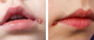
- A large number of ulcers and nodes with a dense structure may appear on the surface of the skin.
- Increased tone of muscle fibers in the back of the head.
During this pathological process, hyperthermia and headaches do not occur. When performing CT and MRI, multiple lesions that resemble embolic infarctions may be noted on the image.
Brain damage determined by various hardware studies
Consequences of meningoencephalitis
In addition to high mortality, the disease is characterized by dangerous conditions leading to disability. Severe consequences of inflammation of the white matter and meninges:
- Paralysis of limbs;
- Epileptic seizures;
- Mental retardation in children;
- Hydrocephalus;
- Psychoses;
- Hallucinosis.
The conditions are irreversible. Early verification and competent therapy prevent negative consequences in bacterial types of encephalitis and meningitis. For other forms of nosology, the prognosis is unfavorable.
Diagnosis of inflammation of the brain and soft membranes
The most accurate laboratory method for verifying nosology at the beginning of development is cerebrospinal fluid analysis. Turbidity in the cerebrospinal fluid that bathes the brain and spinal cord indicates the presence of infection. Determination of additional impurities, accumulations of leukocytes and lymphocytes indicates a bacterial infection. With pathology, an increase in the content of glucose and protein occurs.
Clinical and instrumental methods for examining the cerebral parenchyma - radiography, CT, MRI, electroencephalography (EEG). Neuroimaging determines the spread of inflammation, the depth of the lesion, and concomitant pathology.
Diagnostic features
Since this pathology is quite dangerous and the patient’s death can occur at almost any time, it is important to diagnose this disease immediately after the first symptoms appear.
Features of examination at the primary stage of amoebic meningitis:
- to identify primary amoebic meningoencephalitis, bacterioscopy of fluid in the spinal cord and bacteriological blood culture are performed;
- if the patient has this pathology, then an increased level of red blood cells, protein, leukocytes will be found in his cerebrospinal fluid, and a reduced concentration of glucose will also be noted;
- Microscopic diagnostics of the liquid is required to determine the presence of mobile microorganisms.
During amoebic meningitis, the greatest damage occurs to the gray matter of the brain. Necrosis of nerve tissue is also observed; a large number of vegetative forms of parasites may be present around the arteries.
The main affected areas may be the diencephalon region and its periphery, or the posterior cranial fossa, which is located in the brain stem area.
Additionally, a lumbar puncture is performed during diagnosis. Increased cerebrospinal fluid pressure is usually noted during this test. The fluid may have a hemorrhagic structure with pronounced neutrophilic pleocidosis.
When collecting information when taking an anamnesis, you must tell the doctor whether you have recently swum in bodies of water. If patients have immunodeficiency, the damage may affect the lung area and skin.
However, in most cases, an accurate diagnosis is usually established after the death of the patient.