Normal basal ganglia
The basal nuclei (also basal ganglia, lat. nuclei basales) are several accumulations of gray matter located in the white matter lateral to the thalamus at the level of the base of the telencephalon hemispheres.
The basal ganglia are part of the forebrain, located on the border between the frontal lobes and above the brain stem. Traditionally, the basal nuclei included the striatum (Latin corpus striatum), in turn consisting of the caudate nucleus (Latin nucleus caudatus), the putamen (Latin putamen) and the globus pallidus (Latin globus pallidus), as well as the fence (Latin . claustrum) and the amygdala (lat. corpus amygdaloideum). The globus pallidus and shell together are called the lentiform nucleus (lat. nucleus lentiformis). The white matter between the thalamus and the lenticular nucleus is called the internal capsule (lat. capsula interna), between the lenticular nucleus and the fence - the outer capsule (lat. capsula externa) and between the fence and the insula - the outermost capsule (lat. capsula extrema). This classification is based on the topography of the anatomical section of the brain, but recently it has increasingly been replaced by a functional one, where the term “basal ganglia” refers to the striatum and several nuclei of the diencephalon and midbrain (subthalamic nucleus (lat. nucleus subtalamicus), substantia nigra (lat. . substantia nigra) and the peduncle-pontine nucleus of the tegmentum (lat. nucleus tegmentalis peduncolopontinus)), which together provide the functional regulation of movements and motivational aspects of behavior. The functions of the fence remain poorly understood, and the structures of the amygdala are classified as part of the limbic system. Anatomy
All basal ganglia are functionally combined into two systems. The first group of nuclei represents the striopallidal system. These include the caudate nucleus, putamen and globus pallidus. The putamen and the caudate nucleus have a layered structure and are therefore combined under the name “striatum”. The globus pallidus is lighter than the striatum and does not have a layered structure. The putamen and globus pallidus are united into a lenticular nucleus. The shell forms the outer layer of the lenticular nucleus, and the globus pallidus forms its inner parts. The globus pallidus, in turn, consists of an outer and an inner sheath. The fence and amygdala are part of the limbic system of the brain.
Radiological anatomy
The basal ganglia are usually isodense or isotensive with the cerebral cortex. The globus pallidus contains more myelin compared to the putamen, so the nucleus pallidus appears more hypointense on T2WI, GRE, and SWI images. With aging, calcium is deposited in the globus pallidus, which leads to a decrease in the signal, and with calcification of more than 40%, the signal is lost on all sequences. With aging, iron is also deposited in the shell, which leads to an increase in the T2 signal. This phenomenon is often detected in patients aged 70 to 80 years.
Functions of the basal ganglia
The main functions of the basal ganglia are to regulate and maintain the functioning of all life support systems and organs. But since the basal ganglia also interact with other brain structures, as well as various systems, the functional orientation is complemented by a regulatory set.
General functionality of the basal ganglia in the brain:
- Control and regulation of motor skills, including fine motor skills and complex postures;
- Regulation of the autonomic nervous system;
- Participation in the processes of higher nervous activity;
- Activation of neurotransmitters during concentrated work (impulse transmission).
The basal ganglia in the brain have one feature - when a person is in a state of complete rest (sleep), they suspend their work. Some scientists correlate this ability as a connection with consciousness, but there has been no confirmation so far.
Symptoms of basal ganglia dysfunction
The subcortical ganglia, along with other brain structures, are subject to various primary and secondary pathologies. And since their functionality mainly consists of maintaining the autonomic nervous system, the general condition of a person directly depends on the healthy functioning of the nerve connections.
Symptoms of the pathology increase gradually and quite often patients do not pay attention to them at the initial stage. Calcification of the basal ganglia is considered a sluggish process, since a long period of time is required for the accumulation of calcifications on the surface of the nucleus.
Progressive pathologies include corticobasal degeneration. The clinical picture increases gradually, with an increase in the number of self-destruction of nerve cells.
All clinical manifestations of pathology of the basal ganglia are divided into two groups: hyper or hypokinetic disorders.
General symptoms:
- Various movement disorders - tremor, impaired sense of balance, atony or hypertonicity of muscles, erratic movements, pathological postures;
- Changes in the emotional sphere - apathy or depression;
- Feeling tired (broken);
- Disturbances in the functioning of vital centers (blood circulation, breathing);
- Speech center disorder;
- Poor facial expressions;
- Inadequate assessment of one's behavior.
Symptoms can also be supplemented by general cerebral manifestations, since the basal ganglia of the brain function in conjunction with other structures and systems.
Pathological states of nuclei
The causes of nuclear pathology in the brain are numerous infections and traumatic brain injuries, and congenital anomalies cannot be ruled out.
In patients with pathologies of the basal ganglia of the brain, there are signs of irritation - the damaged nervous tissue does not respond to various stimuli (external or internal), as a result of which spontaneous outbreaks of irritation occur in the cerebral cortex without an obvious source.
The most common diseases:
- Cortical paralysis - the striopallidal system is affected, resulting in convulsive syndrome. A characteristic feature is the proboscis symptom and involuntary head movements;
- Parkinson's disease is a progressive degenerative disease where neurons die and dopamine production stops. The muscles become rigid, as a result of which the patient suffers from various movement disorders;
- Hethington's disease is a genetic disease that, in addition to muscular manifestations, is combined with mental personality disorders.
To treat diseases, complex therapy is selected, which also includes correctional work with a psychologist and speech therapist. Thanks to complex work, the irritation of the nervous system is restored and the correct physiological reaction is formed.
Diagnosis and prognosis of pathology
An initial examination based on the patient’s complaints is carried out by a neurologist. To differentiate the pathology, the patient is sent for functional diagnostics:
- CT or MRI of brain structures.
- Angiography or ultrasound examination of the vessels of the brain and neck.
- EEG.
The prognosis for life with pathologies of the basal ganglia depends on many factors (gender, concomitant diseases, age), including the compensatory capabilities of the body, which are individual.
For positive dynamics during rehabilitation, it is important not only the timeliness of the diagnosis, but also the characteristics of the disease. Most pathologies, unfortunately, cannot be treated and it is only possible to carry out a course of maintenance therapy.
According to statistics, depending on the type of pathology, patients have a 50/50 chance, that is, half of the cases have an unfavorable prognosis for life, the other part of the patients has a chance of returning to normal life after long-term rehabilitation.
Accompanying illnesses
The basal ganglia perform important functions in the brain. Thus, changes in both the anatomy and the functioning of these structures are usually associated with the appearance of pathologies.
In fact, several diseases have been discovered that are etiologically related to the condition of the basal ganglia. Most of them are serious and degenerative pathologies.
The main diseases associated with the basal ganglia are: Parkinson's disease, Huntington's disease, cerebral palsy and PAP syndrome.
Parkinson's disease
Parkinson's disease is a degenerative disorder characterized by tremors, muscle stiffness, and difficulty moving quickly and smoothly while standing or walking.
Likewise, as the disease progresses, Parkinson's disease typically causes mood disorders, depression, apathy, anxiety, memory loss, cognitive decline, and dementia.
It usually appears in old age, although there are cases of early onset. This disease has its origin in the death of cells in the substantia nigra of the basal ganglia.
The neurons in this area of the brain are destroyed and gradually die, and they cause a gradual loss of dopamine and cerebral melanin, a fact that motivates the appearance of symptoms.
Huntington's disease
Huntington's disease is also a degenerative pathology. It is characterized by progressive memory loss and the appearance of strange and sudden movements known as "Koreas".
This is a hereditary disease, the etiology of which is associated with the death of neurons in the caudate nucleus. It usually begins at age 30, although it can begin at any age.
There is currently no cure for this disease, as no intervention has been able to eradicate the destruction of the caudate nucleus, which causes the pathology.
Cerebral paralysis
Cerebral palsy causes severe motor problems such as spasticity, paralysis, or even stroke.
Spasticity occurs when the body's muscles are constantly tense, preventing normal movement and posture.
This disease appears to be associated with the generation of brain damage during pregnancy. Causes may include intrauterine infection, environmental toxins, or lack of oxygen, and the damage usually affects the basal ganglia among other brain structures.
PAP syndrome
PAP syndrome is a pathology characterized by an unusual lack of motivation.
Due to the importance of the caudate nucleus in the development of this type of senses, some studies suggest that the etiology of the disorder is related to changes in the functioning of this region of the brain.
links
- Calabresi P, Pisani A, Mercuri NB, Bernardi G. Corticostriatal projection: from synaptic to basal ganglia disorders. Trends Neurosci 1996; 19: 19-24.
- Deniau JM, Mailly P, Maurice N, Charpier S. Pars reticulata of the substantia nigra: window to the basal ganglia output. Prog Brain Res 2007; 160: 151-17.
- Helmut Wicht, “The Basal Ganglia,” Mind and Brain, 26, 2007, pp. 92–94.
- Groenewegen HJ. Basal ganglia and motor control. Neural Plasticity 2003; 10: 107-120.
- Graybil A.M. Basal ganglia: learning new tricks and loving them. Curr Op Neurobiol 2005; 15: 638-644.
- Herrero MT, Barcia C, Navarro JM. Functional anatomy of the thalamus and basal ganglia. Childs Nerve Syst. 2002; 18: 386-404.
Spinal cord, its position, structure, functions. Sheaths of the spinal cord.
The spinal cord lies in the spinal canal and in adults is a long (45 cm in men and 41-42 cm in women), somewhat flattened from front to back cylindrical cord, which at the top (cranially) directly passes into the medulla oblongata, and at the bottom (caudally) ends in a conical point, conus medullaris, at the level of the II lumbar vertebra. Knowledge of this fact is of practical importance (in order not to damage the spinal cord during a lumbar puncture for the purpose of taking cerebrospinal fluid or for the purpose of spinal anesthesia, it is necessary to insert a syringe needle between the spinous processes of the III and IV lumbar vertebrae). From the conus medullaris, the so-called filum terminale extends downwards, representing the atrophied lower part of the spinal cord, which below consists of a continuation of the membranes of the spinal cord and is attached to the II coccygeal vertebra.
Litigation is important: how this syndrome manifests itself and is treated
WITH
The spinal cord along its length has two thickenings corresponding to the nerve roots of the upper and lower extremities: the upper one is called the cervical thickening, intumescentia cervicalis, and the lower one is called the lumbosacral thickening, intumescentia lumbosacralis. Of these thickenings, the lumbosacral one is more extensive, but the cervical one is more differentiated, which is associated with a more complex innervation of the hand as an organ of labor.
Outer dura mater of the spinal cord
separated from the spinal column by the epidural space. The middle, arachnoid, shell is separated from the hard shell by the subdural space, and from the soft shell by the subarachnoid space. The latter forms below the spinal cord (in the area of the roots of the spinal nerves - the so-called cauda equina) a terminal ventricle filled with cerebrospinal fluid.
Notes
- ↑ 12
Foundational Model of Anatomy https://wikidata.org/Track:Q1406710″>https://wikidata.org/Track:P1402″> - Gray's Anatomy, 2021, p. 247.
- Anatomy of the central nervous system: A textbook for university students // N. V. Voronova, N. M. Klimova, A. M. Mendzheritsky. - M.: Aspect Press, 2005. - 128 p. ISBN 5-7567-0388-8
- Khudayberdiev, Kh. Kh. Neurosurgical anatomy of the substantia nigra of the brain: abstract. diss. ...cand. medical sciences / Kh. Khudaiberdiev. - Leningrad, 1970. - 15 pages
- Dudiev V.P.
Psychomotorics: dictionary-reference book. - M: Vlados, 2008. - 366 p. — 50,000 copies. — ISBN 978-5-691-01684-4. - Pigarev I.
The main paradox of the sleep state and its experimental resolution
(undefined)
. polit.ru (May 4, 2014). - How attention helps you remember
- Memory formation linked to auxiliary brain cells
The importance of the subcortical nodes for the body
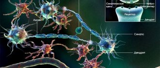
The functions of the basal ganglia are determined by their interaction with other areas of the central nervous system. They form neural loops connecting the thalamus and the most important areas of the cerebral cortex: motor, somatosensory and frontal. In addition, the subcortical nodes are connected to each other and to some areas of the brain stem.
The caudate nucleus and putamen perform the following functions:
- control of direction, strength and amplitude of movements;
- analytical activity, learning, thinking, memory, communication;
- control of eye, mouth, and face movements;
- maintaining the functioning of internal organs;
- conditioned reflex activity;
- perception of sensory signals;
- control of muscle tone.
Functions of the globus pallidus:
- development of an orienting reaction;
- control of arm and leg movements;
- eating behavior;
- facial expressions;
- display of emotions;
- providing auxiliary movements and coordination abilities.
The functions of the fence and amygdala include:
- speech;
- eating behavior;
- emotional and long-term memory;
- development of behavioral reactions (fear, aggression, anxiety, etc.);
- ensuring social integration.
Thus, the size and condition of individual basal ganglia affects emotional behavior, voluntary and involuntary movements of a person, as well as higher nervous activity.