Diencephalic syndrome and the hypothalamus
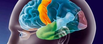
The hypothalamus (lat. hypothalamus) or hypothalamus is the part of the brain that suffers from diencephalic syndrome. This is the highest vegetative center that controls the work of all endocrine glands: the pituitary gland, adrenal glands, ovaries, thyroid and pancreas.
The hypothalamus controls the respiratory, cardiovascular, digestive and excretory systems. It is responsible for regulating body temperature, sleep and wakefulness rhythms, feelings of thirst and hunger, as well as human emotions and behavior.
Reasons for the development of diencephalic syndrome
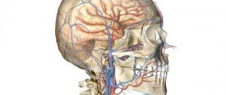
The vessels involved in the blood supply to the hypothalamus are characterized by increased permeability. This makes them vulnerable to various damaging factors, the impact of which causes the development of diencephalic syndrome. The function of the hypothalamus may be affected for the following reasons:
- traumatic brain injury;
- previous neuroinfection;
- the presence of tumors that put pressure on the hypothalamus;
- severe diseases of internal organs;
- hormonal changes during pregnancy;
- birth trauma or postpartum hemorrhage;
- insufficient protein nutrition, starvation, anorexia nervosa;
- stress or mental trauma;
- the presence of foci of chronic infection of the ENT organs, genitourinary system, gastrointestinal tract;
- intoxication (drinking alcohol, smoking, drug use, occupational hazards, environmental pollution).
Considering the multifaceted function of the hypothalamus in the body’s activities, the clinical picture of its lesions is extremely diverse.
Various clinical picture of diencephalic syndrome
Due to the many symptoms, doctors of various specialties often encounter diencephalic syndrome: endocrinologists, therapists, gynecologists, neurologists, surgeons, psychiatrists, dermatologists, etc.
With diencephalic syndrome, the following types of disorders are noted:
Autonomic-vascular disorders are manifested by crises, during which the following occur: suffocation, weakness, drowsiness, sweating, nausea, and also a rare pulse, a drop in blood pressure, pallor, and decreased motor activity. Autonomic-vascular crises are often replaced by sympatho-adrenal crises, which are characterized, on the contrary, by an increase in blood pressure.
Violation of thermoregulation is characterized by the appearance during a crisis of chills, increased sweating, an increase in body temperature to 38-39°C and often involuntary urination.
Neuromuscular disorders are expressed in asthenia, general weakness and adynamia, accompanied by low-grade fever, feelings of hunger and thirst, insomnia and unpleasant sensations in the heart area. The course of the disease is often paroxysmal.
Neurotrophic disorders are manifested by itching, dryness, the occurrence of neurodermatitis and bedsores, ulcers of the gastrointestinal tract, as well as softening of bones (osteomalacia). Against this background, the following are noted: drowsiness, general weakness, adynamia, tremor and a feeling of thirst. The course of the disease is critical.
Neuropsychic disorders are characterized by asthenia, sleep disturbances, and a decrease in the level of mental activity. In this case, hallucinations, a state of anxiety and fear, frequent mood swings, hypochondriacal disorders, and delusional states occur.
Hypothalamic epilepsy is a special form of epileptic seizures in which the primary focus is located in the hypothalamus. From it, excitation is transmitted to the cortical and subcortical motor centers. During attacks, the patient experiences palpitations with a rise in temperature and blood pressure (BP), tremors, respiratory distress, and fears. The electroencephalogram (EEG) records epileptic outbreaks in the form of single waves.
Neuroendocrine disorders are associated with dysfunction of not only the hypothalamus, but also other endocrine glands: thyroid, adrenal glands, pituitary gland. Isolated forms of endocrine dysfunction are often observed, such as diabetes insipidus, hypothyroidism, Itsenko-Cushing's disease, Sheehan's syndrome. The last two are often encountered in the practice of a gynecologist, so we will talk about them in more detail.
NSICU.RU neurosurgical intensive care unit website of the intensive care unit of the N.N. Research Institute Burdenko
Research Institute of Neurosurgery named after N.N. Burdenko, RAMS, Moscow
Introduction
Diencephalic structures (DS) include the thalamus, hypothalamus, epithalamus, subthalamus and pituitary gland [1]. The thalamus, located below the lateral ventricles, is a cluster of nuclei, has an oval shape, forms the lateral wall of the third ventricle and occupies 4/5 of the space of the diencephalic region. The main function of the thalamus is the primary analysis and transmission of information to the cerebral cortex from all sense organs with the exception of the olfactory analyzer. The hypothalamus is a complex of nuclei that form the bottom and side walls of the third ventricle.
There are paraventricular, supraoptic, preoptic, suprachiasmatic, ventromedial, arcuate, mammillary, posterior and other nuclei. The nuclei of the hypothalamus have extensive intracranial connections. The hypothalamus synthesizes peptides - liberins and statins. The hypothalamus is the highest center of endocrine regulation, ensures the constancy of the internal environment of the body, regulates the functioning of organs and systems of the body, coordinates the work of the nervous, endocrine and immune systems. The epithalamus - the pineal body, the leash, its nuclei and commissures, the posterior commissure - form the roof of the third ventricle, to which the choroid plexus is adjacent.
Melatonin is synthesized in the pineal gland. The epithalamus regulates the sleep-wake cycle, takes part in the regulation of emotions and the functioning of the autonomic nervous system. The subthalamus includes the subthalamic nucleus, the rostral parts of the red nucleus and the substantia nigra, is located between the thalamus and the midbrain tegmentum and is involved in the extrapyramidal regulation of movements. The pituitary gland is located in the sella turcica of the sphenoid bone and has two lobes - the anterior (adenohypophysis) and posterior (neurohypophysis). The neurohypophysis is connected to the hypothalamus by the pituitary stalk, which is the axons of the neurons of the nuclei of the hypothalamus and epithalamus and through which vasopressin, oxytocin and melatonin are transported. The anterior lobe of the pituitary gland synthesizes tropic hormones (adrenocorticotropic, thyroid-stimulating, gonadotropic, somatotropic hormones and prolactin), the release of which is regulated by hypothalamic peptides. Liberins and statins enter the adenohypophysis from the hypothalamus through a branched system of arterioles, capillaries and venules. Endocrine regulation is the main function of the pituitary gland.
Syndromes that develop as a result of damage to the DS are described: diencephalic dysthermia (hyperthermia, hypothermia, poikilothermia), hypothalamic obesity, diencephalic syndrome in children, hypothalamic cachexia in adults, anorexia, bulimia, diencephalic glycosuria, acromegaly, Cushing's disease, hypothalamic hypogonadism, pituitary dwarfism, gigantism, syndrome Pradera-Willi and others [2,3]. In neurocritical care patients with traumatic brain injury, signs of damage to the DS are detected in 15-33% of cases [4 – 6]. The range of clinical manifestations that these authors consider to be a consequence of damage to the DS in patients with traumatic brain injury is wide and includes impaired consciousness, dilated pupils, increased intracranial pressure, arterial hypertension, tachycardia, bradycardia or other cardiac arrhythmias, fever, increased muscle tone, opisthotonus, hyperhidrosis, tachypnea. Considering the mechanisms of brain damage during traumatic brain injury, it is difficult to talk about isolated damage to the DS in this category of patients. A number of the above symptoms are a consequence of primary damage to brainstem and subcortical structures or dislocation of the brainstem.
In patients with tumors of the chiasmal-sellar region (CSR) and a complicated course of the postoperative period, the critical condition is caused by isolated damage to the DS. In this case, a characteristic syndrome is formed - diencephalic dysfunction syndrome (DDS).
Purpose of the study
Description of DDS in intensive care patients. In connection with this goal, it was necessary to solve the following tasks: determine the structure of the DDS; identify SDD options; to draw a correlation between the variant of DDS and the outcome of the disease.
Materials and research methods
The study included patients who underwent surgery at the Institute from 2006 to 2009, inclusive. Inclusion criteria were: Age over 18 years; XSO tumor; complicated course of the early (7 days) postoperative period; the patient's stay in the intensive care unit for more than 48 hours. Exclusion criteria were: patient stay in the intensive care unit for less than 48 hours; intracranial complications not associated with damage to diencephalic structures (meningeal hematomas, intracerebral hematomas at a distance from the bed of the removed tumor, ischemic brain damage in the basin of the great vessels of the brain); atonic coma that developed within 7 days after surgery; pulmonary embolism and acute myocardial infarction that developed within 7 days after surgery; sepsis that developed within 7 days after surgery. Manifestations of multiple organ dysfunction were recorded. For this purpose, the criteria of the SOFA scale were used [7]. The exceptions were the classification of altered consciousness (AC) and dysfunction of the respiratory system. Consciousness was assessed using the RASS scale (Table 1).
The RASS scale was chosen because the patient population studied develops both depression of consciousness and psychomotor agitation. Under these conditions, the use of other generally accepted scales assessing the level of consciousness, including the Glasgow Coma Scale, did not cover all changes in consciousness characteristic of patients with CSO tumors during a complicated postoperative period. The absence of IS corresponded to a RASS score of 0; with any other RASS score, consciousness was considered altered.
The criteria for dysfunction of the respiratory system were a decrease in the pO2/FiO2 index below 400, as in the SOFA scale, and the need for prolonged mechanical ventilation (more than 24 hours after surgery). This is due to the fact that the cause of respiratory dysfunction in neurocritical care patients is not only parenchymal damage to the lungs, which reflects the pO2/FiO2 index, but also other mechanisms, for example, disruption of the central mechanisms of respiratory regulation or depression of consciousness [8]. Other somatic organ dysfunctions (SOD) were classified strictly according to the SOFA scale. Cardiovascular system dysfunction was defined as the development of arterial hypotension (a decrease in mean blood pressure below 70 mmHg) or the need to use sympathomimetic drugs. Renal dysfunction was defined as an increase in creatinine levels above 110 μmol/L or a decrease in urine output less than 500 ml/day. Hepatic dysfunction was defined as an increase in bilirubin levels above 20 μmol/L. Hematological dysfunction was defined as thrombocytopenia (less than 150 thousand/μl).
Fluid-electrolyte disturbances (WED) and intestinal dysfunction often develop in patients with CSO tumors and a complicated course of the postoperative period. The SOFA scale does not include assessment of these disorders, so the following criteria were introduced to qualify them. VEN was defined as dysnatremia (increase above 145 mmol/L or decrease below 135 mmol/L) due to diabetes insipidus, syndrome of inappropriate vasopressin secretion, or salt-wasting syndrome. Intestinal dysfunction was defined as gastrointestinal paresis. Fever was defined as an increase in body temperature above 38.5ºC, hypothermia - a decrease in body temperature below 36ºC. To prevent the development of hormonal deficiency, all patients after surgery received hormone replacement therapy at a dose that is used in patients with hormonal deficiency who are in a state of severe stress: hydrocortisone 2.5 - 3 mg/kg/day, L-thyroxine 2-2.5 mcg/kg/day Data are presented as medians with 25th and 75th percentiles indicated. The Mann-Whitney test and risk ratio were used in data analysis. Differences were considered significant at p<0.05, and the risk was considered significant if the lower limit of the confidence interval exceeded one. Statistical analysis was carried out using the Statistica 7.0 program.
Research results and discussion
According to the inclusion criteria, 83 patients were included in the study. 7 patients were excluded from the analysis in accordance with the above criteria: 2 patients with epidural hematoma, 3 patients with the formation of atonic coma within 48 hours, 1 patient with acute myocardial infarction and 1 with PE. Thus, 76 patients with a complicated course of the early postoperative period aged from 19 to 77 years (median 49 years) were analyzed.
There were 43 (56.6%) women, 33 (43.4%) men. 46 patients had a pituitary adenoma, 17 had a craniopharyngioma, 8 cases had meningioma in the CSR, and one observation each included patients with glioma and ependymoma of the third ventricle, hemangiopericytoma, epidermoid cyst, and cancer metastasis in the CSR.
All 76 patients (100%) had IS and dysnatremia, developing, respectively, on the first and second days after surgery (Table No. 2).
Taking into account the anatomy and physiology of the DS, this is logical. Since vasopressin is synthesized in the paraventricular and supraoptic nuclei, deposited in the neurohypophysis, and transported there along the pituitary stalk, there is a high probability of perioperative damage to any one or more of these anatomical structures. This immediately leads to dysnatremia. IP is also easy to explain. The thalamus, which occupies 4/5 of the DS, isolates important information coming from all sensory analyzers and transmits only it to the cerebral cortex. When this limiting mechanism is damaged, the cortex receives a huge amount of unnecessary information, which it is not able to analyze, and an obvious overload occurs, leading to a change in consciousness [9]. The epithalamus synthesizes melatonin, which is then deposited in the neurohypophysis. Melatonin is of paramount importance for the adequate functioning of the circadian sleep-wake rhythm. When the epithalamus, pituitary stalk or pituitary gland are damaged, sleep disturbances develop, which lead to changes in consciousness [10]. In addition, various DS are associated with stem structures, the limbic system, and the cerebral cortex. Morphological damage to these connections can also cause IS [11].
Dysfunction of the respiratory and cardiovascular systems and water and electrolyte disturbances developed on the second day after surgery, and paresis of the gastrointestinal tract - on the third. The most common SODs were cardiovascular dysfunction, intestinal dysfunction, and respiratory dysfunction. Dysfunction of the cardiovascular and respiratory systems developed on the second day, respectively, in 52 (68%) and 50 (66%) patients. Intestinal failure developed on the third day in 51 (67%) patients. The cardiovascular, respiratory systems and gastrointestinal tract are those structures on which DS have a pronounced and direct effect. Based on this, the frequent development of these SODs in patients with a complicated course of the postoperative period is logical. Thrombocytopenia developed much less frequently and later – in 21 (28%) patients from 4 days after surgery. This is because platelet levels are regulated by other mechanisms. Renal and hepatic dysfunction developed in the early stages after surgery, but were detected very rarely - in three (4%) and one (1%) cases, respectively. DS do not directly regulate glomerular filtration, so they cannot have a direct effect on creatinine clearance [12]. In diabetes insipidus, renal failure develops only with the development of decompensated hypovolemia, which can be easily avoided with adequate infusion and hormone replacement therapy with vasopressin preparations. In addition, renal failure can develop as a result of resistant arterial hypotension, which occurs extremely rarely in the studied group of patients [13]. DS probably also lack a direct regulatory effect on liver function, which, however, may suffer due to hypoperfusion due to resistant arterial hypotension. Another possible cause of the development of renal and liver failure may be abdominal compartment syndrome, which develops as a result of paresis of the gastrointestinal tract [14]. However, resistant arterial hypotension and compartment syndrome develop quite rarely [13], which is reflected in the frequency of these dysfunctions in the studied category of patients.
Fever developed in 18 (23.7%) patients; hypothermia was not detected in any of the observations. Thus, dysthermia, previously described as a symptom characteristic of damage to the DS, rarely developed in our patients.
When the DS is damaged, dysfunction of the adrenal glands and thyroid gland is inevitable. Clinical manifestations of adrenal and thyroid insufficiency are arterial hypotension, brady- or tachycardia, hypothermia or fever, paresis of the gastrointestinal tract, increased membrane permeability, leading to polyserositis and interstitial edema [2,15]. In accordance with accepted intensive care protocols, all patients included in the study received glucocorticosteroid and thyroid hormones in doses that completely replaced their production by the adrenal glands and thyroid gland under conditions of severe stress. It follows from this that we could exclude adrenal and thyroid insufficiency from among the causes of the development of the above organ disorders.
It is obvious that if the posterior lobe of the pituitary gland or its stalk is damaged, VEN will inevitably appear [2]. In which type of damage to the DS the patient will obligately develop IS and SOD still remains unknown. The analysis also failed to answer this question. This means that accurate preoperative and intraoperative predictors of the development of IS and SOD are still lacking.
It should be emphasized that before the operation, all patients included in the study were in a compensated state, and they had no manifestations of somatic organ dysfunction, and VEN were less pronounced or completely absent. Thus, when describing the structure of postoperative DDS, dysnatremia, IS and SOD that developed after surgery should be taken into account. To determine the variants of DDD, patients were divided into groups depending on the combination of identified dysfunctions. There were 6 groups (Table No. 3).
Note. Gr – group; IS – altered consciousness; VEN – water and electrolyte disturbances; SD – somatic dysfunction; ICU – intensive care unit; GOS – Glasgow Outcome Scale; DS – diencephalic structures; SS – stem structures; PE – pulmonary embolism.
Group 1 consisted of 12 patients. They showed an isolated combination of IS and VEN without SOD. The median age of these patients was 42.5 years, the median length of stay in the ICU was 8.5 days. In 11 (91.7%) patients, according to the Glasgow Outcome Scale (GOS), a favorable outcome was recorded (GOS=4), one patient (8.3%) had an unfavorable outcome (GOS=3), there were no deaths.
Group 2 consisted of 11 patients. They had one SOD in addition to IS and dysnatremia. Most often, 6 (54.5%) patients developed dysfunction of the cardiovascular system. Intestinal paresis developed in two patients, and respiratory failure developed in two cases. In one observation, thrombocytopenia was detected. The median age was 51 years, and the median length of stay in the ICU was 13 days. A favorable outcome (GOS=4) was in 5 (45.5%) patients, an unfavorable outcome (GOS=3) was in 6 patients (54.5%).
Group 3 consisted of 12 patients. They had two SODs against the background of IS and dysnatremia. Intestinal failure developed in 9 (75%) patients, respiratory failure - in 8 (66.7%) patients, cardiovascular failure - in 7 (58.3%) cases. The median age in this group was 47 years, and the median length of stay of patients in the ICU was 23.5 days. 3 (25%) patients had a favorable outcome: GOS = 4 in two observations; GIS=5 in one observation. 9 patients (75%) had an unfavorable outcome. Of these, 5 patients developed severe disability (GOS = 3), and in 4 cases (33%) a fatal outcome was recorded (GOS = 1). The cause of death in 3 cases was sepsis, in one case it was PE.
Group 4 consisted of 25 patients. They had three SODs in addition to IS and dysnatremia. Respiratory failure developed in 24 (96%) cases, cardiovascular failure - in 23 (92%) patients, intestinal failure - in 24 (96%) patients. Thrombocytopenia was noted in 4 (16%) patients. The median age in this group was 53 years, the median length of stay in the ICU was 22 days. A favorable outcome was observed in 6 (24%) patients: GOS = 5 in two patients, GOS = 4 in four patients, unfavorable outcome in 19 patients (76%), of which 8 developed profound disability (GOS = 3). Death occurred in 11 patients (44%). In 4 patients who died, the cause of death was sepsis, in 2 – meningoencephalitis, in another 2 – hemotamponade of the ventricular system, dislocation and herniation, 2 patients died due to confirmed morphological damage to diencephalic structures, and 1 – due to PE.
Group 5 consisted of 12 patients. They were simultaneously diagnosed with four SODs with IS and dysnatremia. All patients developed respiratory, cardiovascular, intestinal failure and thrombocytopenia. The median age in this group was 57.5 years, the median length of stay in the ICU was 40.5 days. A favorable outcome (GOS=4) was recorded in 4 (33.3%) patients in this group. An unfavorable outcome developed in 8 (66.7) cases. However, only one of the 8 patients with an unfavorable outcome survived (ORG=3). The remaining 7 (58.3%) patients had a fatal outcome (GOS=1). In 5 observations, the cause of death was sepsis, in 2 observations – damage to diencephalic structures.
Group 6 consisted of 4 patients. They simultaneously developed 5 SOD with IS and dysnatremia. All patients developed respiratory, cardiovascular, intestinal failure and thrombocytopenia. In 3 patients, renal dysfunction was detected, and in one observation, hepatic dysfunction. The median age in this group was 52 years, the median length of stay in the ICU was 7.5 days. There were no patients in this group with a favorable outcome; in all observations, death occurred. The cause of death in one patient was sepsis, in three – morphologically confirmed damage to diencephalic and structures.
The outcomes of patients in the first group with an isolated combination of IS and dysnatremia were better than in all other groups. Their duration of stay in the ICU was the shortest. The differences between the first and second groups were not significant (p=0.5). However, significant differences were obtained in the length of stay of patients in the ICU between the first and third, first and fourth groups (p = 0.04 and p = 0.01, respectively). The analysis did not take into account the length of stay in the ICU of patients in group 5, since these were the most severe patients in whom death occurred early after surgery. Favorable outcomes in group 1 developed more often than in other groups, and unfavorable ones - less often (Table No. 3). The risks of developing unfavorable and fatal outcomes in groups 2 – 6, compared with the first group, are shown in Table No. 4.
There were no deaths in groups 2 and 3, and in group 6 all patients had a death, so it was impossible to calculate risks in these groups.
It is known that a number of patients with isolated dysnatremia, isolated IS, or even a combination of IS and dysnatremia do not require ICU stay in the postoperative period [16]. In some patients, these disorders may persist for a long time after they are discharged from the hospital, and adequately selected therapy allows patients to return to their daily activities.
Obviously, patients in group 1 had more severe forms of IS and dysnatremia than patients with similar disorders, but who did not require a stay in the ICU. Compared with the latter, the distinguishing feature of patients in group 1 was their instability. This was manifested by the need to change the intensive therapy during the day. However, the fact that the same manifestations can occur both in intensive care patients and in those who have already returned to their daily activities does not allow us to distinguish the isolated combination of IS and dysnatremia into one of the forms of DDS.
In group 2, the number of unfavorable outcomes significantly exceeded the number of favorable ones (Table No. 3). This gives grounds to consider patients in group 2 as patients of the first stage of DDS. Therefore, DDS is a syndrome that includes altered consciousness, dysnatremia, and at least one somatic organ dysfunction. The outcomes of patients in each subsequent group were more severe than the outcomes of patients in the previous group (Table No. 4). The risk of developing an unfavorable outcome compared to the first group was higher in each subsequent group: 13.2; 33; 34.8, respectively (Table No. 4). The lower limit of the confidence interval exceeded one in all groups, which indicates the reliability of the identified risk. It is not clear why the risk of developing an unfavorable outcome in group 5 compared to group 1 was 22, while in groups 3 and 4 it was 33 and 34.8, respectively. However, in this group the risk of developing a lethal outcome compared to the first group was significantly higher, and for group 4 the risk of developing a lethal outcome compared to the first group did not reach a significant level (Table No. 4). It follows from this that SDD has 5 degrees of severity. Stage 1 DDS has IS, dysnatremia and one SOD. At each subsequent degree, one SOD is added.
DDS is essentially a variant of multiple organ dysfunction syndrome (MODS). However, unlike SOD, which develops against the background of systemic inflammatory response syndrome, SDS has a number of fundamental differences. First, DDS necessarily includes IS and dysnatremia, which do not always develop in SPOD [17]. Secondly, renal and hepatic dysfunction, typical of DSOD, is not typical for DDS [18]. Thirdly, a significant difference is the timing and order of development of SOD. With DDD, early and virtually simultaneous development of respiratory, cardiovascular and intestinal dysfunction occurs within 1–3 days. Even renal dysfunction, which is not typical for DDD, developed within a day and a half after the operation. SOD is characterized by a slower and gradual emergence of SOD [19]. Fourth, thrombocytopenia has different prognostic significance for DDS and SPOD. In our study, it was detected in 21 patients, which is significantly more common than renal or hepatic failure, but significantly less common than cardiovascular, respiratory or intestinal dysfunction. This does not provide a basis for judging whether thrombocytopenia is a typical or atypical phenomenon in patients with DDS. While thrombocytopenia is typical for SPOD [17,18]. We found no differences in the frequency of its occurrence in different groups. In other words, if the appearance of any other organ dysfunction means a worsening of the patient's condition and a worsening of his prognosis, then the detection of thrombocytopenia meant little to the outcome. However, the most important fact is that in none of our observations did thrombocytopenia reach a level of 50 thousand per µl, and almost always the platelet count exceeded 100 thousand per µl. This meant that thrombocytopenia in DDS does not require correction, unlike SPOD.
conclusions
- In patients with neurosurgical pathology CSO, diencephalic dysfunction syndrome in the postoperative period is manifested by a combination of altered consciousness, water and electrolyte disturbances in the form of dysnatremia and at least one somatic organ dysfunction.
- The presence of altered consciousness and dysnatremia in a patient is not sufficient to diagnose diencephalic dysfunction syndrome.
- The severity of diencephalic dysfunction syndrome and outcome are determined by the number of somatic organ dysfunctions that have developed.
Literature
- Beer M, Frotscher M. Topical diagnosis in neurology according to Peter Duus. Anatomy, physiology, clinic. Per. from English edited by BEHIND. Suslina. 4th ed. M: Practical Medicine, 2009. 478 pp.
- Endocrinology. DeGroot LJ, Jameson JL Elsevier Saunders, Philadelphia, 2006. p. 3336.
- Russell A. Diencephalic syndrome of emaciation in infancy and childhood. // Arch Dis Child, 1951. V.26 p274.
- Baguley IJ, Heriseanu RE, Cameron ID, et al. A critical review of the pathophysiology of dysautonomia following traumatic brain injury. // Neurocrit Care. 2008. V. 8 p. 293-300.
- Boeve BF, Wijdicks EF, Benarroch EE Paroxysmal sympathetic storms (“diencephalic seizures”) after severe diffuse axonal head injury. // Mayo Clin. Proc. 1998. V. 73 p. 148-152.
- Clifton GL, Robertson CS, Kyper K. Cardiovascular response to severe head injury. // J Neurosurg. 1983. V. 59 p. 447-54.
- Vincent JL, Moreno R, Takala J, et al. The SOFA (Sepsis-related Organ Failure Assessment) score to describe organ dysfunction/failure. On behalf of the working group on sepsis-related problems of the European society of intensive care medicine.// Intensive Care Med. 1996. V. 22 p. 707-710.
- Popugaev K.A., Savin I.A., Goryachev A.S. and others. Scale for assessing the severity of respiratory failure in neurosurgical patients. // Anest. i rek., 2010. No. 4 p. 42-50.
- Stewart JT, Quijije N., Sheyner I., Stover KT Delirium without focal signs related to a thalamic stroke.// J. Am. Geriatr. Soc. 2010. V. 58 p. 2433-2434.
- Figueroa-Ramos MI, Arroyo-Novoa CM, Lee KA, et al. Sleep and delirium in ICU patients: a review of mechanisms and manifestations.// Intensive Care Med. 2009. V. 35 p. 781–795.
- Frontera JA Delirium and sedation in the ICU.// Neurocrit Care 2011. V. 14 p. 463-474.
- Legrand M., Payen D. Understanding urine output in critically ill patients. // Ann Intensive Care. 2011. V. 24 p.13.
- Popugaev KA, Savin IA, Goriachev AS, Kadashev BA Hypothalamic injury as a cause of refractory hypotension after sellar region tumor surgery.// Neurocrit Care. 2008. V. 8 p. 366-373.
- Cheatham ML: Intra-abdominal hypertension and abdominal compartment syndrome.// New Horiz. 1999. V. 7 p. 96–115.
- Popugaev K.A., Savin I.A., Goryachev A.S. and others. Secondary abdominal compartment syndrome in the complicated course of the postoperative period in patients with tumors of the chiasmal-sellar localization. // Anest. i rek., 2011. No. 4 p. 37-42.
- Pituitary adenomas: clinical picture, diagnosis, treatment / Ed. B.A. Kadasheva. M.-Tver: Triada Publishing House LLC, 2007. 368 p.
- Knaus WA and Wagner DP Multiple systems organ failure: epidemiology and prognosis.// Crit. Care Clin. 1989. V. 5 p. 221–232.
- Cerra FB Multiple organ failure syndrome./ In: Bihari DS, Cerra FB (eds). New Horizons. Multiple Organ Failure. Fullerton, CA: Society of Critical Care Medicine. 1989. p 1–24.
- Murray MJ, Coursin DB Multiple Organ Dysfunction Syndrome.// Yale J. Biol. Med. 1993. V. 66 p.501-510.
Diencephalic syndrome: Itsenko-Cushing's disease
Itsenko-Cushing's disease is a severe neuroendocrine disease in which, due to damage to the hypothalamus, the production of its specific factor increases, causing excessive synthesis of adrenocorticotropic hormone (ACTH) by the pituitary gland and, as a consequence, glucocorticoids by the adrenal glands.
This disease often develops during puberty, after childbirth and abortion, which is explained by the vulnerability of the hypothalamic parts of the central nervous system during these periods, and can also occur as a result of brain injury or neuroinfection.
Patients with Cushing's disease experience increased blood pressure and blood sugar. With this pathology, fat deposition is observed in the neck, face, abdomen and thighs. The face becomes moon-shaped, the cheeks become red. Purple stripes (striae) form on the skin, rashes and boils appear on the body.
In women suffering from Cushing's disease, the menstrual cycle is disrupted until the complete disappearance of menstruation (amenorrhea), infertility occurs, libido decreases, and anorgasmia is noted.
It should be noted that a similar clinical picture develops in the presence of pituitary and adrenal tumors (Itsenko-Cushing syndrome).
Examination and treatment of patients with Itsenko-Cushing's disease is carried out by a gynecologist-endocrinologist. The diagnosis is established on the basis of laboratory research methods that determine an increase in the level of ACTH and corticosteroids in the urine and blood, as well as using special tests with dexamethasone.
Computed tomography (CT) or magnetic resonance imaging (MRI) data can exclude tumors of the pituitary gland and adrenal glands.
A comment
The subject of discussion in the presented work was a rather rare pathology - diencephalic cachexia (DC), the pathogenesis of which remains unexplored to date. The article presents a unique clinical observation of DC in a young woman with papillary craniopharyngioma. A detailed presentation of clinical manifestations, laboratory and MRI data clearly demonstrates the primary role of diencephalic lesions in weight loss and the development of cachexia. The long follow-up period gives particular value to this observation. It is important to emphasize that the regression of cachexia after surgery is probably associated only with compression of the diencephalic structures, and not with their destruction. Based on the analysis of the literature carried out by the authors, the issues of etiology, clinical picture, diagnosis of DC against the background of various tumors of the chiasmal-sellar region and modern approaches to their treatment are also considered. It is emphasized that weight loss may be the first manifestation of craniopharyngioma. The rapid course of the disease with the development of cachexia and the lack of effective therapeutic agents emphasize the importance of early diagnosis and timely surgical treatment, as well as the need for a multidisciplinary approach to the management of such complex patients. All this determines the exceptional relevance of this problem (article).
The article is written in good literary language and is read with great interest.
L.Ya. Rozhinskaya (Moscow)
Diencephalic syndrome after childbirth (Simmonds-Schien syndrome)
Diencephalic syndrome can form after childbirth. Pregnancy is accompanied by an increase in the size and mass of the pituitary gland, the main “subordinate” of the hypothalamus. If a woman has bleeding during the postpartum period, vascular spasm occurs as a response, including in the brain. This contributes to the development of ischemia, and subsequently necrosis of the enlarged pituitary gland, as well as the nuclei of the hypothalamus. This condition is called Simmonds-Sheehan syndrome (hypothalamic-pituitary cachexia, postpartum hypopituitarism).
This may disrupt the functioning of all endocrine glands: thyroid, ovaries, adrenal glands. Characteristic signs of the disease are: lack of lactation after childbirth and sudden weight loss. There may also be complaints of headache, fatigue, low blood pressure, symptoms of anemia (dry skin, brittle hair, pain in the heart, etc.).
Due to dysfunction of the ovaries, a woman’s menstruation disappears and the genitals atrophy. Hypothyroidism is manifested by hair loss, swelling, disturbances in the gastrointestinal tract, and memory impairment.
Diagnosis of Simmonds-Sheehan syndrome is based on the study of the hormonal profile, which reveals a decrease in the blood level of the following hormones: somatotropic (STT), thyroid-stimulating (TSH), follicle-stimulating (FSH), luteinizing (LH) and adrenocorticotropic (ACTH).
In order to assess the functional state of the hypothalamic-pituitary-adrenal system, special tests are performed with ACTH and a load of metapyrone.
According to CT and MRI data, in Simmonds-Schien syndrome, structural changes in the sella turcica, the bone at the base of the skull on which the pituitary gland lies, can be detected.
Discussion
Weight loss is widespread in the pathogenesis of many diseases (chronic heart and kidney failure, diabetes mellitus, Addison's disease, rheumatoid arthritis, Alzheimer's disease, malignant neoplasms, etc.). Regardless of etiology, cachexia is a serious medical problem. Even a slight decrease in body weight in patients determines an unfavorable prognosis for the course of the disease and is a powerful predictor of high mortality [24–26]. The most life-threatening condition is a loss of more than 40% of ideal body weight [26, 27]. Since ancient times, the term “cachexia” (Greek kachexia, kakos
"bad" + hexis "condition) was synonymous with rapid death. “The flesh disappears... shoulders, collarbones, chest, fingers seem to melt. This state is the face of death,” wrote Hippocrates [28]. I.S. Turgenev in 1846, in his story “Living Relics,” describes cachexia in a young woman who was “the first beauty, tall, plump, white, ruddy...”. “Before me lay a living human being, but what was it? The head is completely dry, one-color, bronze - like an icon of an ancient letter; the nose is narrow, like a knife blade; You can barely see your lips—only your teeth and eyes are white, and from under your scarf, thin strands of yellow hair spill out onto your forehead. At the chin, on the fold of the blanket, two tiny hands, also bronze-colored, are moving, slowly moving their fingers, like sticks... the face is not only not ugly, even beautiful, but terrible, extraordinary” [29].
Despite the fact that the first description of cachexia was made more than 2000 years ago, questions of its pathogenesis in various diseases are still the subject of debate and scientific research. Body weight loss in CSR tumors remains unstudied. We describe a rare case of diencephalic cachexia in a young woman with CF.
CF are benign epithelial tumors (WHO I), which develop from the remains of embryonic cells of Rathke's pouch, located along the pharyngeal-pituitary tract. The incidence of CF in adults is about 2.1–4.6% of all intracranial neoplasms [30–32].
Histologically, there are two main types of CF: adamantine-like and papillomatous. The papillomatous variant occurs, as a rule, only in adults. CF are characterized by the presence of a solid and cystic component, which is detected in approximately 80% of cases [33, 34]. The occurrence of papillomatous CF, in contrast to adamantine-like ones, is associated with metaplasia of epithelial cells. Papillomatous K.F. predominantly localized in the region of the third ventricle; one of the names of these tumors is papillomas of the third ventricle.
Embryogenesis and topography of CF determine the high frequency of endocrine and metabolic disorders. The result of damage to the hypothalamic region can be an imbalance in energy homeostasis, leading to the development of obesity or cachexia. It is known that CF is often associated with the development of diencephalic (hypothalamic) obesity [35, 36]. DC is less common and is represented by descriptions of only isolated cases. Thus, in a large retrospective study among children with CF, out of 485 cases, the development of DC was observed in only 7 patients [37]. It is assumed that the development of obesity or cachexia in CF is associated with invasive tumor growth in contrast to its compression of the hypothalamic region, respectively [6, 37]. To date, the pathophysiology of DC remains unknown. Modern ideas about the central mechanisms of metabolic control suggest that weight loss in DC is caused by dysfunction of hypothalamic neurons involved in the regulation of energy homeostasis and body weight. The lack of clinical and laboratory data on secondary hypofunction of peripheral glands, primarily hypocortisolism, clearly demonstrates different mechanisms of weight loss in DC and Symonds and Sheehan syndromes.
Weight loss is a key symptom and the main reason for seeking medical help. When examining such patients, at the first stage of diagnosis, malnutrition, anorexia nervosa and conditions associated with malabsorption (impaired absorption of nutrients in the small intestine) are excluded. Further, the diagnostic search should be aimed at excluding organic pathology of the brain, even in the absence of general cerebral or focal neurological symptoms.
In our clinical observation, the manifestation of the disease coincided with the postpartum period; secondary amenorrhea was not regarded by the patient as a reason to seek medical help. Only a rapid decrease in body weight led her to be examined by doctors, and the appearance of neuro-ophthalmological symptoms in the form of chiasmal syndrome played a decisive role in conducting an MRI of the brain.
By the time the tumor was detected, the body weight deficit was about 20%, and by the time of hospitalization - more than 40% of the initial body weight, which is critical for the patient’s life.
Until the stage of surgical treatment or the impossibility of surgery, such patients require constant nutritional support. Currently, there is no generally accepted pharmacological therapy for the treatment of cachexia of any etiology.
From laboratory data, attention was drawn to the increase in basal GH levels characteristic of DCs against the background of a decrease in IGF-1. Similar changes can be observed with cachexia of any etiology, for example with anorexia nervosa [38].
The patient also showed a significant increase in prolactin levels, which was not previously described in D.K. This change is most likely due to blockade of the dopaminergic system when the tumor is located suprasellar. Laboratory tests confirmed secondary hypogonadism. No other clinically significant biochemical abnormalities were detected. All these changes are not the cause of weight loss, but develop secondary.
It should be noted that despite the pronounced lack of body weight, the patient remained in good health and physical activity, and was in an “elevated” mood, which created a contrast with her appearance. The presence of elevated mood and even euphoria has often been described in children with DC by authors of previously published cases.
This clinical observation is also of interest because the development of DC is typical for children in the first 2-3 years of life and is extremely rare in adults. Development of D.K. most often cause astrocytomas of the diencephalic region of varying degrees of malignancy. Other types of tumors, including CF, as in our case, are rather an exception. The development of cachexia is more likely a sign of irritation of intact hypothalamic structures, since regression of symptoms was noted after surgery.
The main treatment method for CF remains surgical. The anatomical proximity of the tumor to vital structures (the floor of the third ventricle, the optic nerves and chiasm, the great vessels of the base of the brain) determines the significant complexity of surgical treatment and creates difficulties for their radical removal [39–44]. As a rule, removal of CP leads to an increase in neuroendocrine disorders [44, 45]. In the observation we presented, the patient developed additional disorders after the first operation - diabetes insipidus, central hypocortisolism and hypothyroidism, and secondary hypogonadism persisted (see table). The patient required lifelong replacement therapy with L-thyroxine, hydrocortisone, desmopressin and sex steroids. However, it is important to emphasize that the patient began to quickly gain weight, despite the fact that there were tumor remnants in the area of the bottom of the third ventricle. Thus, we can assume a significant role of compression, and not destruction of diencephalic structures for the development of cachexia in the observation we described.
To date, the control of tumor growth after non-radical removal of CF remains an unsolved problem [46]. This tumor has a high recurrence rate, reaching 30% within 10 years with total resection [34, 39, 47].
Incomplete removal of CF is a prerequisite for tumor recurrence and determines the indications for stereotactic irradiation [39]. Our observation was also indicative in this regard. Continued tumor growth was detected in the patient 8 months after non-radical surgery. According to MRI of the brain, the size of the recurrent tumor exceeded the original ones, which characterizes CF as a tumor with an aggressive course. Considering the localization of the tumor, the goal of treating such patients is not only radical removal of the tumor, but also preventing the development of severe complications associated with damage to the hypothalamus. Such operations should be performed in institutions that have a multidisciplinary team of specialists, including a neurosurgeon, neurologist, radiologist, endocrinologist, psychiatrist and ophthalmologist.
Thus, in each case, the treatment strategy is determined individually based on the growth pattern and histology of the tumor, as well as the age and condition of the patient. Often, only partial removal or biopsy of the tumor is possible, followed by the use of non-surgical methods. In such cases, radiation and/or chemotherapy may be the only chance to save the patient [8, 9, 48, 49]. It is difficult to explain why, but even partial removal of the tumor can lead to regression of cachexia, which is what happened in our observation. Sometimes there is a restoration of body weight followed by the development of obesity [8, 23]. A detailed study of the mechanisms that control energy metabolism may shed light on the pathogenesis of these disorders and determine a targeted approach in the treatment of patients with metabolic disorders.
All photographic materials are posted with the voluntary written consent of the patient
.
The authors declare no conflict of interest.