Svetlana Yastrebova “Popular Mechanics” No. 6, 2018
Decades of discussions, long-established sayings, experiments on mice and sheep - but still, can the adult brain form new neurons to replace the lost ones? And if so, how? And if it can’t, why?
A cut finger will heal in a few days, a broken bone will heal. Myriads of red blood cells replace each other in short-lived generations, muscles grow under load: our body is constantly renewed. For a long time it was believed that there was only one outsider left in this celebration of rebirth: the brain. Its most important cells, neurons, are too highly specialized to divide. The number of neurons is falling year by year, and although they are so numerous that the loss of several thousand does not have a noticeable effect, the ability to recover from damage would not hurt the brain. However, scientists have not been able to detect the presence of new neurons in the mature brain for a long time. However, there were not enough sophisticated tools to find such cells and their “parents”.
The situation changed when, in 1977, Michael Kaplan and James Hinds used radioactive [3H]-thymidine, which could be incorporated into new DNA. Its chains actively synthesize dividing cells, doubling their genetic material and at the same time accumulating radioactive labels. A month after administering the drug to adult rats, the scientists obtained slices of their brains. Autoradiography showed that the marks were located in the cells of the dentate gyrus of the hippocampus. Still, they reproduce, and “adult neurogenesis” exists.
Survival Challenges
It is not enough for a nerve cell to simply be born; it must survive and acquire the functionality of mature neurons. And they die more often than you might think: almost half of the hippocampal cells produced during adult neurogenesis in rats die within a month of their appearance. In mice, losses reach 75% - but for those who lasted this period, death is no longer in danger, and by the end of it, new neurons are already fully operational. In macaques, with their larger brains, the maturation of new neurons and their integration into data processing structures takes much longer, about six months.
About Men and Mice
During this process, mature neurons do not divide, just as muscle fiber cells and red blood cells do not divide: various stem cells that retain their “naive” ability to reproduce are responsible for their formation. One of the descendants of the divided progenitor cell becomes a young specialized cell and matures into a fully functional adult state. The other daughter cell remains a stem cell: this allows the population of progenitor cells to be maintained at a constant level without sacrificing the renewal of the surrounding tissue.
Neuronal precursor cells were found in the dentate gyrus of the hippocampus. Later they were found in other parts of the rodent brain, in the olfactory bulb and the subcortical structure of the striatum. From here, young neurons can migrate to the desired area of the brain, mature in place, and integrate into existing communication systems. To do this, the new cell proves its usefulness to its neighbors: its ability to excite is increased, so that even a weak impact causes the neuron to produce a whole volley of electrical impulses. The more active a cell is, the more connections it forms with its neighbors and the faster these connections stabilize.
Adult neurogenesis in humans was confirmed only a couple of decades later with the help of similar radioactive nucleotides - in the same dentate gyrus of the hippocampus, and then in the striatum. Our olfactory bulb, apparently, is not renewed. However, how active this process is and how it changes over time is not exactly clear today.
For example, a 2013 study showed that until old age, approximately 1.75% of cells in the dentate gyrus of the hippocampus are renewed each year. And in 2018, results emerged showing that the formation of neurons here stops already in adolescence. The first measured the accumulation of radioactive tracers, and the second used dyes that selectively bind to young neurons. It is difficult to say which conclusions are closer to the truth: it is difficult to compare rare results obtained by completely different methods, and even more so to extrapolate work done on mice to humans.
The first studies of neurogenesis were carried out on planarian flatworms, and the first vertebrate models were axolotls. Today, experiments most often take place with zebrafish and laboratory rodents
NERVE CELLS ARE RESTORED
The catchphrase “Nerve cells do not regenerate” has been perceived by everyone since childhood as an immutable truth. However, this axiom is nothing more than a myth, and new scientific data refute it.
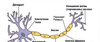
Schematic representation of a nerve cell, or neuron, which consists of a body with a nucleus, one axon and several dendrites.
Neurons differ from each other in size, dendritic branching, and axon length.
The concept of 'glia' includes all cells of nervous tissue that are not neurons.
Neurons are genetically programmed to migrate to one or another part of the nervous system, where, with the help of processes, they establish connections with other nerve cells.
Dead nerve cells are destroyed by macrophages that enter the nervous system from the blood.
Stages of formation of the neural tube in the human embryo.
‹
›
Nature builds a very high margin of safety into the developing brain: during embryogenesis, a large excess of neurons is formed. Almost 70% of them die before the child is born. The human brain continues to lose neurons after birth, throughout life. This cell death is genetically programmed. Of course, not only neurons die, but also other cells of the body. Only all other tissues have a high regenerative capacity, that is, their cells divide, replacing dead ones. The regeneration process is most active in epithelial cells and hematopoietic organs (red bone marrow). But there are cells in which the genes responsible for reproduction by division are blocked. In addition to neurons, these cells include cardiac muscle cells. How do people manage to maintain intelligence until very old age if nerve cells die and are not renewed?
One possible explanation: in the nervous system, not all neurons “work” at the same time, but only 10% of neurons. This fact is often cited in popular and even scientific literature. I have repeatedly had to discuss this statement with my domestic and foreign colleagues. And none of them understands where this figure came from. Any cell simultaneously lives and “works”. In each neuron, metabolic processes occur all the time, proteins are synthesized, and nerve impulses are generated and transmitted. Therefore, leaving the hypothesis of “resting” neurons behind, let us turn to one of the properties of the nervous system, namely, its exceptional plasticity.
The meaning of plasticity is that the functions of dead nerve cells are taken over by their surviving “colleagues,” which increase in size and form new connections, compensating for the lost functions. The high, but not unlimited, effectiveness of such compensation can be illustrated by the example of Parkinson's disease, in which gradual death of neurons occurs. It turns out that until about 90% of the neurons in the brain die, the clinical symptoms of the disease (trembling of the limbs, limited mobility, unsteady gait, dementia) do not appear, that is, the person looks practically healthy. This means that one living nerve cell can replace nine dead ones.
But the plasticity of the nervous system is not the only mechanism that allows one to maintain intelligence into old age. Nature also has a backup option - the emergence of new nerve cells in the brain of adult mammals, or neurogenesis.
The first report on neurogenesis appeared in 1962 in the prestigious scientific journal Science. The article was titled “Are New Neurons Forming in the Adult Mammalian Brain?” Its author, Professor Joseph Altman from Purdue University (USA), used an electric current to destroy one of the structures of the rat’s brain (the lateral geniculate body) and injected it with a radioactive substance that penetrates into newly emerging cells. A few months later, the scientist discovered new radioactive neurons in the thalamus (a region of the forebrain) and the cerebral cortex. Over the next seven years, Altman published several more papers demonstrating the existence of neurogenesis in the brain of adult mammals. However, then, in the 1960s, his work aroused only skepticism among neuroscientists; their development did not follow.
And only twenty years later, neurogenesis was “discovered” again, but in the brain of birds. Many songbird researchers have noticed that during each mating season, the male canary Serinus canaria
performs a song with new “knees”. Moreover, he does not adopt new trills from his brothers, since the songs were updated even in isolation. Scientists began to study in detail the main vocal center of birds, located in a special part of the brain, and discovered that at the end of the mating season (for canaries it falls in August and January), a significant part of the neurons of the vocal center died, probably due to excessive functional load . In the mid-1980s, Professor Fernando Notteboom from Rockefeller University (USA) was able to show that in adult male canaries the process of neurogenesis occurs in the vocal center constantly, but the number of neurons produced is subject to seasonal fluctuations. Peak neurogenesis in canaries occurs in October and March, that is, two months after the mating seasons. That is why the “record library” of male canary songs is regularly updated.
In the late 1980s, neurogenesis was also discovered in adult amphibians in the laboratory of the Leningrad scientist Professor A.L. Polenov.
Where do new neurons come from if nerve cells do not divide? The source of new neurons in both birds and amphibians turned out to be neuronal stem cells from the wall of the ventricles of the brain. During the development of the embryo, it is from these cells that the cells of the nervous system are formed: neurons and glial cells. But not all stem cells turn into nervous system cells—some of them “lurk” and wait in the wings.
New neurons have been shown to arise from adult stem cells in lower vertebrates. However, it took almost fifteen years to prove that a similar process occurs in the mammalian nervous system.
Advances in neuroscience in the early 1990s led to the discovery of “newborn” neurons in the brains of adult rats and mice. They were found mostly in evolutionarily ancient parts of the brain: the olfactory bulbs and the hippocampal cortex, which are mainly responsible for emotional behavior, the response to stress and the regulation of sexual functions in mammals.
Just as in birds and lower vertebrates, in mammals neuronal stem cells are located close to the lateral ventricles of the brain. Their transformation into neurons is very intense. In adult rats, about 250,000 neurons are formed from stem cells per month, replacing 3% of all neurons in the hippocampus. The lifespan of such neurons is very high - up to 112 days. Neuronal stem cells travel a long distance (about 2 cm). They are also able to migrate to the olfactory bulb, turning into neurons there.
The olfactory bulbs of the mammalian brain are responsible for the perception and primary processing of various odors, including the recognition of pheromones - substances that are close in their chemical composition to sex hormones. Sexual behavior in rodents is regulated primarily by the production of pheromones. The hippocampus is located under the cerebral hemispheres. The functions of this complex structure are associated with the formation of short-term memory, the realization of certain emotions and participation in the formation of sexual behavior. The presence of constant neurogenesis in the olfactory bulb and hippocampus in rats is explained by the fact that in rodents these structures bear the main functional load. Therefore, the nerve cells in them often die, which means they need to be renewed.
In order to understand what conditions affect neurogenesis in the hippocampus and olfactory bulb, Professor Gage from Salka University (USA) built a miniature city. The mice played there, did physical exercise, and looked for exits from the mazes. It turned out that in “city” mice new neurons appeared in much greater numbers than in their passive relatives, mired in routine life in the vivarium.
Stem cells can be extracted from the brain and transplanted into another part of the nervous system, where they turn into neurons. Professor Gage and his colleagues conducted several similar experiments, the most impressive of which was the following. A piece of brain tissue containing stem cells was transplanted into the destroyed retina of a rat's eye. (The light-sensitive inner wall of the eye has a “nervous” origin: it consists of modified neurons - rods and cones. When the light-sensitive layer is destroyed, blindness occurs.) The transplanted brain stem cells turned into retinal neurons, their processes reached the optic nerve, and the rat regained its sight! Moreover, when brain stem cells were transplanted into an undamaged eye, no transformations occurred with them .
Probably, when the retina is damaged, some substances are produced (for example, so-called growth factors) that stimulate neurogenesis. However, the exact mechanism of this phenomenon is still unclear.
Scientists were faced with the task of showing that neurogenesis occurs not only in rodents, but also in humans. To this end, researchers led by Professor Gage recently carried out sensational work. In one of the American oncology clinics, a group of patients with incurable malignant tumors took the chemotherapy drug bromodyoxyuridine. This substance has an important property - the ability to accumulate in dividing cells of various organs and tissues. Bromodioxyuridine is incorporated into the DNA of the mother cell and is retained in the daughter cells after the mother cell divides. A pathological study showed that neurons containing bromodyoxyuridine are found in almost all parts of the brain, including the cerebral cortex. This means that these neurons were new cells that arose from the division of stem cells. The finding unconditionally confirmed that the process of neurogenesis also occurs in adults. But if in rodents neurogenesis occurs only in the hippocampus, then in humans it can probably involve larger areas of the brain, including the cerebral cortex. Recent research has shown that new neurons in the adult brain can be formed not only from neuronal stem cells, but from blood stem cells. The discovery of this phenomenon caused euphoria in the scientific world. However, publication in the journal Nature in October 2003 largely cooled the enthusiastic minds. It turned out that blood stem cells actually penetrate the brain, but they do not turn into neurons, but merge with them, forming binucleate cells. Then the “old” nucleus of the neuron is destroyed, and it is replaced by the “new” nucleus of the blood stem cell. In the rat's body, blood stem cells mainly merge with the giant cells of the cerebellum - Purkinje cells, although this happens quite rarely: only a few fused cells can be found in the entire cerebellum. More intense fusion of neurons occurs in the liver and heart muscle. It is still completely unclear what the physiological meaning of this is. One hypothesis is that blood stem cells carry with them new genetic material, which, when entering the “old” cerebellar cell, prolongs its life.
So, new neurons can arise from stem cells even in the adult brain. This phenomenon is already quite widely used for the treatment of various neurodegenerative diseases (diseases accompanied by the death of brain neurons). Stem cell preparations for transplantation are obtained in two ways. The first is the use of neural stem cells, which in both embryos and adults are located around the ventricles of the brain. The second approach is the use of embryonic stem cells. These cells are located in the inner cell mass at the early stage of embryo formation. They can transform into almost any cell in the body. The greatest difficulty in working with embryonic cells is getting them to transform into neurons. New technologies make this possible.
Some medical institutions in the United States have already formed “libraries” of neural stem cells obtained from embryonic tissue and are transplanting them into patients. The first attempts at transplantation give positive results, although today doctors cannot solve the main problem of such transplants: uncontrolled proliferation of stem cells in 30-40% of cases leads to the formation of malignant tumors. No approach has yet been found to prevent this side effect. But despite this, stem cell transplantation will undoubtedly be one of the main approaches in the treatment of neurodegenerative diseases such as Alzheimer's and Parkinson's diseases, which have become the scourge of developed countries.
“Science and Life” about stem cells:
Belokoneva O., Ph.D. chem. Sci. A ban on nerve cells. - 2001, No. 8.
Belokoneva O., Ph.D. chem. Sci. The mother of all cells. - 2001, No. 10.
Smirnov V., academician RAMS, corresponding member. RAS. Rehabilitation therapy of the future. - 2001, No. 8.
Model problems
Most studies of adult neurogenesis are conducted in laboratory animals, which reproduce quickly and are easy to maintain. This combination of characteristics is found in those who are small in size and live very short lives - mice and rats. But in our brains, which only finish maturing by age 20, things can happen completely differently.
The dentate gyrus of the hippocampus is part of the cerebral cortex, albeit a primitive one. In our species, like in other long-living mammals, the cortex is noticeably more developed than in rodents. Perhaps neurogenesis covers its entire volume, being realized by some of its own mechanisms. There is no direct evidence of this yet: studies of adult neurogenesis in the cerebral cortex have not been performed either in humans or in other primates.
But such work has been carried out with ungulates. A study of sections of the brain of newborn lambs, as well as slightly older sheep and sexually mature individuals, did not find dividing cells - the precursors of neurons in the cerebral cortex and subcortical structures of their brains. On the other hand, in the cortex of even older animals, young neurons that were already born but immature were found. Most likely, they are ready to complete specialization at the right moment, forming full-fledged nerve cells and taking the place of the dead. Of course, this is not exactly neurogenesis, because new cells are not formed during this process. However, it is interesting that such young neurons are present in those areas of the sheep brain that in humans are responsible for thinking (cerebral cortex), integration of sensory signals and consciousness (claustrum), and emotions (amygdala). There is a high probability that we will also find immature nerve cells in similar structures. But why might an adult, already trained and experienced brain need them?
How to train your brain correctly?
Today it is fashionable to go to the gym for training, but it is important to remember that such training does not train the brain, it only trains the body. To train your brain, you need to do something that is not familiar and new to you. Yes, it's not easy. It's about getting out of your comfort zone to find yourself in a growth zone. But just as it is not comfortable to do exercises with increased weight, when the muscles are stretched, the brain should be boiling when we reach peaks in unfamiliar areas of development. Special brain simulators are very suitable for this.
- Memory trainer. Used with challenges to remember objects in a picture or words.
- The attention trainer has become more relevant today than ever, because due to many modern factors the brain has become distracted. The Schulte table will help you develop your brain 5 out of 5.
- The speed of thinking simulator is used if you want to increase the speed of decision making.
All exercise equipment can be found on our site and used every day. It is very important to do training regularly with enough time and constantly adding loads. The brain develops only when there are difficulties and unusual actions. Nerve cells are not only restored, but also developed. Good luck to you!
Memory hypothesis
The number of neurons is so large that some of them can be safely sacrificed. However, if a cell has switched off from working processes, this does not mean that it has died. The neuron may stop generating signals and responding to external stimuli. The information he has accumulated does not disappear, but is “canned.” This phenomenon has led Carol Barnes, a neuroscientist at the University of Arizona, to theorize that this is how the brain stores and shares memories from different periods of life. According to Professor Barnes, from time to time a group of young neurons appears in the dentate gyrus of the hippocampus to record new experiences. After some time - weeks, months, and maybe years - they all go into a state of rest and no longer send signals. This is why memory (with rare exceptions) does not retain anything that happened to us before the third year of life: access to this data is blocked at some point.
Considering that the dentate gyrus, like the hippocampus as a whole, is responsible for transferring information from short-term memory to long-term memory, this hypothesis even seems logical. However, it still needs to be proven that the adult hippocampus actually produces new neurons, and in fairly large numbers. There is only a very limited set of possibilities for conducting experiments.
Stress story
Typically, human brain specimens are obtained during autopsy or neurosurgery, such as for temporal lobe epilepsy, where seizures are not treatable with drugs. Both options do not allow us to trace how the intensity of adult neurogenesis affects brain function and behavior.
Such experiments were carried out on rodents: the formation of new neurons was suppressed by targeted gamma radiation or by turning off the corresponding genes. This exposure increased the animals' susceptibility to depression. Mice incapable of neurogenesis were almost not happy about sweetened water and quickly gave up trying to stay afloat in a water-filled container. The content of cortisol, the stress hormone, in their blood was even higher than in mice stressed by conventional methods. They were more likely to become dependent on cocaine and had poorer recovery from stroke.
One important note worth making about these results: it is possible that the shown connection “fewer new neurons - a sharper reaction to stress” closes on itself. Unpleasant life events reduce the intensity of adult neurogenesis, which makes the animal more sensitive to stress, so the rate of formation of neurons in the brain decreases - and so on in a circle.
Why are new neurons important?
They are especially important for learning and memory. Studies of adult brains with a blocked ability to produce new neurons have found that the absence of new neurons invariably affects memory.
This memory ability is especially important for spatial orientation. For example, thanks to it you can find your way around a familiar city. Therefore, people who experience neurodegeneration (for example, due to Alzheimer's disease) have problems with orientation and often cannot find their way home.
Recent discoveries have shown that the creation of new neurons is important not just for the ability to remember, but also for the quality of memory. Neurogenesis helps us distinguish between memories that may appear the same at first glance. For example, when you park your car in the evening, you have the habit of leaving it in a certain part of the parking lot, but in different places each time. It is thanks to neurogenesis that you will be able to retain a new place in memory.
Discovering the production of neurons in the hippocampus allowed us to make real discoveries. For example, there is a direct link between neurogenesis and depression. A person who is depressed or in a state of emotional burnout always has a lower level of production of new neurons.
That is why, in order to improve memory, mood, and avoid problems in the brain caused by aging and stress, it is necessary to study neurogenesis in more detail.
Business on nerves
Despite the lack of accurate information about adult neurogenesis, businessmen have already appeared who are ready to build a profitable business on it. Since the early 2010s, a company that sells water from springs in the Canadian Rockies has been producing Neurogenesis Happy Water
.
It is claimed that the drink stimulates the formation of neurons due to the lithium salts it contains. Lithium is indeed considered a drug beneficial for the brain, although there is much more of it in tablets than in “happy water”. The effect of the miracle drink was tested by neuroscientists from the University of British Columbia. They gave rats “happy water” for 16 days, and a control group - plain water from the tap, and then examined slices of the dentate gyrus of their hippocampus. And although the rodents that drank Neurogenesis Happy Water
had as much as 12% more new neurons, their total number was small and there is no statistically significant advantage.
For now, we can only state that adult neurogenesis clearly exists in the brain of representatives of our species. Perhaps it continues until old age, or maybe only until adolescence. It's actually not that important. What is more interesting is that the birth of nerve cells in the mature human brain generally occurs: from the skin or from the intestines, the renewal of which occurs constantly and intensively; the main organ of our body differs quantitatively, but not qualitatively. And when the information about adult neurogenesis forms a coherent, detailed picture, we will understand how to translate this quantity into quality, forcing the brain to “repair”, restore the functioning of memory, emotions - everything that we call our life.
How does nutrition affect neurogenesis?
What and how we eat also affects the brain’s production of new neurons.
Calories and diet
I'll tell you one more thing that may seem completely incredible. Restricting the daily diet by 20–30% of calories enhances neurogenesis. Regular short fasts and stretching the time between meals also contribute to the production of new neurons.
Dark chocolate and blueberries
This information should especially appeal to women: consuming dark chocolate and blueberries increases neurogenesis. The flavonoids contained in these products are responsible for this.
Fats
All foods that contain omega-3 fatty acids (such as fatty fish) help neurogenesis. On the other hand, a diet rich in saturated fats (eg red meat, palm oil, etc.) has a very negative effect on the production of new nerve cells.
Alcohol
The ethanol contained in alcoholic drinks literally kills brain neurons.
A small detail. If you drink wine, there's good news for you. It contains a substance that supports the life of neurons. Therefore, wine can be enjoyed in small quantities, keeping in mind the harmfulness of ethanol.
Hard and crunchy foods
I'll tell you about the latest study conducted in Japan. The Japanese simply love combining a variety of textures in food. For example, they can combine something sticky and chewy with something hard and crunchy, etc. So, scientists from this country have found that soft foods slow down neurogenesis, while hard and crunchy foods, which have to be chewed thoroughly, speed it up.
Healthy eating
Healthy foods and raw foods are good for the body and stimulate neurogenesis. Poor nutrition and foods with excessive acid content not only provoke the development of depressive conditions, but also worsen memory and mood, and slow down the production of neurons in the hippocampus.