The role of venous circulation disorders in the origin and course of cerebrovascular diseases has been underestimated for a long time. This is explained by the previously existing difficulties of intravital assessment of cerebral venous hemodynamics when using traditional methods of recording venous blood flow in the vessels of the brain, as well as insufficient attention from researchers to this section of angiology.
The emergence of modern neuroimaging methods, in particular MRI diagnostics, has greatly facilitated the identification of such diseases.
Let's consider several examples of pathological processes detected by MRI diagnostics using MR venography.
THROMBOSIS OF THE VEINS AND SINES OF THE BRAIN
The cause of the development of thrombosis of the veins and sinuses of the dura mater can be septic lesions, trauma, compression of the sinus by a tumor, and systemic lesions of the connective tissue.
In addition, sinus thrombosis can develop due to thrombophlebitis of the extremities or inflammatory foci in the body (in the postpartum period, after an abortion, with infectious diseases, as well as diseases of the ears and paranasal sinuses).
Taking into account the patient’s age, the degree of development of collateral circulation, as well as the localization of the pathological process, the clinical manifestations of venous thrombosis are quite variable and nonspecific.
It is very difficult to identify typical clinical manifestations of sinus thrombosis, but the most common initial symptoms are the following: 1. headache 2. papilledema (a sign of intracranial hypertension) 3. focal neurological deficit 4. disturbances of consciousness (appear in case of damage to the brain substance in the form of progressive edema, developing infarction or hemorrhage).
In case of damage to the sinuses, general cerebral symptoms depend on the size and rate of increase of thrombosis.
Focal symptoms develop when brain matter is involved in the process, i.e. with the development of cortical venous infarction. Cortical motor deficits, cortical symptoms, and seizures appear according to the location of the affected sinus.
When a clinical picture of thrombosis of the veins and sinuses of the brain occurs, MRI in most cases reveals signs of extensive areas of ischemia and hemorrhage. However, in some cases, standard neuroimaging methods fail to detect changes in the brain parenchyma.
The method of choice in such cases is magnetic resonance imaging (MRI) of the brain using MR venography.
Thrombosis of the right transverse sinus - hypointense areas on T2 (intracellular deoxyhemoglobin).
To confirm venous sinus thrombosis and determine the exact location and extent of the thrombus, MR venography is necessary.
MR venography – lack of visualization of blood flow in the right transverse sinus and jugular vein.
MRI of the brain: on the right (green arrow) the T2-weighted image shows the normal phenomenon of “empty flow” from the right sigmoid sinus and jugular vein. On the left (orange arrow) there is an abnormally high signal, most likely resulting from thrombosis. To confirm sinus thrombosis and definitively determine the location and extent of thrombosis, MR venography is necessary.
MR venography: thrombosis of the left transverse sinus. There is loss of MR signal from the left transverse sinus. The presence of sinus visualization on “raw” data or brain MRI confirms sinus thrombosis and excludes its hypoplasia.
MR venography: thrombosis of the right transverse sinus. There is loss of MR signal from the right transverse sinus. The presence of sinus visualization on “raw” data or MRI of the brain confirms sinus thrombosis and excludes its hypo- and aplasia.
Thrombosis of the right transverse sinus. Absence of the phenomenon of “empty flow” from the right transverse sinus on MRI of the brain. Absence of visualization of the right transverse sinus on MR venography.
As mentioned above, in cases of a clinical picture of cerebral venous thrombosis along the veins and sinuses, an MRI of the brain in some cases reveals an area of ischemia and hemorrhage.
MRI of the brain: a combination of vasogenic (orange arrow), cytotoxic edema and hemorrhage (green arrow) is noted. This MR picture, as well as the location of the pathological zone in the projection of the temporal lobe, makes us think about hemorrhagic venous cerebrovascular accident due to thrombosis of the vein of Labbe. Confirmation requires MR venography or contrast-enhanced MRI.
STENOSIS, AREAS OF PATHOLOGICAL EXTENSION AND HYPOPLASIA OF VENOUS STRUCTURES OF THE BRAIN
MRA picture of pronounced asymmetry of the venous network with a predominance and mild dilatation of the veins of the right hemisphere (transverse, sigmoid sinuses and jugular vein on the right); hypoplasia of the left transverse and sigmoid sinus. Single areas (2) of local dilatation of veins in the parasagittal parts of the left hemisphere, the great vein of the brain. Asymmetrical, dilated and clearly convoluted venous structure of the extracranial sections on the right.
MRA signs of slight dilatation of the superior sagittal sinus, local decrease in blood flow and narrowing of the lumen of the distal straight sinus; asymmetry of the lumen of the transverse, sigmoid sinuses and internal jugular veins.
VASCULAR MALFORMATIONS
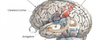
Venous system of the brain, lesion syndromes
The veins of the brain are divided into superficial and deep. Superficial veins, located in the pia mater, collect blood from the cortex and white matter, deep veins - from the white matter of the hemispheres, subcortical nodes, ventricular walls and choroid plexuses. The veins of the dura mater pass together with the arteries in the thickness of the shell and form a significant venous network. All veins carry blood to the venous blood collectors - the venous sinuses of the dura mater, located between its two leaves. The main ones are: the superior longitudinal sinus, passing along the upper edge of the large falciform process; the inferior longitudinal sinus, located along the lower free edge of the large falciform process with the tentorium of the cerebellum; transverse sinus - the widest of all, located on the sides of the internal occipital bone thickening; cavernous sinus located on the sides of the sella turcica. Between the left and right cavernous sinuses, the intercavernous sinuses, anterior and posterior, pass transversely, thus forming a circular sinus around the pituitary gland. The outflow of blood from the cranial cavity occurs through the internal jugular vein, partly through the vertebral vein and emissaries - venous graduates located inside the flat bones of the skull and connecting the venous sinuses of the dura mater with the diploic veins and with the external veins of the head. When the normal outflow of blood from the brain is disrupted, the development of picture of venous stagnation. Its causes may be cardiac and pulmonary failure, compression of extracranial veins - internal jugular, innominate and superior cava, brain tumors, traumatic brain injury, thrombosis of the veins and sinuses of the brain, compression of veins with craniostenosis and cerebral hydrocele, hanging, asphyxia of newborns. Thrombosis cerebral veins can occur without previous inflammation (phlebothrombosis) or against the background of inflammation (thrombophlebitis), but distinguishing them by clinical signs is difficult. Thrombosis of superficial cerebral veins
.
The clinical picture is characterized by a combination of neurological symptoms with signs of an inflammatory, usually infectious process (fever, inflammatory reaction of the blood, and sometimes of the cerebrospinal fluid). The disease almost always begins with a headache
, which is often accompanied by nausea and vomiting. Quite often, consciousness is impaired (sometimes with psychomotor agitation), and against this background, focal brain symptoms arise (paresis or paralysis of the limbs, aphasia, focal or general epileptic seizures, etc.). Lability of focal symptoms is characteristic, due to the migration of the process from one venous trunk to another. The morphological substrate causing the symptoms described above are hemorrhagic infarctions in the gray and white matter of the brain, intracerebral and subarachnoid hemorrhages, ischemia and cerebral edema.
The overwhelming majority of cases of thrombophlebitis of the superficial cerebral veins are observed in the postpartum period. It should be borne in mind that in such cases, hemorrhagic cerebrospinal fluid can be obtained with a lumbar puncture. Thrombosis of the deep veins of the brain and the great cerebral vein (veins of Galen). The clinical picture is particularly severe.
Patients are usually in a comatose state, general cerebral phenomena, signs of dysfunction of brainstem and subcortical structures are pronounced, intravital diagnosis is extremely difficult; importance should be given to the development of brain symptoms against the background of thrombophlebitis of the extremities or inflammatory foci in the body, for example, in the postpartum period, after an abortion, with diseases of the ears and paranasal sinuses, and with various infectious diseases.
Venous sinus thrombosis
characterized by a sharp headache, meningeal symptoms, swelling of the subcutaneous tissue of the face or scalp, sometimes fever, changes in consciousness (from stupor to coma). There is congestion in the fundus. In the blood there is leukocytosis, the cerebrospinal fluid is clear or xanthochromic, sometimes with slight pleocytosis. Focal neurological symptoms correspond to the location of the affected sinus.
Thrombosis of the sigmoid transverse sinus
occurs most often and is a common complication of purulent otitis or mastoiditis. It is characterized by pain and swelling of the soft tissues in the mastoid area, pain when chewing and turning the head in a healthy direction. Usually accompanied by severe septic symptoms. When the inflammatory process moves to the jugular vein, there are signs of damage to the IX, X and XI nerves.
Cavernous sinus thrombosis
(the most common variant of cerebral venous pathology) is usually a consequence of a septic condition, complicating purulent processes in the face, orbit, ear and paranasal sinuses. Against the background of pronounced inflammatory symptoms, there are clear signs of impaired venous outflow: swelling of the periorbital tissues, increasing exophthalmos, chemosis, swelling of the eyelids, congestion in the fundus, sometimes with the development of optic nerve atrophy. Most patients experience external ophthalmoplegia due to damage to the III, IV, VI cranial nerves, ptosis, pupillary disorders, corneal opacity are observed, due to damage to the superior branch of the trigeminal nerve - pain in the area of the eyeball and forehead, sensitivity disorders in the area of the supraorbital nerve.
Thrombosis of the cavernous sinus can be bilateral; in such cases, the disease is especially severe, and the process can spread to adjacent sinuses. There are cases of cavernous sinus thrombosis with a subacute course and cases of aseptic thrombosis, for example, in hypertension and atherosclerosis.
Thrombosis of the superior longitudinal sinus
. The clinical picture varies depending on the etiology, rate of development of thrombosis, its localization within the sinus, as well as the degree of involvement of the veins flowing into it. Cases of septic thrombosis are especially difficult. With thrombosis of the superior longitudinal sinus, overflow and tortuosity of the veins of the eyelids, root of the nose, temples, forehead, crown (“head of the jellyfish”) with swelling of this area is observed; Nosebleeds often develop, and pain is noted upon percussion of the parasagittal region. The neurological syndrome consists of symptoms of intracranial hypertension, convulsive seizures, often starting from the foot; sometimes lower paraplegia with urinary incontinence or tetraplegia develops.
Marantic sinus thrombosis occurs in debilitating diseases, usually in young children and the elderly.
Infectious thrombosis of the cerebral veins and sinuses can be complicated by purulent meningitis, encephalitis, and brain abscess.
Treatment.
Antibiotics, sulfonamides, anticoagulants, diuretics. In all cases, sanitation of the primary source of infection is necessary. In case of thrombosis and purulent inflammation of the sigmoid sinus, urgent surgical intervention is indicated, which is performed in the area of the primary lesion (in case of mastoiditis - on the mastoid process) and in the sinus area (opening it, removing the blood clot); in cases complicated by a brain abscess (usually in the cerebellum and temporal lobe), the abscess cavity is emptied. The prognosis is especially serious for septic sinus thrombosis. When treating cavernous sinus thrombosis, it is very important to promptly open ulcers located in the face, orbit, nasal cavity, paranasal sinuses, etc.
Venous malformation (venous angioma).
It occurs relatively frequently and is not a true malformation; it is more a variant of the structure of the venous outflow.
The course is usually asymptomatic. Convulsions rarely occur.
Venous malformation. Scheme. Small dilated venules in the form of an “umbrella”, “jellyfish head” are identified, draining into a large transcortical vein, which, in turn, flows into the superior sagittal sinus.
a) T1 with intravenous contrast. Arrows indicate dilated deep white matter veins draining into the dilated transcortical vein; b) Contrast-enhanced MR venography shows venous dysplasia draining into a dilated internal cerebral vein. Venous malformation.
Classification of variants of arteries and variants of the arterial circle of the human brain
The characteristics of the structure of the arteries of the human brain have been known for a long time. In fact, T. Willis (1664), having described for the first time a typical arterial anastomosis, began the scientific chronicle of variations in the structure and topography of the arteries of the human brain. Since the 19th century the characteristics of the variants of the structure of the arterial bed of the brain began to be of a systemic nature, and numerous authors began to consider the Circle of Willis not only as an anastomosis consisting of individual arteries, but as a single arterial circle of the cerebrum.
Anastomoses of the arterial circle of the cerebrum (ACCA) are more often observed in the child’s brain; with age, their number decreases. Variants of the structure of the ABM are reduced to three main groups: 1) open variants of the circle - its front and rear semi-rings; 2) variants of atypical origin of cerebral arteries; 3) options with an asymmetrical diameter of the vessels of the right and left parts of the circle. It is important to remember that the identification of arteries as a variant or anomaly depends, among other things, on imaging capabilities. There are types of branching of the internal carotid artery (ICA): fetal, transitional, anterior trifurcation, posterior trifurcation. The following variants of the ACBM structure are of greatest importance during operations: partial and complete anterior trifurcation of the ICA; partial and complete posterior trifurcation of the ICA; partial and complete quadrifurcation of the ICA; aplasia of the posterior communicating artery (PCA).
The classic anatomical or “typical” structure of the arterial bed of the human brain is quite clearly reflected in the “International Anatomical Terminology”. Among the significant attempts to classify variants of the structure and topography of the arterial bed of the brain, taking into account individual arteries from well-known classical and modern literary sources and available Internet resources, perhaps only the classification of R.M. Belenkaya (1979). However, taking into account the knowledge accumulated over the past 40 years, an updated approach to the assessment and interpretation of variations in the structure and topography of both individual arteries and the arterial bed of the brain as a whole is needed. In this regard, we propose for consideration a classification that, on the one hand, distinguishes between simpler and more complex variants of structure and topography, and on the other, more systematically characterizing the morphology of the arterial bed of the brain.
So, the options for the structure and topography of the arterial bed of the human brain are quite numerous and diverse. The very concept of “variant of the structure and topography of the arterial bed of the brain” combines both features of the structure and/or topography of one vessel that differ from generally accepted concepts, and features of the structure and/or topography of two or more vessels simultaneously. In order to distinguish between options according to the degree of complexity, we consider it advisable to introduce the elementary concept of “the phenomenon of the structure and topography of a cerebral artery.” From our point of view, the “phenomenon of the structure and topography of a cerebral artery” should be understood as features of the structure or topography of one vessel, or sometimes two (with the participation of both arteries in the substrate of the phenomenon), that differ from generally accepted ideas. The presence of one or a combination of two or more phenomena forms the actual “variant of the structure and topography of the arterial bed of the brain.”
We have universalized all variants of human cerebral arteries that we found in the available literature into the following sets:
- options for the structure and topography of individual arteries of the brain (options for the structure and topography of each artery separately are considered);
- variants of the structure and topography of the anterior part of the arterial circle of the cerebrum (variants of the structure and topography of two or more arteries of the anterior part of the arterial circle of the cerebrum are considered);
- options for the structure and topography of the posterior part of the arterial circle of the cerebrum (variants of the structure and topography of two or more arteries of the posterior part of the arterial circle of the cerebrum and intracranial parts of the vertebral arteries are considered);
- variants of the arterial circle of the cerebrum as a whole.
According to the magnetic resonance angiography (MRA) data we obtained, the typical structure of the arterial bed of the brain was identified in 166 (51.6%) of 322 practically healthy individuals, and variations in the structure and topography of individual arteries of the brain, the anterior part of the arterial circle of the cerebrum, the posterior part of the arterial circle of the cerebrum and the arterial circle of the cerebrum as a whole were identified in 156 (48.4%) of 322 practically healthy individuals.
According to MRA of the brain, from the phenomena of the structure and topography of the ICA, hypoplasia of the ICA was verified in 2 (1.3%) patients out of 156 practically healthy people. The following phenomena of the structure and topography of the anterior cerebral arteries (ACA) were verified: anterior trifurcation of the ICA in 3 (1.9%), hypoplasia of the ACA in 2 (1.3%), as well as a combination of two phenomena of the structure and topography of the ACA: bending of both ACAs in 3 (1.9%) people. The phenomena of the structure and topography of the anterior communicating artery have not been verified. The phenomena of the structure and topography of the middle cerebral artery have not been verified. Of the structural and PCA phenomena, hypoplasia of one or both PCAs was verified in 7 (4.4%) patients. The following phenomena of the structure and topography of the posterior cerebral arteries (PCA) were verified: posterior trifurcation of the ICA in 8 (5.1%) patients, deviation of the PCA in 1 (0.6%) patient, hypoplasia of the PCA in 1 (0.6%) patient, as well as a combination of two phenomena of the structure and topography of the PCA: posterior trifurcation of the ICA and curvature of the PCA in 10 (6.3%) patients, posterior trifurcation of both ICAs partial and complete in 3 (1.9%) patients, bends of both PCAs in 1 (0 .6%) of the patient. The following phenomena of the structure and topography of the basilar artery (BA) were revealed: BA deviation in 7 (4.5%) patients, BA bending in 5 (3.2%) patients, BA tortuosity in 3 (1.9%) patients, doubling ( non-fusion) BA in 3 (1.9%) patients, BA bifurcation in 1 (0.6%) patient, as well as a combination of two phenomena of the structure and topography of the basilar artery: BA deviation and BA bending in 7 people (2.1%) out of 156 practically healthy people. The following phenomena of the structure and topography of the intracranial parts of the vertebral arteries (ICVA) were verified: hypoplasia of one of the VCPA in 17 people (10.9%), excessive tortuosity of one of the VCPA in 1 (0.6%) patient, S-shaped VCPA in 1 ( 0.6%) of the patient, as well as a combination of two phenomena of the structure and topography of the ICPA: tortuosity and hypoplasia of one HCPA in 4 (2.7%) patients, hypoplasia of both HCPA in 2 (1.3%) patients, tortuosity of both HCPA in 9 (5.8%) patients, as well as a combination of four phenomena of the structure and topography of the ICPA was identified in 3 (1.9%) people: hypoplasia and tortuosity of both HCPA.
We verified the following variants of the structure and topography of the anterior part of the ACBM from 156 practically healthy people:
- hypoplasia of both ICAs and hypoplasia of the PCA in 2 (1.3%) patients;
- hypoplasia of both PCA and hypoplasia of the ACA in 1 (0.6%) patient;
- anterior trifurcation of the ICA, tortuosity and hypoplasia of both MCAs, hypoplasia of the basilar artery.
Variants of the posterior part of the ACBM were verified from 156 practically healthy people:
- hypoplasia of one PCA and bending of the BA in 1 (0.6%) patient;
- hypoplasia of one HFPA and hypoplasia of one PCA in 2 (1.3%) patients;
- hypoplasia of both ICPA and hypoplasia of the PCA in 1 (0.6%) patient;
- hypoplasia of both PCAs and deviation of the BA in 2 (1.3%) patients.
AKBM variants as a whole have been verified:
- hypoplasia of the ACA and hypoplasia of both PCAs in 3 (1.8%) patients;
- anterior trifurcation of the internal carotid artery and curvature of the posterior cerebral artery in 2 (1.3%) patients;
- PCA deviation and PCA hypoplasia in 3 (1.9%) patients;
- hypoplasia of both PCAs and deviation of the BA in 1 (0.6%) patient;
- posterior trifurcation of both ICAs and hypoplasia of both ICAs in 2 (1.3%) people;
- hypoplasia of both PCAs and hypoplasia of the ICPA on one side in 2 (1.3%) people;
- posterior trifurcation of the ICA and hypoplasia of the ICA on one side in 1 (0.6%) patient;
- posterior trifurcation of both ICAs and angulation of the BA in 6 (3.9%) patients;
- hypoplasia of both PCA and hypoplasia of the BA in 3 (1.9%) patients;
- hypoplasia of both PCAs and hypoplasia of both ICPAs in 2 (1.3%) patients;
- posterior trifurcation of the ICA and hypoplasia of both ICAs in 2 (1.3%) patients;
- ACA hypoplasia and HFPA hypoplasia in 5 (3.2%) patients;
- ACA hypoplasia and BA deviation in 4 (2.6%) patients;
- hypoplasia of the ACA and tortuosity of both ICPA in 4 (2.6%) patients;
- anterior trifurcation of the ICA, posterior trifurcation of the ICA and S-shaped BA in 3 (1.9%) patients;
- hypoplasia of both PCAs and deviation of both ICAs in 1 (0.6%) patient;
- hypoplasia of the PCA and S-shaped BA in 2 (1.3%) patients
Thus, among the variants of ACBM, 24 phenomena of the structure and topography of the cerebral arteries were verified, the combination of which determined 49 variants of the cerebral arteries. All identified phenomena of the structure and topography of the arterial bed of the brain, the totality of which makes it possible to systematize and clarify the number of structural variants in close relationship with each other, create a critical need to reflect them in a classification scheme.
- Variants of the structure, topography and branches of individual arteries of the brain (variants of each artery are considered separately).
- Variants of the structure and topography of the arterial circle of the cerebrum (variants of two or more ACBM and VCPA are considered):
- options for the structure and topography of the front part of the battery;
- variants of the structure and topography of the posterior part of the ACBM and variants of the intracranial parts of the vertebral arteries;
- variants of the structure and topography of the arterial circle of the cerebrum as a whole and variants of the intracranial parts of the vertebral arteries.
- Atypical and abnormal (including so-called persistent embryonic anastomoses) arteries as variants of the structure and topography of the arterial bed of the brain.
- Notes:
- a variant of the structure and topography is represented by one phenomenon or a set) of phenomena of the structure and topography of an artery or arteries;
- list of probable phenomena of the structure and topography of the artery or arteries (the phenomenon is indicated as a single event):
- aplasia – absence of an artery;
- hypoplasia - a sharp decrease in the diameter of the artery;
- hyperplasia - a sharp increase in the diameter of the artery;
- bend, curvature, tortuosity, deviation, S-shaped artery - a change in the typical (generally recognized) course or direction of the artery;
- elongation;
- shortening;
- duplication - non-fusion of connecting arteries;
- fusion of paired arteries into a common trunk;
- insular structure - division of an artery in a limited area;
- plexiform structure - division of the artery over a long section;
- anterior trifurcation of the ICA – the origin of both ACAs from one ICA;
- partial posterior trifurcation of the ICA – equality in diameter of the posterior communicating artery and the posterior cerebral artery of one side;
- posterior trifurcation of the ICA is complete – superiority in diameter of the PCA over the proximal segment of the PCA on one side.
Medicine/8. Morphology
Ph.D. Rybakov A.G., Ph.D. Loshkarev I.A., Ph.D. Machinsky P.A.,
Ph.D. Tishkov S.V.
Mordovian State University named after. N.P. Ogareva, Russia
Variant anatomy of the anterior communicating artery of the circle of Willis
The circle of Willis is an arterial anastomosis located at the base of the brain in the subarachnoid space between the anterior part of the optic chiasm and the pons. The circle of Willis has two parts: front and back. The anterior part is formed by the initial sections of the two anterior cerebral arteries, which arise from the internal carotid arteries and are connected by the anterior communicating artery. The posterior part is formed by two posterior cerebral arteries, which, with the help of paired posterior communicating arteries, connect to the internal carotid artery on their side. Thus, the branches of the internal carotid and vertebral arteries take part in the formation of the circle of Willis. The connecting arteries serve as a connecting link between the sections of the circle of Willis.
Along with the typical structure of the arteries of the base of the brain, there are atypical varieties of the circle of Willis (1, 2, 8, 9). Variants and anomalies of the vessels of the arterial circle of the brain are important in neurological and neurosurgical practice (3, 4, 5, 7). Among the various variants of the structure of the arteries of the base of the brain, the most hemodynamically unfavorable are aplasia and hypoplasia of the communicating arteries. In this case, anatomical and functional disconnection of the circle of Willis occurs, and when cerebral circulation is impaired, extensive foci of ischemic brain damage develop (1, 6).
The purpose of our study was to study the structural options of the anterior communicating arteries of the circle of Willis.
The study was carried out on 124 preparations of the arteries of the base of the human brain. The vessels of the arterial circle were isolated by dissection.
Research results. The typical structure of the anterior communicating artery was found in 94 cases (75.8%), various variants of the structure of the anterior communicating artery were observed in 29 cases (23.4%), and in 1 case (0.8%) we found an aneurysm of the anterior communicating artery.
In typical cases, the anterior communicating artery is a stem 2-4 mm long, 1.5-2.5 mm in diameter, which is located between the initial sections of the anterior cerebral arteries (Fig. 1).
Rice. 1.
Typical structure of the anterior communicating artery (indicated by an arrow)
Among the variants, the most common was the median artery of the corpus callosum, which was found in 10 cases (8.0%). The median artery of the corpus callosum originated from the anterior communicating artery and was directed between the right and left anterior cerebral arteries along the corpus callosum (Fig. 2).
Rice. 2.
Median artery of the corpus callosum (indicated by arrow)
In 8 cases (6.5%), a double anterior communicating artery was observed, which in the form of two parallel trunks connected the right and left anterior cerebral arteries (Fig. 3a). In 3 cases (2.4%) partial bifurcation of the anterior communicating artery was found. In this case, the artery began with one stem from the anterior cerebral artery on one side, and then forked fork-shaped and connected with the anterior cerebral artery of the opposite side (Fig. 3b).
Rice. 3 A.
Double anterior communicating artery (indicated by arrow)
Rice. 3 B.
Partial bifurcation of the anterior communicating artery (indicated by an arrow)
the median artery of the corpus callosum is also visible
In 1 case (0.8%) a triple anterior communicating artery was observed (Fig. 4a) and in 1 case (0.8%) a vascular network was found at the site of the anterior communicating artery (Fig. 4b).
In 4 cases (3.2%), hypoplasia of the anterior communicating artery was noted, which was represented by a thread-like branch located between the right and left anterior cerebral arteries (Fig. 5a). In 5 cases (4.0%), the anterior communicating artery was absent, and the anterior cerebral arteries were connected to each other using a side-to-side anastomosis (Fig. 5b). The specified connection was located vertically over a distance of 4-5 mm.
In 1 case (0.8%), an aneurysm of the anterior communicating artery was found, which had a round shape and a diameter of about 2.5 mm (Fig. 6).
Rice. 4 A.
Triple anterior communicating artery (indicated by arrow)
Rice. 4 B.
Anterior communicating artery in the form of a vascular network (indicated by an arrow)
Rice. 5A.
Hypoplasia and partial bifurcation of the anterior communicating artery
(indicated by arrow)
Rice. 5 B.
The anterior communicating artery is absent, the anterior cerebral arteries are connected to each other using a side-to-side anastomosis (indicated by an arrow)
Rice. 6.
Aneurysm of the anterior communicating artery (indicated by arrow)
Literature
1. Belenkaya R.M. Stroke and variants of the cerebral arteries. M.: Medicine, 1979. – 176 p.
2. Gladilin Yu.A. Anatomical features of the internal carotid arteries and the arterial circle of the cerebrum. – Saratov: Publishing House of Saratov Medical University, 2008. – 242 p.
3. Krylov V.V., Tkachev V.V., Dobrovolsky G.F. Microsurgery of aneurysms of the polygon of Willis. – M.: Antidor, 2004. – 160 p.
4. Pucillo M.V., Vinokurov A.G., Belov A.I. Neurosurgical anatomy. Ed. A.N. Konovalova. – M.: Antidor, 2002. – 206 p.
5. Alawad AH, Hussein MA, Hassan MA Morphology and normal variations of the cerebral arterial circle of Willis in Khartoum Diagnostic Center // Khartoum Medical Journal. – 2009. – Vol. 2, No. 2. – P. 215-219.
6. De Silva K., Silva R. et al. Prevalence of typical circle of Willis and the variation in the anterior communicating artery: a study of a Sri Lankan population // Ann. Indian. Acad. Neurol. –2009. – Vol. 12, No. 3. – P. 157-161.
7. Gurdal E., Cakmak O. et al. Two variations of the anterior communicating artery: a clinical reminder // Neuroanatomy. – 2004. – Vol. 3. – P. 32-34.
8. Kapoor K., Singh B., Dewan LI Variations in the configuration of the circle of Willis // Anat. Sci. Int. – 2008. – Vol. 83, No. 2. – P. 96-106.
9. Nayak SB, Somayaji SN, Soumya KV Variant arteries at the base of the brain //
International Journal of Anatomical Variations. – 2009. – Vol. 2. – P. 60-61.