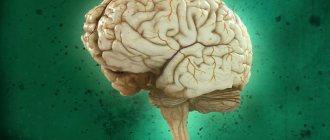
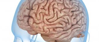
General information
This part of the brain arose long before the front part became responsible for the functions of smell.
It consists of various parts of the entire analyzer, which is responsible for the functions of human senses. It is worth noting that the fish brain is almost entirely composed of ancient structure. But in humans, the limbic system, part of which is the olfactory brain, is responsible for these functions. It is localized at the junction of the inferior and medial surfaces of both hemispheres of the brain. If we consider its phylogenetic path of development, we can distinguish 3 main stages:
- Paleopallium, is one of the components of the temporal lobe of the cerebral cortex;
- Archipallium is located next to the so-called bulbus olfactorius area, covered with bark of old origin;
- Neopallium is a modern cloak; the highest centers of olfactory reflexes are formed in it - these are unique analyzers of the cortex.
Animals in ancient times used only this system to operate their olfactory functions. As we evolved, the olfactory brain became part of the limbic system, which is responsible for human sensory sensations.
Anatomy
The structure of a living individual is not easy to describe. Especially such a component as the brain. This universe that exists in everyone continues to hide its secrets. But this does not mean that they are not worth understanding.
Development
The forebrain is formed at 3-4 weeks of prenatal development. By the end of the 4th week of embryogenesis, the telencephalon, diencephalon, and the cavity of the third ventricle are formed from the forebrain.
Diencephalon
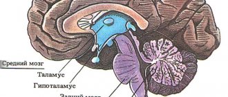
It consists of the thalamic and hypothalamic regions, which are located on the sides of the third ventricle between the hemispheres and the midbrain.
The thalamic region unites:
- The thalamus is an ovoid formation located deep under the cerebral cortex. The oldest, largest (3-4 cm) formation of the diencephalon;
- The epithalamus is located above the thalamus. It is famous for the fact that it contains the pineal gland. Previously, it was believed that the soul lived here. Yogis associate the pineal gland with the seventh chakra. By awakening the organ, you can open the “third eye”, becoming clairvoyant. The gland is tiny, only 0.2 g. But the benefits for the body are enormous, although previously it was considered a rudiment;
- subthalamus - a formation located below the thalamus;
- metathalamus - bodies located in the posterior part of the thalamus (previously considered a separate structure). Together with the midbrain, they determine the work of the visual and auditory analyzers;
The hypothalamic region includes:
- hypothalamus. Located under the thalamus. Weighs 3-5 g. Consists of specialized groups of neurons. Connected with all departments. Controls the pituitary gland;
- the posterior lobe of the pituitary gland is the central organ of the endocrine system, weighing 0.5 g. Located at the base of the skull. The posterior lobe, together with the hypothalamus, forms the hypothalamic-pituitary complex, which controls the activity of the endocrine glands.
Finite brain
Unites:
- cortical hemispheres. The bark appeared late in the development of the animal world. Occupies half the volume of the hemispheres. Its surface can exceed 2000 cm2;
- corpus callosum - a nerve tract connecting the hemispheres;
- striped body. Located on the side of the thalamus. On a section it looks like repeating stripes of white and gray matter. Promotes regulation of movements, motivation of behavior;
- olfactory brain. Unites structures that differ in purpose and origin. Among them is the central section of the olfactory analyzer;
Structure
This part of the human brain consists of many structures.
Conventionally, it can be divided into two parts: peripheral and central. Let's look at the structure of each of them. The peripheral part is the olfactory lobe, it includes the bulb, tract and sulcus. The grooves have a triangle at their end, which in turn consists of 2 olfactory stripes:
- lateral, which bends around the lateral sulcus, its end is located in the temporal region;
- medial. It passes into the area of the subcallosal gyrus and the peri-olfactory field of the brain, which in turn are located in the area of the corpus callosum.
The central section consists of many cortical gyri: parahippocampal, dentate and vaulted. The latter has the shape of a ring; it is responsible not only for olfactory functions, but also for the functioning of all internal organs and systems. This gyrus is formed by the cingulate and parahippocampal regions, which are connected to each other by a special septum. The dentate gyrus is a small hook, it is directed towards the medial part of the brain. It also includes the hippocampus, fornix, and nuclei of the transparent part of the septum.
Two main parts create a unique system, it is responsible for the processes of smell. Most of them form the so-called limbic system. It was she who took over all the functions responsible for the human subconscious.
Structure functions
The main functions of the limbic system are:
- motivational processes. Development of certain needs, creation of functions for their implementation, assessment of the possibility of performing these actions, etc.;
- emotions. In this case, the structures in question are responsible for the formation of basic emotions in a person;
- memorization. This is one of the important points. This part of the brain is responsible for the so-called short-term memory function (it is created after a person’s full wakefulness cycle). further functioning is ensured by conservation of impulses;
- long-term memory;
- formation of higher centers of nervous regulation.
Among the emotions that originate in the olfactory brain are:
- feeling of fear. In this case, there is strong stimulation of the hypothalamus and tonsils;
- feeling of anger or complete calm;
- the feeling of pleasure or dependence on something is based on impulses passing through the brain's amygdala, nucleus accumbens and hippocampus.
Also, the olfactory brain, as a component of the limbic system, provides a person with functions responsible for sleep, appetite, sexual desire, memory, fear, etc.
If the functioning of one of its parts is disrupted, a person is diagnosed with serious pathologies.
Functions
Thanks to the ability to distinguish odors, a person receives information about the surrounding biochemical environment. Other functions of the olfactory brain are associated with the formation of instinctive reactions when interacting with the outside world. The sense of smell in the context of the formation of reflex behavioral patterns in response to external stimuli covers the nutritional, protective, sexual, and orientation spheres of the body’s life. The olfactory brain and limbic system are structures that are involved in the regulation of processes:
- Formation of emotions.
- Behavioral, motivational reactions.
- Sleep-wake mode.
- Learning, intuition and memory.
Odorants are volatile substances that have an odor. Once in the nasal passages, odorants reach the olfactory epithelium (Regio olfactorius), where they interact with chemoreceptors. The olfactory zone is located on the mucosal surface in the upper region of the nasal passages, covers an area of about 2.5 cm2, and contains about 50 million sensory neurons.
Axonal branches of nerve cells pass through the ethmoid bone towards the olfactory bulb within the brain. Here the processes form synaptic connections with the dendrites of second-level nerve cells and unite into glomeruli. Level 2 neurons include mitral (the largest cellular elements) and tufted cells. Their branches emerge from the bulb, which is a multilayer neural network.
The axons of nerve cells (mitral, fascicular) extending from the bulb transmit information to the primary zones of the cortex, where the stimulus is decoded, the information is interpreted and an adequate reflex response is formed. The signal is then transmitted to the center of smell, located in the cortical layer of the brain, which regulates the higher functions of the psyche, where a conscious sensation of aroma arises.
At the same time, the stimulus enters the limbic system, where emotions and conscious behavioral reactions are formed that are adequate to the perceived smell. Odorants are transmitted predominantly along unmyelinated (not covered with a protective layer of myelin) nerve fibers. Impaired smell function worsens a person's quality of life. For example, many people with such disorders have impaired digestive function, which is due to the close relationship between the smell and taste of food.
Pathologies
A neurologist treats problems associated with dysfunction of the olfactory brain. First of all, neurodegenerative processes should be attributed to pathological problems with this area. They cause serious disturbances in the hippothalamus, as a result of which the patient is diagnosed with Alzheimer's disease. Other diseases include:
- increased feeling of anxiety, which entails damage to the tonsils of the brain;
- epileptic seizures. They occur in both adult patients and children. In this case, partial sclerosis of the hippocampus occurs;
- depression;
- in case of disruption of the tonsils and some olfactory gyri, patients are diagnosed with autism or Asperger syndrome;
- the presence of malignant tumors that pinch the nerve endings in this area;
- problems with the functioning of the circulatory system, which are accompanied by insufficient nutrition of blood vessels in the area of the olfactory brain;
- schizophrenia;
- increased aggressiveness and excitability;
- congenital pathologies;
- impairment of olfactory functions, which may be accompanied by loss of taste, vision or smell.
In the latter case, the patient experiences a delay in the functions of transporting odors to the neuroepithelium in the olfactory area. They can occur against the background of chronic inflammatory or infectious processes, injuries, or exposure to chemical substances on the mucous membranes that can impair the function of the sense of smell.
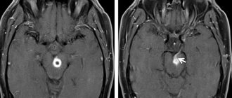
To diagnose pathologies, MRI and CT examinations are used. They help to fully assess the state of all parts of the structure. Based on the research results, an effective treatment regimen is selected for the patient. For depression and increased excitability, tranquilizers and sedatives are used. Many disorders in the functioning of this area cannot be treated; the patient is given supportive therapy.
In older people, due to changes in this part of the brain, partial or complete memory loss occurs, tremors of the limbs occur, nerve cells die, etc. Unfortunately, such pathologies cannot be cured. In this case, the patient is constantly monitored by a specialized specialist.
Characteristic
In anatomy, the olfactory brain is closely interconnected with the limbic system, which determines the joint regulation of many body functions, for example, the formation of emotions and the perception of odors. The olfactory system in the cortex is projected in the area of the anterior segment of the hippocampal gyrus. The olfactory analyzer perceives and analyzes odor. The system supports the following functions:
- Determining the suitability of food by smell.
- Regulation of eating behavior. The pleasant smell of food stimulates appetite.
- Stimulates the activity of the digestive tract. The smell of food, according to the principle of a conditioned reflex, triggers the mechanisms of digestion and food processing.
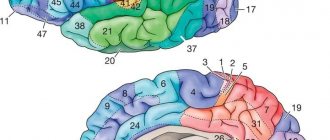
The structure of the olfactory brain involves close interaction between the departments - the amygdala within the limbic system, the hippocampus, the orbitofrontal and temporal (medial part) areas of the cortical layer. The vaulted gyrus (gyrus fornicatus) consists of parts - the cingulate, parahippocampal gyrus, and isthmus.
The central section within the olfactory brain includes the olfactory bulb, located in the area of the forebrain where the primary processing of information coming from the external environment occurs. The olfactory center within the brain is located in the lower segment of the temporal and frontal cortical layers covering the cerebral hemispheres.
The cortical sections perform secondary processing - they identify the smell, comparing it with stored data formed as a result of the accumulation of life experience. After identification, a general reaction of the body is formed that is adequate to the situation. As part of the central section of the combined structures - the olfactory brain and the limbic system, there are elements:
- Gyri – parahippocampal, cingulate, dentate, orbital.
- Hippocampal (seahorse) hook.
- Areas of the cortex in the frontal, parietal part, temporal pole, insular zone.
- Reticular formation.
- Basal ganglia (nuclei formed by gray matter inside white matter).
- Hypothalamus (a region of the intermediate part of the brain involved in the regulation of homeostasis - maintaining a constant environment in the body).
- Transparent septum (triangular, thin membrane separating the horns of the lateral ventricles).
The peripheral section within the olfactory brain includes bulbs, triangles, and pathways. The peripheral section also includes the perforated substance (a section of the cerebral hemisphere pierced with holes for blood vessels, located on its lower surface posterior to the triangle) and olfactory stripes (branches of the olfactory sulcus). The part of the brain responsible for smell interacts with the analyzer, which is represented by the following departments:
- Receptor. Contains receptors located in the mucous membrane of the upper nasal passages. In total, humans have about 10 million receptors that perceive odors.
- Conductive. The olfactory nerve, consisting of 15-20 nerve filaments passing through the ethmoid bone in the direction of the anterior fossa of the cranium.
- Bulbs (bulbus olfactorius), pathways, triangles and other subcortical structures of the system.
- Cortical area. Located in the lower segment of the temporal lobe and uncus in the region of the parahippocampal gyrus.
The olfactory triangle is a part of the brain that regulates the function of smell, which is represented by the expansion of pathways that transmit signals from receptors to the cortical analyzer. The olfactory triangle (trigonum olfactorium) borders the perforated substance. The mechanism of odor perception is realized through the comparison of odorous molecules with receptors located in the villi of neurosensory cells.
Separate groups of receptors perceive certain aromatic molecules. Several groups of receptors are involved in the recognition of one odor, the total number of which can reach several thousand. Information entering the brain along the pathways is summarized and compared with data stored in memory.
The process of adapting information about an aromatic substance lasts several seconds or minutes. The duration of the process depends on the concentration of the aromatic substance in the air and the speed of air flow in the area of the olfactory epithelium. The sensitivity of human receptors is quite high. The receptor responds to a single molecule of aromatic substance.
