Postherpetic neuralgia (PHN) is a complication of herpes zoster, caused by the same virus as chicken pox (lat. herpes zoster virus). This disease affects the skin and nerve fibers, which manifests itself as severe burning pain after the herpetic blisters have healed.
The risk of postherpetic neuralgia increases with age; Most often this disease affects people over 60 years of age. This condition occurs in approximately 10–20% of patients who have had herpes zoster. There is no cure, but treatment for postherpetic neuralgia can relieve symptoms and relieve pain. In more than half of patients, the pain goes away on its own after some time.
At CELT you can get advice from a specialist algologist.
Make an appointment
Symptoms of postherpetic neuralgia are usually limited to the area of the skin where the rash first appeared - most often they surround the torso or appear on the side, but the disease can also affect the face.
Pain may last for 3 months or longer after the herpetic blisters have healed. Patients describe it as burning, sharp, piercing or deep, and occurs with even the slightest touch of the skin in the affected area, including the light touch of clothing (allodynia). Much less often, patients feel itching and numbness.
Causes of postherpetic neuralgia
Once a person has had chickenpox, the virus remains in the body for life. With age, the immune system weakens (particularly from taking medications or chemotherapy) and the virus can become more active, resulting in shingles.
Damage to nerve endings during rashes leads to postherpetic neuralgia. When damaged, nerve fibers cannot send signals from the skin to the brain as they normally do. Instead, they transmit amplified signals, and the patient feels excruciating pain that can last for months or even years.
Risk factors
Only those who have had chickenpox can get shingles. But there are certain groups that are more susceptible to postherpetic neuralgia:
- Age – the chance of postherpetic neuralgia is higher in the older age group. (30% of people over 60 years of age who have had herpes zoster subsequently develop postherpetic neuralgia) and only 10% of the younger group develop postherpetic neuralgia.
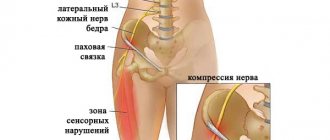
- Localization of rashes - symptoms of neuralgia are more pronounced if the rashes were in the forehead or eyes.
- Concomitant pathology – The presence of a suppressed immune system (after chemotherapy or taking immunosuppressants) or diseases such as AIDS.
Early treatment of herpes zoster is important. Treatment started within 2-3 days after the rash appears can help reduce symptoms and avoid the risk of postherpetic neuralgia.
Drug therapy
Drug therapy involves the use of the following groups of drugs:
- Anticonvulsants (Lyrica, Neurontin, others). These drugs have shown good effectiveness against neuropathic pain. However, they have a number of side effects, the most common of which are decreased concentration, drowsiness and swelling of the legs;
- Antidepressants. Some antidepressants (Amitriptyline, Duloxetine) affect the production of special brain substances that are responsible for both depressive states and the perception of pain. Doctors often prescribe minimal doses of antidepressants in the complex treatment of postherpetic neuralgia. Their main side effects include: drowsiness, dry mouth, dizziness;
- In cases of severe pain and insufficient effectiveness of other drugs, patients may be prescribed narcotic analgesics.
Diagnosis
If pain occurs after an episode of rashes or sensory disturbances, you should consult a doctor. Diagnosis is based on medical history, examination and laboratory tests (necessary to exclude other diseases). Instrumental diagnostic methods (CT, MRI, EMG, ultrasound) are prescribed only if there is a need for differential diagnosis.
Forecast
Exercise therapy, physiotherapy and drug treatment in most cases help reduce symptoms and restore quality of life. Especially if treatment is carried out in a timely manner.
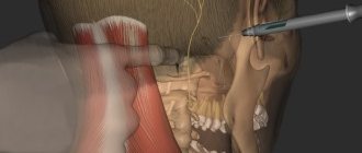
Epidural administration of anesthetics and steroid hormones
Spinal epidural block involves the injection of drugs into the epidural space, which leads to loss of sensation in the area of impact. This means that this procedure involves introducing the active substance directly into the source of pain. Achieving a therapeutic effect in this case is possible due to the following factors:
- analgesic properties of the drug;
- the maximum possible concentration in the affected area;
- reflex action at all levels of the nervous system.
Treatment
Drug treatment:
- Analgesics such as acetaminophen (Tylenol, Panadol, Tempra), and nonsteroidal anti-inflammatory drugs (NSAIDs) such as aspirin, ibuprofen, naproxen, and Celebrex.
- Opioids – Your doctor may prescribe opioid analgesics for severe pain that is not controlled by regular analgesics, but these medications should be used with caution due to the risk of serious side effects. For example: This group includes tramadol or oxycodone. Some studies suggest that oxycodone may also help reduce allodynia.
- Antidepressants – These medications are effective in treating depression. In addition, they improve sleep (for example, amitriptyline, Cymbalta, etc.).
- Anticonvulsants – Drugs in this group are intended mainly for the prevention of convulsive conditions. But sometimes they are quite effective for postherpetic neuralgia (Neurontin, Lyrica, Topamax, carbamazepine).
- Blocks - Injections of corticosteroid into paravertebral points sometimes lead to a significant reduction in pain.
- Local painkillers - ointments, gels containing analgesics or anesthetics (lidocaine). Helps temporarily reduce symptoms.
Kinds
In medical practice, it is important to subdivide the disease depending on the type of pain and area of the rash.
Based on the duration of the pain syndrome, the following types of neuralgia are distinguished:
- Spicy. Manifestations are observed no more than a month from the moment the vesicles appear.
- Subacute. This name is appropriate when pain persists from one to four months.
- Chronic. The pain syndrome can last for years, then subside, then appear again.
The localization of the rash can be in the chest (from a fifth to half of all cases), head (about 12–20%), neck (up to 11%), much less often - the lower back, extremities.
ICD-10 code
It is known that the ICD (International Classification of Diseases) is the main document for processing statistical data in all healthcare institutions; diseases are classified by different codes. In the Russian Federation, the transition to the modern ICD, tenth revision (ICD-10) occurred in 1999, and outpatient, inpatient and statistical institutions of the Ministry of Health still work with this document, namely ICD-10.
Neuralgia in ICD-10
Neuralgia is presented in ICD-10 in two classes (codes): G (diseases of the nervous system, class 6) and M (diseases of the musculoskeletal system and connective tissue, class 13). This division is justified, since very often the cause of neuralgia is large muscles that cause compression or compression of one or another nerve. This situation may be accompanied by myofascial muscular-tonic syndrome.
“Pure” neuralgia, in which muscles and compression with their help have no role, are described in class G. An example is trigeminal neuralgia.
Examples of class G neuralgia:
- 1Lesions of the glossopharyngeal nerve;
- 0 Trigeminal neuralgia;
- 1 Atypical facial pain;
- 0*Neuralgia after herpes zoster.
Examples of class M neuralgia:
- 1 Radiculopathy (this includes brachial, lumbar, lumbosacral and thoracic variants);
- 2 Cervicalgia;
- 3 Sciatica;
- 4 Lumbago with sciatica.
M79.2 Neuralgia and neuritis, unspecified
This diagnosis can be made, for example, due to lack of time. It is known that in hospitals, MES, or medical and economic standards, “rule the show.” In the event that a patient has been “over-lying” without reason, the department’s internal expert points out the shortcomings, and an external examination or unscheduled inspection may assign penalty points, which will affect funding.
Therefore, in extreme cases, you can be discharged with this diagnosis, and clarification can be done on an outpatient basis.
Another heading, which is sometimes interpreted in an extremely broad way, is the following:
- R52 Pain not elsewhere classified. This refers to pain that cannot be attributed either to the mammary gland, ear, pelvis, spine or any other part of the body. For example, “wandering pain.”
Pain not elsewhere classified (R52)
As you can see, it belongs to class R. It is referred to as “Symptoms, signs and abnormalities identified by clinical and laboratory tests, not classified elsewhere.”
This allows the doctor to “get out”, but such encryption may cause dissatisfaction with the hospital administration for the reason that this class is non-core. In other words, this diagnosis says: “we found something, but what we found does not fit into anything.”
In fact, there should be no non-core diagnoses in the department: three days are given to establish a diagnosis, when it changes from a “diagnosis upon admission” to a working one. If it turns out that the patient is a “stranger,” he must be transferred or discharged. If he spent time in the department with a non-core diagnosis and was discharged, then this is a sign of incompetent management of the department.
Therefore, the correct use of ICD-10 in coding certain types of neuralgia can not only help process large amounts of statistical data, but also save significant funds for a medical institution.
Prevention
Certain antiviral medications may help prevent or reduce the effects of shingles, thereby reducing the risk of postherpetic neuralgia:
- Chickenpox vaccine – The varicella zoster virus vaccine (Varivax) is now a routine childhood vaccine, but may also be recommended for older children and adults who have never had chickenpox. This vaccine does not guarantee that a person will not get chickenpox or shingles, but it may reduce the duration and severity of symptoms and the risk of complications such as postherpetic neuralgia.
- Shingles vaccine - (Zostavax) can be given to people over 60 years of age (who have had chickenpox but not shingles). Zostavax is not recommended for use in certain groups of people (for example, those undergoing cancer treatment or those who are immunocompromised).
- Antiviral medications – Antiviral medications such as acyclovir, valocyclovir, famciclovir, when taken within the first 72 hours after the shingles rash appears, can help shorten the duration of shingles and reduce the chance of developing postherpetic neuralgia.
Physiotherapy helps reduce pain and relieve inflammation. Various techniques are used (including transcutaneous electrical stimulation).
Exercise therapy helps restore the elasticity of ligaments and muscles. Exercises can be carried out both on simulators and in the form of gymnastics.
Acupuncture. This method is quite effective in restoring conductivity and reducing pain.
Survey
If you suspect postherpetic neuralgia, it is advisable to consult a neurologist.
The diagnosis is not difficult in cases where pain occurs after the appearance or extinction of vesicles. The difficulty is presented by patients whose pain has developed before the skin manifestations of herpes. During the examination, the neurologist (in his absence, the therapist) notes the following abnormalities in the patient:
- hyperalgesia - excessive pain with slight impact - pressure, gathering the skin into a fold;
- allodynia – the appearance of pain from contact with irritants that normally do not cause it (touching clothes, patting with the palm of the hand);
- hypoesthesia - a decrease in all types of sensitivity along the nerve fiber.
Laboratory and instrumental studies are prescribed quite rarely. To confirm the GWP, 2 main studies are carried out:
- Tzanck test - scraping from a vesicle, placing the contents on a glass slide, followed by staining. In this case, multinucleated giant cells are revealed.
- Blood test for antibodies to the pathogen - HerpesZoster.
A general blood test, indicating the viral nature of the disease, and a biochemical study, which determines an increase in acute-phase inflammatory proteins, are less informative.
Herpes virus infection does not always manifest as rashes. In such cases, in order to exclude other neurological pathologies, the patient may be referred for ENMG - a study that allows identifying disturbances in conduction along nerve fibers, a change in response when exposed to a stimulus.
Forecast
With timely treatment, the prognosis is favorable - the vast majority of cases end in recovery. When predicting the outcome, it is important to take into account risk factors; the more there are, the higher the likelihood of transition to a chronic form.
The transition to a chronic form can be considered a complication, since intense pain significantly affects the patient’s lifestyle and quality of life.
Other possible complications include changes in skin sensitivity at the site of the rash, the addition of a secondary infection, mycosis,
Statistics
The number of cases is up to 3 diseases per 1000 people per year. According to statistics, older people are more susceptible to pathology - in people 80 years of age and older, the detection rate is already 10 cases per 1000.
If we consider the percentage distribution of pathology within age groups, we can note that people aged 60–75 years account for half of all cases, those aged 76 years and older account for three quarters.
People aged 60+ are at risk of developing constant pain for a long time - about 10% of patients note that the discomfort persisted for up to six months.
6% of people who have suffered postherpetic neuralgia experience relapses, and 10 years or more may pass from the first to the next case.
Herpes zoster and herpes-associated pain
Herpes zoster (HZ) is a sporadic disease that is the reactivation of a latent viral infection caused by the herpes virus type 3 (Varicella zoster virus (VZV)). The disease occurs with primary damage to the skin and nervous system.
VZV is the etiological agent of two clinical forms of the disease - primary infection (varicella) and its recurrence (herpes zoster). After a primary infection (chickenpox), usually in childhood or adolescence, the virus enters a latent state, localizing in the sensory ganglia of the spinal nerves. The commonality of the causative agent of chickenpox and herpes zoster was established even before the isolation of the virus using serological reactions in which liquid obtained from blisters on the skin of patients was used as an antigen. Later, using genomic hybridization, it was proven that in the acute period of herpes zoster, the detection rate of VZV is 70–80%, and in individuals without clinical manifestations, but with antibodies, viral DNA is detected in 5–30% of neurons and glial cells.
The prevalence of herpes zoster in different countries of the world ranges from 0.4 to 1.6 cases per 1000 patients/year in people under 20 years of age and from 4.5 to 11.8 cases per 1000 patients/year in older age groups. The lifetime risk of contracting herpes zoster is up to 20%. The main risk factor for its occurrence is a decrease in specific immunity to VZV, which occurs against the background of various immunosuppressive conditions.
Clinical picture of OH
The clinical picture of OH consists of skin manifestations and neurological disorders. Along with this, most patients experience general infectious symptoms: hyperthermia, enlarged regional lymph nodes, changes in the cerebrospinal fluid (in the form of lymphocytosis and monocytosis). Approximately 70–80% of patients with OH in the prodromal period complain of pain in the affected dermatome, which subsequently develops skin rashes. The prodromal period usually lasts 2–3 days, but often exceeds a week. Rashes with OH have a short erythematous phase, often completely absent, after which papules quickly appear. Within 1-2 days, these papules turn into vesicles, which continue to appear for 3-4 days - the vesicular form of herpes zoster. At this stage, elements of all types may be present on the skin. Elements tend to merge. Pustulation of vesicles begins a week or even earlier after the appearance of the first rash. After 3–5 days, erosions appear at the site of the vesicles and crusts form. If the period of appearance of new vesicles lasts more than one week, this indicates the possibility of an immunodeficiency state. The crusts usually disappear by the end of the 3rd or 4th week. However, peeling and hypo- or hyperpigmentation may remain for a long time after the resolution of OH.
Pain syndrome is the most painful manifestation of OH. While some patients experience rash and pain of relatively short duration, 10–20% of patients experience postherpetic neuralgia (PHN), which can last months or years, significantly reduces quality of life, causes great distress, can lead to loss of independence, and is associated with significant financial costs . Effective treatment of pain associated with OH is an important clinical goal.
Herpes-associated pain
According to modern concepts, pain syndrome in OH has three phases: acute, subacute and chronic. If in the acute phase the pain syndrome is mixed (inflammatory and neuropathic) in nature, then in the chronic phase it is typical neuropathic pain (Fig.). Each of the listed phases has its own treatment features, based on the pathogenetic mechanisms of pain and confirmed by controlled clinical studies.
Acute herpetic neuralgia
Pain in acute herpetic neuralgia usually occurs in the prodromal phase and lasts for 30 days - this is the time required for the rash to resolve. In most patients, the appearance of a rash is preceded by a burning sensation or itching in a specific dermatome, as well as pain, which can be stabbing, pulsating, shooting, paroxysmal or constant. In a number of patients, the pain syndrome is accompanied by general systemic inflammatory manifestations: fever, malaise, myalgia, headache. Determining the cause of pain at this stage is extremely difficult. Depending on its location, differential diagnosis should be made with angina pectoris, intercostal neuralgia, acute attack of cholecystitis, pancreatitis, appendicitis, pleurisy, intestinal colic, etc. The cause of the pain syndrome becomes obvious after the appearance of characteristic rashes. In typical cases, the prodromal period lasts 2–4 days, no more than a week. The interval between the onset of the prodrome and the onset of rash is the time required for reactivated VZV to replicate in the ganglion and be transported along the cutaneous nerve to nerve terminals at the dermoepidermal junction. The replication of the virus in the skin takes some more time, followed by the formation of inflammatory reactions. The immediate cause of prodromal pain is subclinical reactivation and replication of VZV in neural tissue. Experimental studies on animals have shown that at sites of VZV replication, the concentration of neuropeptide Y in nervous tissue, which is a marker of neuropathic pain, increases [1]. The presence of severe pain in the prodromal period increases the risk of more severe acute herpetic neuralgia and the likelihood of subsequently developing postherpetic neuralgia.
In most immunocompetent patients (60–90%), severe, acute pain accompanies the appearance of a skin rash. The severity of acute pain syndrome increases with age. Severe pain is also observed more often in women and in the presence of a prodrome. A characteristic feature of acute herpetic neuralgia is allodynia - pain caused by a non-painful stimulus, such as the touch of clothing. It is allodynia in the acute period that is a predictor of the occurrence of postherpetic neuralgia [2, 3]. The absence of allodynia, on the contrary, is a good prognostic sign and may suggest recovery within three months.
Subacute herpetic neuralgia
The subacute phase of herpetic neuralgia begins after the end of the acute phase and lasts until the onset of postherpetic neuralgia. In other words, this is pain that lasts more than 30 days from the beginning of the prodrome and ends no later than 120 days (Fig.). Subacute herpetic neuralgia can develop into postherpetic neuralgia. Factors predisposing to the continuation of pain include: older age, female gender, the presence of a prodrome, massive skin rashes, localization of rashes in the area of innervation of the trigeminal nerve (especially the eye area) or brachial plexus, severe acute pain, the presence of immunodeficiency [3, 4 ].
Postherpetic neuralgia
The International Herpes Treatment Forum defines PHN as pain lasting more than four months (120 days) after the onset of the prodrome. PHN, especially in older patients, can last for many months or years after the rash has healed. With PHN, three types of pain can be distinguished: 1) constant, deep, dull, pressing or burning pain; 2) spontaneous, periodic, stabbing, shooting, similar to an “electric shock”; 3) pain when dressing or lightly touching in 90%.
Pain syndrome is usually accompanied by sleep disturbances, loss of appetite and weight loss, chronic fatigue, and depression, which leads to social isolation of patients.
PHN is considered to be a typical neuropathic pain resulting from damage or dysfunction of the somatosensory system. Several mechanisms are involved in its pathogenesis.
- Nerve damage disrupts the transmission of pain signals, which leads to increased activity of higher order neurons (deafferentation hyperalgesia) [6–8].
- Nerve fibers damaged by VZV may generate spontaneous activity at the site of injury or other sites along the nerve (spontaneous ectopic activity of injured axons).
- Damage or inflammation of the nerve as a result of virus reactivation leads to a decrease in the threshold for activation of nociceptors, activation of urinary nociceptors—peripheral sensitization [5, 9].
- As a result of these changes in the peripheral parts of the somatosensory system, the activity of central nociceptive neurons increases and new connections are formed between them, provided that the pain continues—central sensitization [10–12]. The systems for recognizing pain and temperature stimuli are characterized by increased sensitivity to minor mechanical stimuli, causing severe pain (allodynia).
In most patients, pain associated with PHN improves within the first year. However, in some patients it can persist for years and even throughout the rest of their lives, causing considerable suffering. PHN has a significant negative impact on the quality of life and functional status of patients, who may develop anxiety and depression.
How to reduce the risk of PHN?
This issue is the most important for any doctor treating a patient with OH, and includes early initiation of etiotropic (antiviral) therapy and adequate pain relief in the acute stage.
Antiviral therapy. The results of many clinical studies have shown that the administration of antiviral drugs reduces the period of viral shedding and the formation of new lesions, accelerates the resolution of the rash and reduces the severity and duration of acute pain in patients with OH. Thus, in controlled studies using recommended dosages, the time to complete cessation of pain when prescribing famciclovir was 63 days, and when prescribing placebo - 119 days. Another study showed greater effectiveness of valacyclovir compared to acyclovir: pain syndrome when prescribed valacyclovir (Valavir) disappeared completely after 38 days, and when prescribed acyclovir after 51 days. Valacyclovir and famciclovir have similar effects on herpes-associated pain in immunocompetent patients [13, 23]. Thus, antiviral therapy is indicated not only for the rapid relief of skin manifestations, but also for the acute phase of the pain syndrome.
In all controlled clinical trials of antiviral therapy (
) it is recommended to start therapy within 72 hours of the onset of rash [1, 14].
The effectiveness of the anti-pain effect of antiviral therapy started at a later date has not been systematically studied, however, numerous clinical data suggest that late-started therapy can also affect the duration and severity of acute pain.
Antipain therapy. Effective relief of acute pain syndrome in OH is the most important stage in the prevention of PHN. It is advisable to stage-by-stage treatment of zoster-associated pain syndrome in all its phases. Thus, in the treatment of acute and subacute herpetic neuralgia, pain therapy consists of three main stages:
- Stage 1: Aspirin, paracetamol, non-steroidal anti-inflammatory drugs (NSAIDs);
- Stage 2: opioid analgesics, including tramadol;
- Stage 3: drugs with a central analgesic effect (tricyclic antidepressants, anticonvulsants).
Considering that in our country there are known organizational difficulties in prescribing opioid analgesics, if simple analgesics and NSAIDs are insufficiently effective, it is necessary to move on to prescribing drugs with central action.
Treatment of postherpetic neuralgia
Currently, there are 5 main groups of medications: anticonvulsants, tricyclic antidepressants, lidocaine patch, capsaicin, opioid analgesics [21].
Anticonvulsants: Gabapentin and pregabalin are the two most commonly used anticonvulsants for the management of neuropathic pain associated with PHN. Drugs are more often used at the beginning of the development of PHN to reduce the acute component of neuropathic pain. In one study [15], in patients taking gabapentin, 43.2% had a decrease in pain perception, compared with 12.1% in the placebo group. In a similar trial [16], pregabalin also reduced the number of patients with PHN, especially in those aged 65 years and older. Gabapentin and pregabalin appear to be equally effective in reducing neuropathic pain [17]. Gabapentin is the drug of first choice for the treatment of any type of neuropathic pain; it is one of the most well-studied and widely used drugs in neurologist practice for the relief of pain in PHN. It is a structural analogue of gamma-aminobutyric acid (GABA). Gabapentin enhances the synthesis of GABA by stimulating the activity of glutamate decarboxylase; modulates the activity of NMDA receptors; blocks a-2-d-subunits of voltage-dependent calcium channels and inhibits the entry of Ca2+ into neurons; reduces monoamine release and sodium channel activity; reduces the synthesis and transport of the excitatory neurotransmitter glutamate; helps reduce the frequency of action potentials in peripheral nerves. The concentration of gabapentin in the blood plasma reaches its peak 2–3 hours after administration, the half-life is 5–7 hours. The dosing interval should not exceed 12 hours, bioavailability is 60%. Eating does not affect the pharmacokinetics of the drug; antacids reduce its concentration in the blood, so gabapentin should be taken no earlier than 2 hours after taking antacids. Excreted in breast milk; The effect of the drug on the child’s body has not been studied. Adverse reactions develop extremely rarely: slight dizziness, drowsiness. Gabapentin enhances the effect of lidocaine and antidepressants. You should refrain from combining it with alcohol, tranquilizers, antihistamines, barbiturates, sleeping pills, and drugs. The drug has important advantages in the treatment of neuropathic pain: safety, low potential for interaction with other drugs, good tolerability, and is not metabolized in the liver. Gabapentin is the drug of choice for the treatment of elderly people during polypharmacotherapy; it is convenient to use and has been proven to be highly effective.
Gabapentin dosage regimen. Initial dose: 1st day 300 mg in the evening; 2nd day 300 mg 2 times (day and evening); Day 3: 300 mg 3 times. Titration: days 4–6 300/300/600 mg; 7–10 days 300/600/600 mg; Days 11–14 600/600/600 mg. Daily therapeutic dose 1800–3600 mg, maintenance dose 600–1200 mg/day.
Pregabalin has a mechanism of action similar to gabapentin, but does not require slow titration and is therefore more convenient for clinical use. The drug is prescribed twice a day. The initial dose is 75 mg twice, the daily therapeutic dose is 300–600 mg. Several randomized clinical studies of the effectiveness of pregabalin in postherpetic neuralgia have been conducted, which showed the rapid development of an analgesic effect (during the first week of administration), good tolerability, ease of use and a reduction in sleep disturbances associated with pain [22].
Antidepressants. Drugs in this group, especially tricyclics (nortriptyline and amitriptyline), are important components in the treatment of pain in PHN. Due to the activation of descending serotonin and norepinephrine antinociceptive systems and the ability to block sodium channels, antidepressants block the perception of pain. In clinical trials of the effectiveness of tricyclic antidepressants in reducing pain in PHN, 47% to 67% of patients reported “moderate to excellent” pain relief, with equivalent effects reported for amitriptyline and nortriptyline [17]. However, nortriptyline does not cause many anticholinergic effects and may therefore be preferable to amitriptyline.
A patch with 5% lidocaine is applied to the affected area at the beginning of chronic pain or immediately after the diagnosis of PHN is made. The patch is applied to intact, dry, non-inflamed skin. It is not used on inflamed or damaged skin (i.e. during active herpetic eruptions). Lidocaine is an antagonist of sodium ion channels, the analgesic effect develops as a result of preventing the generation and conduction of neuronal activity potentials, by binding sodium channels of overactive and damaged nociceptors. A patch with 5% lidocaine has a local effect and has almost no systemic effects. Several studies have shown that the lidocaine patch reduces pain compared with placebo [18]. Comparative studies of the effectiveness of 5% lidocaine and pregabalin showed their equal effectiveness [19]. Capsaicin, which is made from red peppers and is an irritant, is used as an ointment or patch. When applied to the skin, it depletes peptidergic neurotransmitters (eg, substance P) in primary nociceptive afferents. The drug should be applied to the affected area 3-5 times a day to maintain a long-term effect. Despite the fact that a number of studies have shown the effectiveness of capsaicin against PHN, many patients often experienced significant adverse reactions: for example, a third of patients reported the development of an “unbearable” irritant effect of the drug, which significantly limits its clinical use in PHN.
Opioid analgesics (oxycodone, methadone, morphine) can also be used in the treatment of PHN. They reduce neuropathic pain by binding to opioid receptors in the central nervous system or inhibiting the reuptake of serotonin or norepinephrine at peripheral nerve endings - nerve synapses. According to research results, oxycodone, compared with placebo, provides greater pain relief and reduces the severity of allodynia, but causes the development of adverse reactions such as nausea, constipation, drowsiness, loss of appetite, and drug dependence [20]. Comparative studies of the effectiveness of opioids and tricyclic antidepressants have demonstrated their equivalent effectiveness.
In the section “Treatment of postherpetic neuralgia” in the 2009 European guidelines [21] for the treatment of neuropathic pain, first-line therapy is distinguished (drugs with proven effectiveness - class A): pregabalin, gabapentin, lidocaine 5%. Second-line drugs (class B): opioids, capsaicin.
When treating patients with PHN, it is advisable to follow certain steps.
Initially, first-line drugs are prescribed: gabapentin (pregabalin), or TCAs, or local anesthetics (plates with 5% lidocaine). If it is possible to achieve good pain reduction (VAS pain score – 3/10) with acceptable side effects, then treatment is continued. If pain relief is not sufficient, another first-line drug is added. If first-line drugs are ineffective, second-line drugs can be prescribed: tramadol or opioids, capsaicin, non-pharmacological therapy. In the complex therapy of postherpetic neuralgia, non-pharmacological therapy is also used: acupuncture, TENS anesthetic device, the most promising and effective method is neurostimulation.
Treatment of PHN is extremely challenging. Even with the use of various pain medications and referral to a specialist algologist, it is not always possible to achieve the disappearance of the pain syndrome.
Literature
- Dworkin RH Johnson RW, Breuer J., Gnann JW, Levin MJ Recommendation for management of herpes zostritis // Cln Infec Dis. 2007; 44: (Supl 1): S1–S26.
- Dworkin RH, Nagasako EV, Johson RW, Griffin DR Acute pain in herpes zoster: tue famciclovir database project // Pain. 2001; 94: 113–119.
- Hope-Simpson RE Postherpetic neuralgia // JR Coll Gen. Pract. 1975; 157:571–675.
- Choo P., Galil K., Donahue JG Walker et al. Risk factors for postherpetic neuralgia // Arch. Intern. Med. 1997; 157:1217–1224.
- Garry EM, Delaney A, Anderson HA et al. Varicella oster virus induces neuropathic changes in rat dorsal root ganglia and behavior reflex sensitization that is attenuated by gabapentin or sodium channel blocking drugs // Pain. 2005; 118:97–111.
- Yung BF, Johnson RW, Griffin DR, Dworkin RH Risk factors for postherpetic neuralgia in patients with herpes zoster // Neurology. 2004; 62:1545–1551.
- Jonson RW Zoster-associated pain: what is know, who is at risk and how can it be managed? // Herpes. 2001, 14 Supplement; 2:31A–34A.
- Tal. M., Bennett GJ Extra territoiral pain in rats with a peripheral mononeuropathy: mechano-hyperalgesia and mechano-allodenia in the territory of an uninjured nerve // Pain. 1994; 57:375–382.
- Oaklander AL The density of remaining nerve endings in human skin with and without postherpetic neuralgia after shingles // Pain. 2001; 92: 139–145.
- Rowbotham MC, Yosipovitch G., Connoly MK, Finlay D., Forde G., Fields HL Cutaneus innervation density in allodynic from postherpetic neuralgia // Neurobiol. Dis. 1996; 3:205–214.
- Rowbotham MC, Fields HL The relationship of pan, allodynia and thermal sensation in post-herpetic neuralgia // Brain. 1996; 119(Pt2):347–354.
- Scholz J., Broom DC, Youn DH, Mills CD, Kohno T. et al. Blocking caspase activity prevents transsynaptic neuronal apoptosis and the loss of inhibition in lamina 11 of dorsal horn afer peripheral nerve injury // J Neurosci. 205; 25:7317–7323.
- Tyring SK, Beutner KR, Tucker BA et al. Antiviral therapy for herpes zoster. Randomized, controlled clinical trial of vlacyclovir, and farmavir therapy in immunocompetent patients of 50 years and older // Arch Farm Med. 2000; 9:863–869.
- Gross G., Schofer H. et al. Herpes zoster guideline of German Dermatology Society (DDG) // J of Clinical Virology. 2003; 26:277–289.
- Rowbotham M., Harden N., Stacey B. et al. Gabapentin for the treatment of postherpetic neuralgia: a randomized controlled trial // JAMA. 1998. Vol. 280. P. 1837–1842.
- Dworkin R., Young J., Sharma U. et al. Pregabalin for the treatment of postherpetic neuralgia: a randomized, placebocontrolled trial // Neurology. 2003. Vol. 60. P. 1274–1283.
- Stankus S., Dlugopolski M., Packer D. Management of herpes zoster (shingles) and postherpetic neuralgia // Am Fam Physician. 2000. Vol. 61. P. 2437–2444.
- Karly P. Garnock-Jones, Gillin M. Keating/Lidocain 5% Medical Plaster. A review of its use in hjsterpetic neuralgia // Drugs. 2009; 69(15):2149–2165.
- Rehm S., Binder A., Baron R. Post-herpetic neuralgia: 5% lidocain medicated plaster? Pregadflin, or a combination both? A randomized, open/clinical effectiveness study // Cur. Med. Reas. 2010, v. 26, no. 7.
- Watson C., Babul N. Efficacy of oxycodone in neuropathic pain: a randomized trial in postherpetic neuralgia // Neurology. 1998. Vol. 50. P. 1837–1841.
- Attal N. et al. EFNS guidelines of the pharmacological treatment of neuropathic pain: 2009 revision // European Journal of Neurology. 2010.
- Seventer R., Feister H. et al. Efficacy and tolerance of twice-daily pregabalin for treating pain and related sleep interference in postherpetic neuralgia: a 13-week, randomized trial // Curr Med Res Opin. 2006; 22 (2): 375–384.
- Beutner KR et al. Valaciclovir compared with acyclovir from improved therapy for herpes zoster in immunocompetent adults // Antimicrobal agents and chemotherapy. 1995, July, vol. 37, no. 7, p. 1546–1553.
E. G. Filatova, Doctor of Medical Sciences, Professor First Moscow State Medical University named after. I. M. Sechenova, Moscow
Contact information about the author for correspondence
Table.