Hemangioblastomas (syn. angioreticulomas) belong to relatively rare tumors of the central nervous system of the 1st degree of malignancy. They originate from primitive vascular mesenchymal cells or hemocytoblasts. They can be located in the brain and spinal cord, in the retina of the eye. The structure of the tumor in medical sources is described as heterogeneous, since it can include both solid and cystic components. The development of this neoplasm can be sporadic (non-hereditary, for unclear reasons) and familial.
Statistical report
In the general group of all intracranial primary tumors, cerebral hemangioblastomas (CHBs) account for 1%-2.5%, and more often (85%) they are diagnosed in the cerebellar hemispheres. The prognosis after complete (!) surgical removal of a tumor with a sporadic occurrence scenario is positive, they do not recur .
But in 25% of cases, the neoplasm is detected as a manifestation of a hereditary disease - Hippel-Lindau disease. Cerebral tumor associated with this disease often has a poor prognosis. In patients with such a genetic pathology, the tumor process can resurface even after complete resection, and not necessarily in the same place; it can appear in any other part of the central nervous system.
The greatest number of cases with HGM is observed in men, who suffer from them 1.5-2 times more often than women. The peak age range when lesions are detected in patients is 40-50 years. For the first time, a neoplasm combined with Hippel-Lindau disease appears in adolescence and young adulthood (up to 30 years).
Hemangioblastomas are characterized by slow growth and benign biological behavior. However, sometimes (rarely) they are complicated by spontaneous significant hemorrhages in the posterior cranial fossa. Massive hemorrhages usually occur with large tumor sizes (more than 1.5 cm).
Any hemangioblastoma in the brain is a serious problem that requires timely detection and treatment. In every patient, the tumor becomes the cause of progressive neurological deficit and a non-stop decline in quality of life. Mortality is not excluded. Unfortunately, this diagnosis cannot be treated conservatively. The only chance for the patient to survive and recover is purely neurosurgical intervention.
Soon after the birth of a child, parents may notice a formation on the skin of blue, red, pink color, which can quickly grow, capturing new territories on the baby’s tiny body. Most likely, this is a benign vascular tumor, a hemangioma, with all the inherent signs of a benign tumor.
A feature of the course of vascular neoplasms - hemangiomas in young children is the unpredictability of their “behavior”. A punctate hemangioma of the cheek in a one-month-old baby can turn into an extensive and deep tumor with a complex anatomical localization and without a tendency to stop growth within 2-3 weeks. This leads to disruption of the functions of various important organs responsible for vision, hearing, breathing, etc. Due to the unpredictability of the development of hemangiomas in children under one year of age, hoping for independent resorption of large and deep hemangiomas using expectant management means voluntarily taking a conscious risk of the health and life of the baby. It is necessary to distinguish between a vascular tumor and other formations: medial spot, nevi Unna, rosacea, etc.
Therefore, if you find any spots, spots, or blood vessels on the baby’s body, immediately contact pediatric surgeons and oncologists.
Why does hemangioma appear?
It is believed that a tumor as a disease occurs as a result of the body’s reaction to harmful external and internal influences. Therefore, to prevent vascular tumors during pregnancy, simple rules must be followed: contact with harmful carcinogenic substances should be completely avoided; You should not smoke, you should not be in a room where people smoke or have smoked; Uncontrolled use of any medications is strictly prohibited. There is an opinion that the appearance of hemangiomas is associated with complex disorders of intrauterine development of the fetus, especially in the first trimester of pregnancy, when the vascular system of the unborn baby is formed. During this period, emotional and chemical stress, low-frequency electromagnetic influences, injuries, infections, hormonal disorders and any unfavorable factors of a physical or chemical nature that have embryotoxic properties are especially dangerous. Particularly dangerous factors of a physical nature include ultraviolet radiation (solariums!), ionizing radiation, mechanical injuries and elevated temperature in a pregnant woman with long-term exposure. Among the biological factors include viral and bacterial diseases, decreased immunity, and parasitic infections of the mother during pregnancy. Sometimes it is enough to dye your hair in the early stages of pregnancy and the child develops a “stork’s kiss,” as hemangiomas are often called.
Experts are still debating whether hemangiomas are true tumors or a congenital malformation of the vascular system. These tumors have the ability to self-resorb and malignant degeneration. Nowadays, hemangiomas occur in every tenth newborn. In girls it is 2-3 times more common than in boys. If a child already has one hemangioma, then during the first six months of life, 75% of such children may develop hemangiomas in other parts of the body. Recently, the number of children with multiple hemangiomas has increased to 43%. Hemangiomas can grow unevenly. Along with rapid growth, often leading to severe irreversible damage, sometimes there is a complete lack of growth, and in some rare cases even reverse development.
What complications can occur with hemangiomas?
The vast majority of hemangiomas do not have complications. However, in some cases, ulcers and ulcerations occur on them; some hemangiomas are complicated by bleeding. A feature of the course of hemangiomas in the perineum and external genitalia is their tendency to frequent ulceration. Contamination of these areas with urine and feces, friction with diapers or clothing can lead to the formation of ulcers, drip bleeding, and infection. In cases of internal hemangiomas, for example, the larynx or the inside of the nasal passage, the breathing process is complicated. Vascular tumors of the eye can cause occlusion and amblyopia. Hemangiomas of the oral mucosa and lip area disrupt the processes of sucking and digestion. With very large tumors, cardiac dysfunction may occur due to the large amount of blood that must be pumped into the excess blood vessels of the hemangioma. Angiomas adjacent to the bone can also lead to bone erosion. Children with hemangiomas may develop thrombocytopenia due to the death of platelets in the vascular tumor. There are gigantic hemangiomas of skeletal muscles, consisting of thin-walled capillaries infiltrating the entire limb.
How to assess the condition of hemangioma in a child yourself?
Signs of growth, stabilization and regression:
1.Color - a fast-growing tumor is bright red, tense, bulging, the surface is shiny or peeling
2.rapid increase in tumor size: dynamic measurement of the tumor is necessary. You can transfer the contours to cellophane and get a stencil that reflects its size and area. Mark on the stencil the date and overall height of the child at the time of measuring the tumor or the length and circumference of the body part at the measurement site. After 3-4 weeks, take a repeat measurement to create a repeat stencil, observing the same position of the child’s body (standing, lying, sitting). Re-enter the date and information about the child’s height. The assessment is carried out by simple mathematical comparison. If the increase in the size of the hemangioma area in millimeters is equal to or exceeds in digital value the number of centimeters by which the child has grown, then this is an indicator of the progressive development of the neoplasm itself. Such a growing hemangioma requires immediate treatment.
A sign of stabilization of growth or the beginning of regression is its pallor, a decrease in volume, the appearance of areas of whitish coloring in the mass of the tumor - a symptom of “fly agaric”.
If there is a hemangioma, in the interests of the child, taking into account the unpredictability of its development, it is necessary to carry out therapeutic or preventive measures, and the sooner the better.
What treatment methods for hemangiomas exist?
Currently, more than 60 methods of treating hemangiomas are known: surgical, sclerosing, electrocoagulation, radiation, hormonal, laser, cryogenic, medicinal, etc. Each of the methods has certain advantages and disadvantages, and the question is which one is the most rational with in terms of simplicity, accessibility, convenience for the patient, effectiveness in terms of cosmetic and functional results. The choice of treatment method should be based on a comprehensive assessment of each case, taking into account the type, location, clinical form, size, nature of the hemangioma, and the age of the patient. Conservative and invasive methods are used to treat hemangiomas. Of the conservative methods, the most commonly used are hormone therapy, treatment with propranolol, radiation methods, and photochromotherapy. Among the invasive methods, the most popular are surgical excision, cryotherapy (cold treatment), electrocoagulation, laser therapy, sclerotherapy, and embolization of regional vessels. Unfortunately, each of these methods does not provide a guaranteed cure, but has a number of side effects. At the Oncology Research Institute in Rostov-on-Don, a non-invasive, painless method of treating hemangiomas in children was developed and patented: photochromotherapy (a treatment method in which a certain modified red light). This medical technology is protected by Russian Patent No. 2240159 for the invention “Method for treating skin tumors in children under one year of age” and has been widely and successfully used for more than 10 years.
Photochromotherapy - the use of the method is based on the ability of monochrome red light to induce photobioadaptive processes in the child’s body, leading to the activation of humoral factors regulating local blood flow and induction of reparative tissue reactions, which contributes to the stabilization of growth and regression of vascular neoplasms of the skin. This technique is non-invasive and allows it to be used for both therapeutic and preventive purposes. The developed method for treating skin tumors is suitable for children of any age - both up to one year of age and older.
Contraindications:
individual intolerance to light therapy, severe neurological symptoms, acute infectious diseases of unknown origin. Considering the simplicity, harmlessness, painlessness and high efficiency of this method, it can be actively recommended for the treatment of hemangiomas in children.
Errors in the treatment of hemangiomas
The traditional mistake is to wait and see
in the hope of spontaneous resorption. It is not possible to hope for spontaneous regression of large and deep hemangiomas due to their rapid growth or other complications.
Another common mistake is prescribing troxevasin
to the area of hemangiomas. Monitoring of children after the administration of troxevasin in the early stages showed stimulation of tumor growth in 98% of children. This also applies to Lyoton and other heparin-containing drugs. One of the erroneous prescriptions is to apply ice; after such an effect, a phase of sharp vasodilation begins, and the hemangioma grows. There is no point in using Vishnevsky's liniment, and the use of decoctions of St. John's wort, primrose, oats, bee products, acetic and sulfuric acids can significantly worsen the process, lead to dangerous bleeding and tumor growth. The obvious harm is lost time, a neglected process.
What should children with hemangiomas be wary of?
1. Injuries . Danger of bleeding in case of injury - call an ambulance if there is bleeding. It is necessary to apply a hemostatic sponge and press firmly. Cold compress - ice cubes, preferably made from a decoction of oak bark, but you can keep it for no more than 10-15 minutes, then remove it for 15 minutes and repeat the procedure. Hydrogen peroxide 1-2% and antiseptic disinfectants (miramistin or chlorhexidine) are used.
2. Sun. _ You should be wary of direct sunlight. You should take a walk in the summer until 10 a.m. and from 6 p.m. From 11 a.m. to 5 p.m., direct sunlight causes irreparable damage to the skin. Before a walk in the sun, give your baby salted water or tea with lemon. Use sunscreen.
Sunscreens for babies: AVEN: sunscreen for babies spf 50+, Garnier AMBRE SOLER: sunscreen milk-screen baby spf 50, spray, sunscreen color, control for babies, spf30, Vichy Capital Soleil, baby sunscreen spray, spf 50, sunscreen hypoallergenic baby cream, spf 30. La Roche Pose antiheliosdermoKIDs: milk (for babies), spf 50. Mustella sunscreen milk for babies spf 50+, sunscreen spray for children, spf 30. Uriage baby sunscreen milk for hypersensitive skin spf50 spf 30. Floresan, sunscreen milk for babies spf25. Bübchen sunscreen spray for children spf 30, sunscreen milk for children spf 20, sunscreen foam for children spf 15 and 25. Nivea sun sunscreen spray color for children spf 30, children's sunscreen lotion spf 50+. Oriflame children's sunscreen spf 40, spray spf30."Our Mother" Russia children's sunscreen cream spf 15, children's sunscreen mousse with a water-repellent effect, spf 20, children's sunscreen milk spf15."Our Sun", Russia children's sunscreen cream spf 20.1. You need to choose the right protection product: creams are useful for children For those with dry skin, sprays are most convenient.2. Check safety: apply the cream behind the ear, on the elbow and knee bends, observe the skin and the child’s reaction to the smell for 24 hours. 3. Apply the product half an hour before going outside. 4. Distribute correctly over the entire surface of the skin - colored sprays are convenient. 5.Storage sunscreen in the refrigerator for a maximum of six months.
3. Bathing . It is not recommended to bathe in hot water up to 37 degrees, do not bathe in a manganese solution, use neutral soap on the hemangioma area once every 10 days.
4. Before recovery, it is not recommended to do routine preventive vaccinations, since the possibility of provoking the rapid growth of hemangiomas in children after vaccination cannot be ruled out.
Photochromotherapy.
This is a type of treatment with visible electromagnetic radiation (light quanta) with a certain wavelength. The clinical use of red light with a wavelength of 0.67 microns allows for a therapeutic effect due to the photolytic breakdown of biomolecules, which leads to the growth of free forms with high biological activity. This medical technology is protected by RF patent No. 2240159 for the invention “Method for treating skin tumors in children under one year of age.” The technique is non-invasive, which allows it to be used in the treatment of early infants with hemangiomas of critical locations (head and neck, genitals, mammary glands, places of natural skin folds, articular surfaces. The high efficiency of the FCT method lies not only in achieving a local therapeutic result (photo-induced regression of the vascular tumor itself), but also in increasing the overall protective and compensatory potential of the child, achieving an immunostimulating and anti-stress effect on his body.
Causes of neoplasm
What is the trigger for the occurrence of hemangioblastomas, like all existing types of human tumors, has not yet been reliably established. However, experts agree that negative factors such as:
- the body receiving high doses of radiation;
- excessive exposure to the sun, abuse of solariums;
- the effect of intoxication due to work in hazardous industries;
- living in unfavorable environmental conditions;
- brain injuries and infections.
In the case of Hippel-Lindau syndrome, the provoking factor is considered to be an inherited defect in the VHL suppressor gene, which in healthy people is responsible for counteracting tumor transformation of cells.
Terminology
There are many synonyms for this pathology, including the terms cavernous hemangioma , cerebral cavernous malformation , or simply cavernoma . This pathology does not relate to neoplastic processes, therefore the terms hemangioma and cavernoma should be avoided. Additionally, it is important to note that according to the latest ISSVA nomenclature for classifying vascular anomalies, these changes correspond to low-velocity venous malformations. cavernous in the description or conclusion is useful, since this term is best known to most clinicians.
Classification of hemangioblastomas
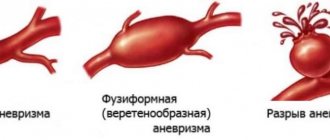
Macroscopically, brain hemangioblastomas are divided into 4 types:
- solid formation - a soft-tissue purple node enclosed in a membrane, on a cut it appears as a spongy mass (occurs in 30% of cases);
- large smooth-walled cyst - a cystic formation with a cavity containing a yellowish transparent liquid, on the wall of which a small nodule (mural nodulus) of the tumor is identified (incidence rate - 60%);
- mixed type - a neoplasm characterized by the presence of a solid node, within which numerous small cysts are found (diagnosed in 4%);
- simple cyst - an ordinary smooth-walled cyst that does not contain a mural node (observed in 6%).
The predominant localization of hemangioblastomas is the posterior cranial fossa (PCF). Typical lesion sites:
- left and right cerebellar hemispheres (85%);
- IV ventricle (6%);
- cerebellar vermis (5%);
- brainstem (3%);
- cerebellopontine angle (1%).
According to the latest data, there is not a single report of the transformation of hemangioblastomas into a malignant form.
Signs of pathology
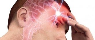
The first manifestations of the tumor process in 90% of patients are general cerebral symptoms characteristic of intracranial hypertension, these are:
- severe headaches (usually in the back of the head);
- nausea, vomiting;
- forced head position (a person intuitively tilts his head to the side to facilitate the conditions for liquor outflow);
- blurred vision.
A feature of clinical signs, as a rule, is increasing or wave-like dynamics with long light intervals. Then, after a few months, focal signs join the symptoms of ICH, these are:
- movement coordination disorder;
- instability, unsteadiness of gait;
- pyramidal insufficiency;
- speech motor disorders;
- involuntary pendulum-like eye movements;
- mental disorders (neurasthenia, depressive neurosis, dysphoria, personality disorientation, etc.).
The listed clinical picture, based on the symptoms of ICH and cerebellar disorders, is characteristic of many cerebral pathologies. Therefore, they cannot be called pathognomonic, that is, specifically for angioreticularis. This fact does not apply only to patients with Hippel-Lindau disease, in whom this symptomatic complex is always combined with retinal angiopathy. In any case, the final diagnosis can be established only after a comprehensive diagnosis, including examination by a neurologist and ophthalmologist, MRI/CT, and selective angiography.
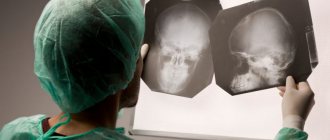
Education on MRI images.
Neurosurgical treatment of GBM
Intracranial hemangioblastomas undergo standard surgical removal. If the tumor is inoperable (mainly with lesions of the brain stem), radiation therapy or non-invasive radiosurgical removal (preferred) is prescribed. Radiosurgery produces significantly better results compared to conventional radiation therapy. In addition, modern stereotactic radiosurgery techniques rarely cause side effects and do not require a long course of radiation (often just 1-2 sessions are sufficient).
Gamma Knife
Types of hemangiomas
- Capillary (ordinary). The tumor is burgundy or blue-red in color. It is characterized by clearly defined boundaries and relatively small size. If pressed, it turns pale, but then returns to its original color.
- Cavernous (cavernous). An inflammatory neoplasm with an uneven, bumpy surface. Very soft, because... the inside is filled not with pus, but with blood.
- Combined. Combines the characteristics of both types listed above.
- Mixed. A tumor affecting vascular, lymphoid, nervous and other tissues. Depending on which areas it affects, it may have different elasticity and different appearance.
Surgery for brain hemangioblastoma
During a conventional operation, which is the “gold standard” for this diagnosis, the tactics of craniotomy are used to create a retrosigmoid approach. Trephination is performed in the area of the foramen magnum; the diameter of the trepanation window is 2-4 cm. The surgical process occurs under the control of an ultra-powerful intraoperative microscope. The operation to completely remove the GBM lasts from 2.5 to 5 hours. Let's consider the main stages of surgical intervention.
- The hair from the scalp in the appropriate area is first shaved.
- In most cases, combined general anesthesia with tracheal intubation and artificial ventilation is chosen for pain relief.
- The patient's position is on his side or stomach, with his head turned to the side. The head is fixed using a special bracket with spikes.
- A longitudinal skin incision is made within the surgical field or a skin flap is cut out and then folded over the mastoid process of the temporal bone.
- Next, a bone hole is formed in the trephinated part of the occipital bone (downward and medial to the sigmoid sinus).
- At the next stage, the neurosurgeon performs a correct opening of the dura mater. After opening the dura mater, access to the surfaces of the cerebellum and other structures of the PCF is freed.
- The specialist begins the process of eliminating the tumor focus with complete preservation of functionally significant brain structures.
- The hemangioblastoma node is removed completely as a single block. All vascular formations feeding the neoplasm are coagulated and intersected as close as possible to its surface.
- If the tumor is a cyst, its contents are carefully aspirated. The walls of the cysts are not removed.
- At the end of the surgical session, the dura mater is sutured, bone fragments are returned to their place and strengthened, and the skin wound defect is closed.
Click on the link to watch a video of the operation to remove hemangioblastoma
The goal of an open operation is to remove the solid component completely, without any residue. It can be completely removed in most cases, in 85-90% of patients. In this situation, we can expect favorable forecasts with a high degree of confidence. But! If partial or subtotal resection is performed, the tumor will continue to grow. It is noted that incomplete removal often leads to the development of hydrocephalus, hemorrhages and hematomas, and death in the very early period.
Radiosurgery
Stereotactic radiosurgery is recommended for a separate group of patients for whom classical surgery is contraindicated due to health conditions or the dangerous location of the tumor. Radiosurgery is also prescribed after incomplete surgical excision of hemangioblastoma in order to eliminate its remaining elements. In the arsenal of neurosurgery, there are 2 methods of stereotactic exposure (without incisions, trepanation and without pain) - Gamma Knife and Cyber Knife.
- Gamma Knife is a radiological procedure specifically designed to specifically destroy abnormal structures within the brain using Cobalt-60 ionizing radiation. Before the operation, a helmet system is put on the patient’s head, which makes it possible to accurately determine and neutralize the focal area. The radiation “works” strictly within the tumor and does not cause harm to nearby healthy brain structures. To install a stereotactic structure on the head, local anesthesia with a long-acting local anesthetic is performed locally (in the places where it is fixed). The course of Gamma Knife therapy consists of 1-3 procedures. Time of 1 session – 30-180 minutes.
- Cyberknife is a procedure for targeted irradiation of pathological tissues using a robotic installation that generates 6 MeV photon radiation. The principle of therapeutic action and effectiveness are approximately identical to the Gamma Knife. But the CyberKnife radiosurgical device, unlike the Gamma installation, is universal. It can be used to treat oncology in different parts of the human body. During this procedure, you do not need to place your head in a frame, and no anesthesia is required.
Contraindications to Gamma Knife and CyberKnife surgeries are tumors that have reached a size of 3.5 cm or more.
Kinds
There are 11 types of benign meningiomas:
- meningothelial meningiomas – 60%;
- transitional meningiomas – 25%;
- fibrous meningiomas – 12%;
- rare types of meningiomas – 3%.
A brain tumor can be located in different areas of the brain:
- convexital tumor – 40%;
- parasagittal – 30%;
- basal location of the tumor – 30%.
Meningioma of the brain of the frontal lobe
Meningioma of the frontal region forms very often, in most cases it does not bother the patient for a long time. If the meningioma is located in the right frontal lobe, symptoms will appear on the opposite side of the body.
The reasons for the development of meningioma of the frontal region are various: traumatic brain injury, inflammatory disease of the meninges, genetic predisposition, food high in nitrates, neurofibromatosis and other reasons. Radiation exposure is considered a proven cause of tumor development; all other causes are considered risk factors.
Meningioma of the frontal region can cause blurred vision, headache, paresis of the facial nerves, arm muscles, lethargy and other symptoms.
Anaplastic meningioma
Anaplastic meningioma is a grade 3 brain tumor; tumor recurrence occurs in all patients within three years after treatment.
Parasagittal meningioma
Parasagittal meningioma is located in the occipital, parietal or frontal part along the longitudinal midline. Often this tumor is accompanied by a pathological increase in the content of bone matter in the bone tissue. Parasagittal meningiomas, growing in the frontal part of the head, cause:
- increased intracranial pressure;
- development of congestive optic discs in the fundus;
- severe nausea and vomiting, headache;
- epileptic seizures.
Parasagittal meningioma of the parietal region of the head is characterized by sensory disturbances and epileptic seizures. Meningioma of the occipital region is characterized by increased intracranial pressure and hallucinations.
Atypical meningioma
Atypical brain meningioma is a grade 2 tumor; tumor recurrence occurs in 30% of patients within 10 years after treatment.
Meningioma falx
A tumor growing from the greater falciform process of the brain is called falx meningioma. Over time, the tumor grows into the sagittal venous sinus, resulting in impaired venous circulation and intracranial hypertension. The growth of the tumor causes the following negative symptoms: epileptic seizures, impaired sensitivity and motor activity of the legs, pelvic disorders.