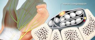
Tunnel neuropathies account for 1/3 of diseases of the peripheral nervous system. More than 30 forms of tunnel neuropathies have been described in the literature [Levin O.S., 2005]. Various forms of compression-ischemic neuropathies have their own characteristics. We will first consider their general characteristics, then we will focus on the most common forms of hand tunnel syndromes (Table 1).
Causes
The anatomical narrowness of the canal is only a predisposing factor in the development of tunnel syndrome. In recent years, evidence has accumulated indicating that this anatomical feature is genetically determined. Another reason that can lead to the development of tunnel syndrome is the presence of congenital developmental anomalies in the form of additional fibrous cords, muscles and tendons, and rudimentary bone spurs. However, predisposing factors alone for the development of this disease are usually not enough. Some metabolic and endocrine diseases (diabetes mellitus, acromegaly, hypothyroidism), diseases accompanied by changes in joints, bone tissue and tendons (rheumatoid arthritis, rheumatism, gout), conditions accompanied by hormonal changes (pregnancy), space-occupying formations can contribute to the development of tunnel syndrome the nerve itself (schwannoma, neuroma) and outside the nerve (hemangioma, lipoma). The development of tunnel syndromes is facilitated by frequently repeated stereotypical movements and injuries. Therefore, the prevalence of carpal tunnel syndrome is significantly higher in people engaged in certain activities and in representatives of certain professions (for example, stenographers have carpal tunnel syndrome 3 times more often).
Clinical manifestations
The full picture of tunnel syndrome includes sensory (pain, paresthesia, numbness), motor (decreased function, weakness, atrophy) and trophic disorders.
Various clinical course options are possible. Most often it starts with pain or other sensory disorders. Less commonly, it begins with movement disorders. Trophic changes are usually expressed insignificantly and only in advanced cases. The most characteristic feature of carpal tunnel syndrome is pain. Typically, pain appears during movement (load), then occurs at rest. Sometimes the pain wakes the patient up at night, which exhausts the patient and forces him to see a doctor. Pain in tunnel syndromes can include both a nociceptive component (pain caused by inflammatory changes occurring in the area of the nerve-canal conflict) and a neuropathic component (due to nerve damage). Tunnel syndromes are characterized by manifestations of neuropathic pain such as allodynia and hyperpathy, a sensation of electric current passing (electrical shooting), and burning pain. In later stages, pain may be due to muscle spasms. Therefore, when choosing pain therapy, it is necessary to be guided by the results of a thorough clinical analysis of the characteristics of the pain syndrome. Motor disorders arise as a result of damage to the motor branches of the nerve and manifest themselves in the form of decreased strength and rapid fatigue. In some cases, the progression of the disease leads to atrophy and the development of contractures (“clawed paw”, “monkey paw”).
With compression of arteries and veins, vascular disorders may develop, which is manifested by pallor, a decrease in local temperature, or the appearance of cyanosis and swelling in the affected area. With isolated nerve damage (in the absence of compression of arteries and veins), trophic changes are most often insignificantly expressed.
Symptoms of a pinched nerve in the hand: pain and numbness in the fingers
Clinical symptoms of a pinched nerve in the hand manifest themselves in the form of impaired sensitivity and motor function. In the first case, the signs are expressed in a feeling of numbness of the skin in a certain part of the hand. Limitation of mobility or the inability to bend fingers or clench them into a fist is also a manifestation of damage to motor axons.
As a rule, pain in the hand due to a pinched nerve is localized at the site of pathological changes and in distant areas that suffer from impaired innervation. A complex pathogenic mechanism is at work here:
- when a nerve is pinched in the elbow joint, two innervation branches are immediately affected;
- pain in the elbow area may be due to the disease that caused the pinching (arthrosis, arthritis, bursitis, tendovaginitis, etc.);
- pain in the elbow area may also be associated with the primary point of nerve damage;
- distant (distal) areas of pain can be located in the fingers, the back of the hand, or the wrist joint;
- this is due to the fact that the innervation of the vascular wall is disrupted, due to which the process of impaired blood microcirculation develops;
- stagnant and trophic changes cause acute pain in those tissues that do not receive an influx of fresh blood;
- compression syndrome with tissue necrosis may develop.
Numbness of the hand due to a pinched nerve develops gradually with increasing amplitude. First, a slight tingling sensation occurs, then a decrease in skin sensitivity occurs, after which complete numbness suddenly develops (the person ceases to feel individual fingers or the entire hand, the back or inner side of the palm.
If numbness in the fingers is observed when a nerve is pinched, then there is a high probability of obstruction of the median or carpal branch. Based on which fingers are numb, a preliminary diagnosis can be made.
To confirm the diagnosis, special examinations must be carried out. If necessary, your doctor may order wrist and elbow x-rays, arthroscopy, or an MRI.
Diagnostics
As a rule, the diagnosis is established on the basis of the characteristic clinical manifestations described above. It is convenient for the clinician to use a number of clinical tests that allow differentiating different types of carpal tunnel syndromes. In some cases, it is necessary to conduct electroneuromyography (the speed of impulses along the nerve) to clarify the level of nerve damage. Nerve damage, space-occupying lesions or other pathological changes causing carpal tunnel syndrome can also be determined using ultrasound, thermal imaging, MRI [Horch RE et al., 1997].
Stop exposure to the pathogenic factor. Immobilization
The first thing to do is to stop physical impact on the affected area. Therefore, immobilization in the affected area is necessary. Recently, special devices have appeared in our country - orthoses, bandages, splints, which allow immobilization in the area of injury. At the same time, they are very convenient to use, they can be put on and taken off very easily, which allows the patient to maintain his social activity (Fig. 1). These funds are widely and successfully used abroad. Studies have appeared on the effectiveness of splinting, which have convincingly shown that it is quite comparable to the effectiveness of hormone injections and surgical operations. In our country, these devices are already used by traumatologists; They are clearly not yet sufficiently introduced into neurological practice.
Change the usual locomotor stereotype and lifestyle
Tunnel syndromes are often the result not only of monotonous activity, but also of ergonomic disorders (improper posture, awkward position of the limb during work). Special exercises and recommendations for optimal organization of the workplace have been developed. To relieve pain and prevent relapse, orthoses and splints using the splinting principle are used. In rare cases, you have to change your profession. Training in special exercises and physical therapy are an important component of the treatment of tunnel neuropathies at the final stage of therapy.
Pain therapy
Physical influences (cold, heat).
In mild cases, ice compresses and sometimes “hot” compresses can help reduce pain. A doctor is usually consulted when these or other “home” methods “do not help.” • Anti-inflammatory therapy. Traditionally, for carpal tunnel syndromes, NSAIDs with a more pronounced analgesic and anti-inflammatory effect (diclofenac, ibuprofen) are used. It should be remembered that with long-term use of drugs in this group there is a risk of gastrointestinal and cardiovascular complications. In this regard, for moderate or severe pain, it is advisable to use a combination of low doses of the opioid analgesic tramadol (37.5 mg) and the safest analgesic/antipyretic paracetamol (325 mg). Thanks to this combination, a multiple increase in the general analgesic effect is achieved with a lower risk of side effects.
• Impact on the neuropathic component of pain. Often, with tunnel syndromes, the use of analgesics and NSAIDs is ineffective (it is in these cases that patients consult a doctor). This may be due to the fact that the dominant role in the formation of pain is played not by the nociceptive, but by the neuropathic mechanism. When pain is the result of neuropathic changes, it is necessary to prescribe drugs recommended for the treatment of neuropathic pain: anticonvulsants (pregabalin, gabapentin), antidepressants (venlafaxine, duloxetine), plates with 5% lidocaine. The choice of a particular drug should be made taking into account the clinical manifestations and individual characteristics of the patient (the possibility of developing side effects). It is important to inform the patient that drugs used for neuropathic pain, unlike “classical painkillers,” do not begin to act immediately (it is necessary to titrate the dose; the effect occurs several days or even weeks after starting the drug).
• Injections of anesthetic + hormones. A very effective and acceptable treatment method for most types of tunnel neuropathies is a blockade with the introduction of an anesthetic (Novocaine) and a hormone (hydrocortisone) into the area of infringement. Special guidelines describe techniques and doses of drugs for various tunnel syndromes [Zhulev N.M., 2005]. This procedure is usually resorted to if other measures are ineffective (cold compresses, the use of analgesics, NSAIDs), but in some cases, if the patient comes at a more advanced stage of the disease and experiences severe pain, it is advisable to immediately offer such a patient this manipulation.
• Other methods of pain relief. Currently, there are reports of the high effectiveness of injection of meloxicam with hydrocortisone into the tunnel area. An effective way to reduce pain and inflammation is electrophoresis, phonophoresis with dimexide and other anesthetics. They can be carried out in a clinic setting. Symptomatic treatment. For tunnel syndromes, decongestants, antioxidants, muscle relaxants, and drugs that improve the trophism and functioning of the nerve (ipidacrine, vitamins, etc.) are also used.
Surgical intervention. Surgical treatment is usually resorted to when other options for helping the patient have been exhausted. At the same time, for certain indications, it is advisable to immediately offer the patient surgical intervention. Surgery usually involves releasing the nerve from compression, “reconstructing the tunnel.” According to statistics, the effectiveness of surgical and conservative treatment does not differ significantly a year later (after the start of treatment or surgery). Therefore, after a successful surgical operation, it is important to remember about other measures that must be followed to achieve a full recovery (prevention of relapses): changing locomotor patterns, using devices that protect against stress (orthoses, splints, bandages), performing special exercises.
Treatment of a pinched nerve in the hand: what to do?
The first thing to do if you have pinched nerves in your hands is to seek immediate medical attention. Long-term compression of the nerve fiber can lead to atrophy and loss of all functions. It is important to visit a doctor immediately after a feeling of numbness or pain appears. If shortly before the onset of unpleasant sensations there was an injury (fall on your arm, bruise, whiplash, dislocation, sprain), then you need to go to the nearest emergency room.
If the feeling of a pinched nerve appears suddenly against the background of general well-being, you should contact a neurologist. An experienced doctor will be able to identify the potential cause of the pathology during the initial examination and palpation. After this, effective treatment will be prescribed.
You can sign up for a free initial consultation at our manual therapy clinic. We see an experienced neurologist. Once diagnosed, you may be offered appropriate treatment.
Usually, to treat a pinched nerve in the hand, it is enough to influence the cause of such a pathological change. If it is cervical osteochondrosis, then traction traction of the spinal column helps well. By restoring normal spaces between the vertebral bodies, compression of the radicular nerves is eliminated. With glenohumeral periarthritis, it is necessary to act on those planes of the joints that are deformed and disrupt the conductivity of the nerve fiber. Osteopathy and massage, kinesitherapy and reflexology can help here.
Carpal tunnel and carpal valve syndromes can also be treated as an emergency with osteopathy and manual therapy. In the long term, an individually developed rehabilitation course is prescribed. It can be based on therapeutic exercises, massage, osteopathy and reflexology.
Carpal tunnel syndrome
Carpal tunnel syndrome (carpal tunnel syndrome) is the most common form of compression-ischemic neuropathy encountered in clinical practice. In the population, carpal tunnel syndrome occurs in 3% of women and 2% of men [Berzins Yu.E., 1989]. This syndrome is caused by compression of the median nerve as it passes through the carpal tunnel under the transverse carpal ligament. The exact cause of carpal tunnel syndrome is not known. The following factors most often contribute to compression of the median nerve in the wrist area: • Trauma (accompanied by local swelling, tendon stretching). • Ergonomic factors. Chronic microtraumatization (often found among construction workers), microtraumatization associated with frequent repeated movements (among typists, with constant long-term work with a computer). • Diseases and conditions accompanied by metabolic disorders, edema, tendon and bone deformities (rheumatoid arthritis, diabetes mellitus, hypothyroidism, acromegaly, amyloidosis, pregnancy). • Massive formations of the median nerve itself (neurofibroma, schwannoma) or outside it in the wrist area (hemangioma, lipoma).
Why does the radial nerve become pinched in the hand?
The causes of nerve conduction disorders can be associated with mechanical, traumatic, inflammatory and trophic (circulatory disorders) effects. Let's look at all of them in more detail.
Why the radial nerve becomes pinched in the arm can only be determined by examination and evaluation of radiographic images. At home, it is very difficult to understand the cause of pathological changes.
Most often, this condition occurs as a result of disruption of nerve fiber conduction against the background of tunnel syndromes:
- narrowing of the carpal valve is accompanied by numbness of the thumb, index and middle fingers;
- Carpal tunnel syndrome is characterized by characteristic numbness of the little and ring fingers.
Tunnel syndromes can have a traumatic, inflammatory and deformational etiology. In case of injury, callus growth may occur, preventing the normal position of the nerve fiber. Inflammatory processes are often associated with arthrosis of the wrist joint. And deformation is the development of arthrosis with the appearance of bone growths and spines.
If a nerve is pinched in the hand, pain and a feeling of muscle weakness may occur. Other reasons include radial fractures in a typical location, tendovaginitis, elbow bursitis, glenohumeral periarthritis, cervical osteochondrosis, etc.
Clinical manifestations
Carpal tunnel syndrome is characterized by pain, numbness, paresthesia and weakness in the arm and hand.
Pain and numbness extend to the palmar surface of the thumb, index, middle and 1/2 ring finger, as well as to the dorsum of the index and middle finger. Initially, symptoms occur when performing any activities using a brush (working on a computer, drawing, driving), then numbness and pain appear at rest, sometimes occurring at night. The following tests are suggested to verify the diagnosis of carpal tunnel syndrome. Tinel test: tapping the wrist (above the median nerve) with a neurological hammer causes a tingling sensation in the fingers or pain radiating (electrical shooting) to the fingers (Fig. 2). Pain may also be felt in the tapping area. A positive Tinel sign is found in 26–73% of patients with carpal tunnel syndrome [Al Zamil M.H., 2008]. Durkan's test: compression of the wrist in the area of the median nerve causes numbness and/or pain in the 1st–3rd, half of the 4th fingers (as with Tinel's symptom). Phalen Test: Wrist flexion (or extension) 90 degrees produces numbness, tingling, or pain in less than 60 seconds (Figure 3). A healthy person may also develop similar sensations, but not earlier than after 1 minute. Opposition test: with severe thenar weakness (which occurs at a later stage), the patient cannot connect the thumb and little finger (Fig. 4); or the doctor (researcher) can easily separate the patient’s closed thumb and little finger.
Carpal tunnel syndrome: treatment and prevention
In carpal tunnel syndrome (also sometimes called carpal tunnel syndrome), pain occurs due to compression of the median nerve, which is responsible for providing sensation to several fingers.
Causes of the syndrome
Increased pressure on the median nerve can be caused by swelling or a number of other problems. As a rule, carpal tunnel syndrome is observed in the fair sex, as well as in those who, by the nature of their profession, are forced to perform monotonous flexion-extension movements of the hand (sign language interpreters, motorcycle racers, drummers, artists and representatives of other specialties). But most often this disease affects those whose work involves typing on a computer keyboard.
Hypothyroidism, rheumatoid arthritis, diabetes, pregnancy, obesity, various injuries and, oddly enough, even smoking can cause wrist pain, as it can reduce blood flow to the median nerve.
Main symptoms
It's easy to tell if you have carpal tunnel syndrome: tingling, numbness, weakness, and pain in your fingers or wrist. The index, middle and partially ring fingers are most often affected. There should be no problem with the little finger in case of carpal tunnel syndrome, since it is not connected to the median nerve.
In the initial stage of the disease, symptoms appear at night. After shaking the brush, the unpleasant sensations disappear - these are the first signs of the development of the syndrome.
Diagnosis and treatment
As soon as pain and discomfort appear, you need to seek help from a specialist. The doctor will examine you, check your health, inquire about your professional activities, and ask about possible injuries to your wrist, arm, or neck (perhaps the cause is simply a sprain). During the test, your doctor will check the sensation, strength, and appearance of your neck, shoulders, arms, wrists, and hands, and you'll also need to take a blood test to see what other issues might be causing the discomfort. To accurately diagnose carpal tunnel syndrome, the Tinel and Phalen tests are used.
Mild symptoms of the syndrome can be treated at home, and the sooner you start doing this, the higher the chances of success and prevention of nerve damage. Here are a few simple rules to follow:
- If discomfort occurs, you should stop working and give your wrist a rest;
- It is necessary to cool the wrist for a few minutes every couple of hours and take special medications to reduce pain;
- Before going to bed, you need to secure your wrist with an elastic bandage: this procedure will reduce pressure on the median nerve.
If the pain does not go away within one to two weeks, and self-treatment does not work, immediately consult a specialist. He will prescribe medication to relieve carpal tunnel syndrome or treat the problem that caused it.
Surgical treatment is also possible, but, as a rule, it is only possible if, due to pain, you cannot work or perform ordinary household manipulations, even after prolonged conservative treatment.
Prevention of carpal tunnel syndrome
To prevent carpal tunnel syndrome, you need to take care of your health. Get on the path to returning to a healthy weight by quitting smoking, exercising, and keeping conditions like arthritis and diabetes under control. In addition, follow these simple preventative tips:
- To hold objects while working, use not only your fingers, but your entire hand;
- When working at a computer, keep your hands slightly above your wrists and your shoulders relaxed;
- Change hands, performing constantly repeating movements.
Author: K.M.N., Academician of the Russian Academy of Medical Sciences M.A. Bobyr
Differential diagnosis
Carpal tunnel syndrome should be differentiated from arthritis of the carpo-metacarpal joint of the thumb, cervical radiculopathy, and diabetic polyneuropathy. Patients with arthritis will show characteristic bone changes on x-rays. In cervical radiculopathy, reflex, sensory and motor changes will be associated with neck pain, while in carpal tunnel syndrome these changes are limited to distal manifestations. Diabetic polyneuropathy is usually a bilateral, symmetrical process involving other nerves (not just the median nerve). At the same time, a combination of polyneuropathy and carpal tunnel syndrome in diabetes mellitus cannot be ruled out.
Treatment
In mild cases of carpal tunnel syndrome, ice compresses and a decrease in load can help. If this does not help, the following measures must be taken: 1. Immobilization of the wrist. There are special devices (splints, orthoses) that immobilize the wrist and are convenient to use (Fig. 1). Immobilization should be carried out at least overnight, and preferably for 24 hours (at least in the acute period). 2. NSAIDs. Drugs from the NSAID group will be effective if the inflammatory process dominates in the pain mechanism. 3. If the use of NSAIDs turns out to be ineffective, it is advisable to inject novocaine with hydrocortisone into the wrist area. As a rule, this procedure is very effective. 4. In outpatient settings, electrophoresis can be performed with anesthetics and corticosteroids. 5. Surgical treatment. For mild or moderate carpal tunnel syndrome, conservative treatment is more effective. In cases where all conservative treatment options have been exhausted, surgical treatment is resorted to. Surgical treatment consists of partial or complete resection of the transverse ligament and releasing the median nerve from compression. Recently, endoscopic surgical methods have been successfully used in the treatment of carpal syndrome.
Treatment of a pinched nerve
Typically, treatment for a pinched nerve begins with therapeutic exercises - simple physical exercises that help restore the position of the nerve. Physiotherapy is also used , which is no less effective, therapeutic massage and osteopathy
Among medications, doctors primarily prescribe painkillers and blockade of the affected nerve with ozone to alleviate the patient’s condition. The analgesic effect of ozone blockade occurs within 10-15 minutes.
When the above methods do not help, the doctor prescribes treatment with anti-inflammatory drugs that can restore the surrounding tissue. If these injections also turn out to be useless, surgical intervention is discussed.
SM-Clinic doctors often meet with the disease in question, make an accurate diagnosis, and prescribe effective treatment . Thanks to many years of experience, specialists do an excellent job of returning patients the ability to full, pain-free movement.
“SM-Clinic” always treats people with care and attention, trying to provide effective assistance .
Pronator teres syndrome (Seyfarth syndrome)
Entrapment of the median nerve in the proximal part of the forearm between the pronator teres fascicles is called pronator syndrome.
This syndrome usually begins to appear after significant muscle activity over many hours involving the pronator and flexor digitorum muscles. Such types of activities are often found among musicians (pianists, violinists, flutists, and especially often among guitarists), dentists, and athletes [Zhulev N.M., 2005]. Long-term tissue compression is of great importance in the development of pronator teres syndrome. This can happen, for example, during deep sleep when the newlywed’s head is positioned for a long time on the partner’s forearm or shoulder. In this case, the median nerve in the pronator snuffbox is compressed, or the radial nerve in the spiral canal is compressed when the partner’s head is located on the outer surface of the shoulder (see radial nerve compression syndrome at the level of the middle third of the shoulder). In this regard, to designate this syndrome in foreign literature, the terms “honeymoon paralysis” (honeymoon paralysis, newlywed paralysis) and “lovers paralysis” (lovers paralysis) have been adopted. Pronator teres syndrome sometimes occurs in nursing mothers. In them, compression of the nerve in the area of the pronator teres occurs when the baby’s head lies on the forearm, he is breastfed, lulled to sleep, and the sleeping person is left in this position for a long time.
Tunnel syndrome: where it’s narrow, it hurts
“Tennis elbow,” “lovers’ palsy,” “park bench” syndrome—metaphors add a romantic charm to the names of tunnel neuropathies. But those who experience their symptoms have no time for lyrical associations: pain from carpal tunnel syndrome not only significantly impairs the quality of life, but can even lead to disability.
What are the most common tunnel neuropathies, how do they manifest and, most importantly, what to do about it?
Bunch of nerves
To transmit signals from the brain, they “entangle” the entire body. Nerve fibers also pass through the so-called anatomical tunnels - narrow spaces, the walls of which are various bones, tendons and muscles.
Normally, there is enough space for nerves in such conditions. But under certain circumstances, the narrow canal can narrow, and then the nerve is compressed. This is how tunnel syndrome or tunnel neuropathy occurs.
Circumstances that predispose to the development of “conflict” may be different. First of all, these are congenital anomalies, for example, additional muscles, tendons, fibrous cords in the tunnel area.
A number of diseases and conditions can contribute to tunnel syndrome, for example, diabetes, osteoarthritis, gout, malignant tumors, edema, pregnancy.
Tunnel neuropathy also occurs in completely healthy people whose professional activities are associated with constant microtrauma.
Programmers who don’t let go of their mouse for days on end, woodcarvers whose hand is used to holding a tool in the same position, and also milkmaids, wrappers, stenographers—representatives of these and some other professions may one day experience “strange” symptoms.
If there is a conflict in the tunnel...
The first sign of pinched nerves passing through the tunnel is usually pain. It appears against the background of inflammatory changes occurring in the “conflict” zone, as well as as a result of damage to the nerve itself.
In the initial stages, pain occurs only after exercise. As it progresses, it begins to occur at rest, including at night.
In this case, pain often appears in response to irritants that usually do not affect pain receptors: light stroking, exposure to moderate temperatures can cause a new attack.
Often the pain has the character of an electric shooting, penetrating the area of “conflict” with lightning speed and sharpness. Classic signs of carpal tunnel syndrome are also numbness and tingling (paresthesia) in the affected area.
Later, difficulties may arise when making “fine” movements, for example, when fastening buttons, as well as increased fatigue of the muscles in the affected area. By the way, this area can be located in a variety of places.
Weak spots
About 30 types of tunnel syndromes are known. They can occur in the neck, shoulder region, arms, legs, and pelvic girdle. The most common tunnel neuropathies include:
• Carpal tunnel syndrome, the most common form of neuropathy, occurs due to compression of the median nerve under the transverse carpal ligament and is accompanied by damage to the hand. Working at a computer, drawing, driving are leading risk factors.
• Radial nerve compression syndrome can occur at various levels. If compression occurs in the middle third of the shoulder, “park bench” syndrome develops. The name comes from the fact that compression occurs when sleeping on hard surfaces. When the deep posterior branch of the radial nerve is compressed, tennis elbow occurs. Compression is typical for athletes who experience regular muscle overload in the area where the nerve passes - tennis players, badminton players, etc.
• Pronator teres syndrome ("lover's palsy") occurs as a result of compression of the median nerve in the forearm. It can occur with prolonged compression of the tissue in the forearm area, for example, if the beloved’s head rests on the forearm of the happy groom all night.
• Scapular notch syndrome develops as a result of compression of the nerve in the gap formed by the scapular notch and is characterized by pain in the depths of the shoulder joint.
• Piriformis syndrome occurs when the sciatic nerve is compressed and manifests as pain along the back of the leg.
Health to the nerves!
Despite the wide “geography” of tunnel syndromes, their diagnosis and treatment have common principles. The diagnosis is most often made on the basis of “telling” clinical manifestations. In addition, there are a number of tests that can distinguish one carpal tunnel syndrome from another.
To carry them out, neither laboratories nor equipment are needed: the sickest and most qualified neurologist who knows how and where to press is enough.
The basic principle of treatment for all tunnel syndromes is to stop exposure to the factor causing the narrowing of the tunnel.
Programmers will have to leave the computer mouse alone, milkmaids will have to take a break from cow cows, and pianists will have to put off playing their favorite instrument until better times.
However, milking cows or playing the piano with a bandage, splint or orthosis is unlikely to work: another necessary therapeutic measure is fixation of the affected area. And, of course, to alleviate the course of neuropathy, pain therapy is prescribed.
Clinical manifestations
With the development of pronator teres syndrome, the patient complains of pain and burning 4–5 cm below the elbow joint, along the anterior surface of the forearm and pain radiating to the 1st–4th fingers and palm.
Tinel's syndrome. In case of pronator teres syndrome, Tinel's sign will be positive when tapping with a neurological hammer in the area of the pronator snuff box (on the inside of the forearm). Pronator-flexor test. Pronating the forearm with a tightly clenched fist while creating resistance to this movement (counteraction) leads to increased pain. Increased pain can also be observed when writing (prototype of this test). When examining sensitivity, a sensitivity disorder is revealed, involving the palmar surface of the first three and a half fingers and the palm. The sensory branch of the median nerve, innervating the palmar surface of the hand, usually passes above the transverse carpal ligament. The occurrence of sensory disturbances on the palmar surface of the first finger, the dorsal and palmar surfaces of the second and fourth fingers, with preservation of sensitivity in the palm, allows one to confidently differentiate carpal tunnel syndrome from pronator teres syndrome. Thenar atrophy in pronator teres syndrome is usually not as severe as in progressive carpal tunnel syndromes.
Supracondylar process syndrome of the shoulder (Strother's band syndrome, Coulomb, Lord and Bedossier syndrome)
In the population, in 0.5–1% of cases, a variant of the development of the humerus is observed, in which a “spur” or supracondylar process (apophysis) is found on its distal anteromedial surface. Due to the accessory process, the median nerve is displaced and stretched (like a bowstring). This makes him vulnerable to defeat. This tunnel syndrome, described in 1963 by Coulomb, Lord and Bedossier, has almost complete similarities with the clinical manifestations of pronator teres syndrome: pain, paresthesia, and decreased flexion strength of the hand and fingers are detected in the zone of innervation of the median nerve. In contrast to pronator teres syndrome, when the median nerve is damaged under Strather's ligament, mechanical compression of the brachial artery with corresponding vascular disorders is possible, as well as severe weakness of the pronator teres (teres and minor). The following test is useful in diagnosing supracondylar process syndrome. When extending the forearm and pronation in combination with formed flexion of the fingers, painful sensations are provoked with localization characteristic of compression of the median nerve. If it is suspected that the compression is caused by a “spur” of the humerus, an x-ray examination is indicated. Treatment involves resection of the supracondylar process (“spur”) of the humerus and ligament.
Cubital tunnel syndrome
Cubital tunnel syndrome (Sulcus Ulnaris Syndrome) is compression of the ulnar nerve in the cubital canal (Mouchet's canal) in the area of the elbow joint between the internal epicondyle of the humerus and the ulna bone and is the second most common after carpal tunnel syndrome. Cubital tunnel syndrome develops for a number of reasons. Cubital tunnel syndrome can be caused by repetitive bending of the elbow joint. Therefore, cubital tunnel syndrome is classified as a disorder called accumulated trauma disorder (overuse syndrome). Those. the disorder may occur during normal, repetitive movements (most often associated with a specific occupational activity) in the absence of obvious traumatic injury. Direct trauma can also contribute to the development of cubital tunnel syndrome, such as leaning on the elbow while sitting. Patients with diabetes and alcoholism are at greater risk of developing cubital tunnel syndrome.
Ulnar nerve compression or cubital syndrome
MRI of the hand
- Cost: 7,000 rub.
More details
Occurs in those who often bend their arm at the elbow - cyclists, programmers and others. The ulnar nerve is often affected in very thin women. In this case, pain and numbness begins with the little finger, spreading to the entire hand. The muscles on the back of the hand between the thumb and index finger become thinner and begin to work less well. In damp, cold weather the pain becomes almost unbearable.
Clinical manifestations
The main symptoms of cubital tunnel syndrome are pain, numbness and/or tingling. Pain and paresthesia are felt in the lateral part of the shoulder and radiate to the little finger and half of the fourth finger. At first, discomfort and pain occur only when pressure is applied to the elbow or after prolonged bending. In a more severe stage, pain and numbness are felt constantly. Another sign of the disease is weakness in the arm. It manifests itself as a loss of “confidence” in the hand: suddenly objects begin to fall out of it during some habitual actions. For example, it becomes difficult for a person to pour water from a kettle. In advanced stages, the hand on the sore arm begins to lose weight, and pits appear between the bones due to muscle atrophy.
Causes of pain in the hand with osteochondrosis of the cervical spine
At first glance, pain in the arm and osteochondrosis are not related in any way. But the fact is that such a symptom as a painful sensation in the upper extremities can accompany cervical osteochondrosis due to the fact that the peripheral nerves that are located in the spine also regulate their activity.
What is cervical osteochondrosis
Osteochondrosis is a disease affecting the articular-cartilaginous system. The disease itself is very insidious, since pain is felt only during the transition to the active stage.
As a rule, intervertebral discs are affected. Their dystrophy is the cause of smoothing. The movements, in turn, compress the nerve roots from the spine, and then there is inflammation of the muscles, which further compresses the nerve endings. As a result, weakness and pain in the hand occurs. In some cases, normal blood flow in the upper extremities may even be affected.
Most often, you can find that osteochondrosis of the cervical spine is characteristic of people who often need to sit in an uncomfortable position (for example, with their head bowed).
How does your hand hurt?
The sensations that patients experience in the hand are not the most pleasant:
- Numbness up to loss of sensitivity;
- Trembling;
- Painful symptoms and their increase as you move;
- Pale skin (due to vascular spasms and poor circulation);
- The hands seem to be welded together.
Basically, the above symptoms appear in the morning. It should also be noted that often only one hand is involved in this process - but this is not necessary.
Pain and numbness in the left hand may indicate problems in the heart. If you are also elderly, then the diagnosis is carried out for the presence of disorders specifically in the area of the cardiovascular system. The diagnosis may not reveal any pathology, but the pain and numbness still persist.
The situation is more complicated when a person has both osteochondrosis and heart disease. By the way, the heart can also hurt with thoracic osteochondrosis, but here, too, there are differences.
But pain and numbness in the right hand can be confused with Raynaud's disease. This is nothing more than vasospasm, leading to the above symptoms. No other painful sensations are felt anywhere except the site of inflammation.
Diagnosis and treatment
If symptoms are detected, you should contact a neurologist or vertebrologist. After all the symptoms are described, a correct diagnosis will be required. It is diagnosed on the basis of CT and MRI. These methods make it possible to distinguish a number of diseases such as metastases from intervertebral hernia.
Treatment in this case is conservative and complex, and it must be started as early as possible due to the fact that the disease tends to develop into a chronic one.
Treatment consists of:
- Use of non-steroidal anti-inflammatory drugs (Diclofenac, Ibuprofen, Analgin, Ketorolac);
- Using patches to reduce pain;
- Using medications and vitamins to improve blood supply and strengthen capillary walls (B vitamins, Pentoxifylline);
- Use of blockades;
- Physiotherapy (ozokerite, electro- and magnetic treatment);
- Exercise therapy (but only during the period of retreat of the disease, since it is very important that painful symptoms are not felt);
- The use of such a pleasant procedure as massage also brings very good results.
And only if conservative methods do not provide any tangible improvement, surgical intervention in the body can be applied.
Prevention
Of course, it is always better to take precautions to avoid any problems. Therefore, prevention, which consists of leading a healthy lifestyle, is no less important.
Good prevention is active rest, physical and breathing exercises without excessive stress, a healthy diet, and, in addition, correct and comfortable postures, both during night rest and during the day.
Massage as a preventative measure is also necessary for the body. Author: K.M.N., Academician of the Russian Academy of Medical Sciences M.A. Bobyr
Diagnostics
In the early stages of the disease, the only manifestation (besides weakness of the forearm muscles) may be loss of sensation on the ulnar side of the little finger. If the clinical picture is blurred, the following tests can help verify the diagnosis of Cubital Tunnel Syndrome: Tinel test - the occurrence of pain in the lateral part of the shoulder, radiating to the ring finger and little finger when tapping with a hammer over the area where the nerve passes in the area of the medial epicondyle. Equivalent to Phalen's sign, sudden flexion of the elbow will cause paresthesia in the ring and little fingers. Frohman's test. Because of weakness of the abductor policis brevis and flexor policis brevis, excessive flexion of the interphalangeal joint of the thumb on the affected hand may be detected in response to a request to hold a paper between the thumb and index finger (Figure 5). Wartenberg test. Patients with more severe muscle weakness may complain that when putting their hand in a pocket, the little finger is pulled to the side (does not fit into the pocket) (Fig. 6).
Osteochondrosis: pinched nerve endings
A pinched nerve is a condition in which the nerve roots become compressed. They are connected to the spinal cord, so on their way they may encounter obstacles in the form of a hernia, protrusion, or spasmodic muscles. diseased spine
constantly reminds of itself with severe discomfort, unpleasant sensations in various areas.
Osteochondrosis can be located in any part (rarely in the chest). Most often the sciatic, cervical nerve is pinched. The back begins to hurt in a certain place. It may become numb and less sensitive. In advanced cases, dysfunction of individual internal organs is observed. This is all due to the fact that the nerve root is compressed. By the way, there are 3 types of nerves:
- Sensitive;
- Vegetative;
- Motor.
If the first one is pinched, the person will definitely feel unbearable pain and will try to eliminate the interfering symptom in a timely manner. If compression of the other two nerves occurs, the patient may not experience obvious discomfort. But this only aggravates the situation, because complications develop.
Symptoms
The clinical picture depends on the extent of the affected area, the presence of inflammation, the degree and cause of pinched nerve root.
There are 8 nerves in the cervical region. If at least one of them is compressed, a person feels pain, numbness, tingling in the innervation zone. The shoulder hurts and goes numb, the muscle fibers weaken – the nerve in the area of the C5 vertebra is deformed. A similar picture in C6 provokes numbness and pain in the hands. If we are talking about the C7 vertebra, discomfort is also felt in the middle finger, tingling is possible. In the case of C8, the person cannot move their arms normally.
In the chest area, due to its low mobility, nerves are pinched quite rarely. If this happens, it hurts very much near the heart. Breathing problems begin, and chest tightness appears. If the situation is neglected, the pain reaches the stomach. Take an antispasmodic to rule out gastritis, ulcers and other gastrointestinal diseases. If it helped, the reason is these diseases.
The sciatic nerve is compressed in the lumbar region. The roots are too sensitive. Proteins contained in the disc also increase irritability. Pinching is characterized by pain, although it can be asymptomatic. A burning sensation similar to an electric shock can reach the toes. The lower extremities become numb.
Problems in the lumbosacral region lead to the development of radiculitis. This part of the spinal column bears the heaviest load, especially if a person is overweight.
Issues
The nerve gets pinched for several reasons:
- Muscle hypertonicity, exacerbation of osteochondrosis. The distance between the vertebrae decreases, as a result of which they compress the nerves;
- Spasm of muscle fibers. Not only the nerve endings are pinched, but also the blood vessels. The tissues do not relax, and with prolonged tension, necrosis begins;
- Hernia, disc protrusion;
- Degenerative changes due to age;
- Vegetovascular dystonia. Autonomic nerves suffer;
- The tissues surrounding the spine are inflamed;
- Vertebral displacement;
- Osteochondrosis, especially with injuries, infections, chronic diseases, obesity, poor posture and blood clots, during pregnancy.
Therapy
If a vertebra is displaced, contact a massage therapist or chiropractor. In case of protrusion, the correct position of the disc is restored. If there is another disease, it is eliminated first. If a hernia is diagnosed, surgery is performed.
In all cases, doctors prescribe medications.
To get rid of pain and inflammation, take non-steroidal anti-inflammatory drugs. For severe pain, powerful analgesics are indicated. Muscle relaxants will help eliminate muscle spasms. Corticosteroids are rarely prescribed. Sometimes surgery is needed. Author: K.M.N., Academician of the Russian Academy of Medical Sciences M.A. Bobyr
Treatment
At the initial stages of the disease, conservative treatment is carried out.
Changing the load on the elbow and eliminating elbow flexion as much as possible can significantly reduce pressure on the nerve. It is recommended to fix the elbow joint in an extension position at night with the help of orthoses, hold the car steering wheel with your arms straightened at the elbows, straighten the elbow when using a computer mouse, etc. If the use of traditional drugs (NSAIDs, COX-2 inhibitors, splinting) for 1 week does not have a positive effect, an injection of an anesthetic with hydrocortisone is recommended. If the effectiveness of these measures is insufficient, then an operation is performed. There are several techniques for surgical release of the nerve, but all of them in one way or another involve moving the nerve anteriorly from the internal epicondyle. After surgery, treatment is prescribed aimed at quickly restoring nerve conduction. Guyon's tunnel syndrome Guyon's tunnel syndrome develops due to compression of the deep branch of the ulnar nerve in the canal formed by the pisiform bone, the hook of the hamate, the palmar metacarpal ligament and the palmaris brevis muscle. There are burning pains and sensitivity disorders in the 4th–5th fingers, difficulty in pinching movements, adduction and extension of the fingers.
Tunnel ulnar syndrome is very often the result of prolonged pressure from working tools, for example, vibrating tools, screwdrivers, pliers, and therefore occurs more often in representatives of certain professions (gardeners, leather cutters, tailors, violinists, people working with jackhammers). Sometimes the syndrome develops after using a cane or crutch. Pathological factors that can cause compression also include enlarged lymphatic ganglia, fractures, arthrosis, arthritis, ulnar artery aneurysm, tumors and anatomical formations around Guyon's canal. Differential diagnosis. The difference between Guyon's canal syndrome and ulnar canal syndrome is indicated by the fact that when a nerve is damaged in the hand area, pain occurs in the hypothenar and base of the hand, as well as intensification and irradiation in the distal direction during provoking tests. In this case, sensitivity disorders occupy only the palmar surface of the 4th–5th fingers. On the back of the hand, sensitivity is not impaired, since it is provided by the dorsal branch of the ulnar nerve, which arises from the main trunk at the level of the distal third of the forearm.
When making a differential diagnosis with radicular syndrome (C8), it should be taken into account that paresthesia and sensitivity disorders can also appear along the ulnar edge of the hand. Paresis and hypotrophy of the hypothenar muscles are possible. But with C8 radicular syndrome, the zone of sensory disorders is much larger than with Guyon’s canal, and there is no hypotrophy and paresis of the interosseous muscles. If the diagnosis is made early, limiting activity may help. Patients can be recommended to use fixators (orthoses, splints) at night or during the day to reduce trauma. If conservative measures fail, surgical treatment is performed aimed at reconstructing the canal in order to free the nerve from compression.
Radial nerve compression syndrome
Three variants of compression damage to the radial nerve can be distinguished: 1. Compression in the armpit area.
Rarely seen. It occurs as a result of the use of a crutch (“crutch paralysis”), and paralysis of the extensors of the forearm, hand, main phalanges of the fingers, abductor pollicis muscle, and supinator develops. The flexion of the forearm is weakened, the reflex from the triceps muscle fades. Sensitivity is lost on the dorsal surface of the shoulder, forearm, and partly the hand and fingers. 2. Compression at the level of the middle third of the shoulder (spiral canal syndrome, “Saturday night paralysis”, “park bench”, “bench” syndrome). It occurs much more often. The radial nerve, emerging from the axillary region, bends around the humerus, where it is located in the bony spiral groove (groove), which becomes the musculoskeletal tunnel, since the two heads of the triceps muscle are attached to this groove. During the period of contraction of this muscle, the nerve is displaced along the humerus and as a result can be injured during forced repeated movements in the shoulder and elbow joints. But most often, compression occurs due to compression of the nerve on the outer-posterior surface of the shoulder. This usually occurs during deep sleep (deep sleep often occurs after drinking alcohol, which is why it is called “Saturday night syndrome”), in the absence of a soft bed (“park bench syndrome”). Pressure on the nerve may be due to the location of the partner's head on the outer surface of the shoulder. 3. Compressive neuropathy of the deep (posterior) branch of the radial nerve in the subulnar region (supinator syndrome, Froese syndrome, Thomson-Kopell syndrome, “tennis elbow” syndrome). Tennis elbow, tennis elbow, or epicondyllitis of the lateral epicondyle of the humerus is a chronic disease caused by a degenerative process in the area of muscle attachment to the lateral epicondyle of the humerus. Compression syndrome of the posterior (deep) branch of the radial nerve under the aponeurotic edge of the short extensor carpi radialis or in the tunnel between the superficial and deep bundles of the supinator muscle of the forearm can be caused by muscle overload with the development of myofasciopathies or pathological changes in perineural tissues. It manifests itself as pain in the extensor muscles of the forearm, their weakness and hypotrophy. Dorsal flexion and supination of the hand, active extension of the fingers against resistance provoke pain. Active extension of the third finger while pressing it and simultaneously straightening the arm at the elbow joint causes intense pain in the elbow and upper forearm. Treatment includes general etiotropic therapy and local effects. Take into account the possible connection of tunnel syndrome with rheumatism, brucellosis, arthrosis of metabolic origin, hormonal disorders and other conditions that contribute to compression of the nerve by surrounding tissues. Anesthetics and glucocorticoids are injected locally into the area of the pinched nerve. Complex treatment includes physical therapy, the prescription of vasoactive, decongestant and nootropic drugs, antihypoxants and antioxidants, muscle relaxants, ganglion blockers, etc. Surgical decompression with dissection of the tissues compressing the nerve is indicated if conservative treatment is unsuccessful. Thus, hand tunnel syndromes are a type of damage to the peripheral nervous system caused by both endogenous and exogenous influences. The outcome depends on the timeliness and adequacy of treatment, correct preventive recommendations, and the patient’s orientation in choosing or changing a profession that predisposes to the development of tunnel neuropathy.
The article uses drawings from the book by S. Waldman. Atlas of commom pain syndromes. – Saunders Elsevier. – 2008.
Symptoms of a pinched nerve
The most common type of nerve entrapment is a sciatic nerve entrapment . There are few symptoms of this disease. Basically, this is a sharp pain that starts from the back, its lower part and spreads along the entire length of the nerve. Limitation of movements, burning sensation, and numbness of the limb are also common.
The feelings are not pleasant, therefore, if signs indicating a pinched sciatic nerve are found, you need to seek professional help from a doctor.