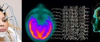
Normal bioelectrical activity of the brain (BEA) may be subject to changes caused by previous illnesses or injuries. These changes may appear localized in a particular area of the brain. Or they can be diffuse in nature - that is, spread to the entire brain as a whole without clearly identifying the source of the change, disrupting the passage of electrical impulses more or less evenly in all areas of the brain. In this case, they talk about disorganization of the bioelectrical activity of the brain. But in order to record diffuse changes in the bioelectrical activity of the brain, confirmation of a number of characteristic symptoms and specific electroencephalogram (EEG) indicators is necessary.
Symptoms and diagnosis of diffuse changes
It is believed that the bioelectrical activity of the brain is disorganized if external signs appear, reflected in the behavior and reaction of the patient, and also if these changes are confirmed or preceded by hardware diagnostics. Often, the bioelectrical activity of the brain is first tested using a hardware method, after which suspicions arise, and only then do patients pay attention to behavioral and cognitive symptoms:
- sudden changes in mood from good to bad - and vice versa,
- decreased self-esteem,
- loss of interest in previous hobbies,
- slower performance of usual work,
- rapid onset of fatigue when performing even basic actions.
In general, the history of cerebral changes in BEA is also typical for other diseases of the central nervous system. A person describes his condition as a general malaise and may not correlate the symptoms with the first signs of diffuse changes in BEA (especially if the symptoms listed above are accompanied by dizziness and headaches, “jumping” blood pressure). Sometimes such changes are accompanied by signs of dysfunction of the diencephalic-trunk structures, which also manifests itself in complaints of poor health.
If diffuse changes are significant, and if a significant decrease in the threshold of convulsive readiness is recorded, then it is considered that the person is predisposed to epilepsy.
EEG
Common causes of changes - atherosclerosis, encephalitis, meningitis, toxic brain damage - are usually reflected in tissue necrosis, inflammation, edema, and scarring. And these pathologies, in turn, are recorded using EEG. In case of a general cerebral lesion, three types of pathological processes are recorded on the EEG, the most significant of which is considered the first, but the diagnosis is made in the presence of all three signs of the pathological process, namely:
- polymorphic polyrhythmic (multiplicity of rhythms) activity in the absence of regular dominant bioelectrical activity,
- disruption of the normal organization of the electroencephalogram, which is expressed in irregular asymmetry with simultaneous disturbances in the distribution of basic EEG rhythms, phase coincidence of waves in symmetrical parts of the brain, amplitude relationships,
- diffuse pathological oscillations (alpha, delta, theta, exceeding normal amplitudes).
Often the EEG is dominated by signs of a symptom complex that appears with lesions of the hypothalamus and pituitary gland (diencephalic syndrome). Decoding the EEG readings does not allow us to see the reason for the appearance of abnormal data. A slight malfunction in the BEA when diagnosed using EEG can also be recorded in a healthy person.
Examples of EEG conclusions:
- “Significant diffuse changes in the BEA of the brain associated with dysfunction of the midline structures. Reducing the threshold of convulsive readiness. The focus of pathological activity, including paroxysmal activity, is in the right frontotemporal region.”
This means that there is a predisposition to epilepsy and seizures. There are lesions in the cerebral cortex that exhibit increased BEA, which can lead to various types of epileptic seizures.
- “The BEA of the brain is somewhat disorganized. During hyperventilation, flashes of pointed theta and alpha waves, deformed single complexes in the frontal leads of the “sharp-slow wave” type are recorded. No pronounced interhemispheric asymmetry was recorded.”
This result, together with the results of the REG, which indicate a continued decrease in pulse blood filling during functional tests, reveals signs of circulatory disorders in the brain.
- “Alpha rhythm over both hemispheres. Amplitude – up to 101 µV on the right and up to 99 µV on the left. Maximum – 57 µV on the right and 54 µV on the left. The dominant frequency is 9.6 Hz with dominance of the alpha rhythm in the occipital leads. Slow theta waves over both hemispheres. In the anterior frontal region – 53 µV, in the frontal – 56 µV, in the parietal – 88 µV, in the central – 81 µV, in the posterior temporal – 55 µV. Signs of a moderate stage of irritation of the midline structures of the brain and cortex. No paroxysmal activity or stable interhemispheric asymmetry was recorded.”
Interpretation: irritation of the cerebral cortex (irritation) may indicate a dysfunction of the cortex; such a change in EEG data is characteristic of circulatory disorders in different parts of the brain. In such a situation, it is advisable to personally consult a neurologist.
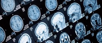
To clarify and identify catalysts for deviation, magnetic resonance imaging (MRI) is used.
Magnetic resonance imaging
When bioelectrical activity is disorganized, the causes of deviations exist, even if they are not immediately obvious. MRI helps identify them. Vascular atherosclerosis is detected by angiography. Tomography demonstrates irritative changes caused by a tumor and helps to establish the nature of the neoplasm.
Alpha Rhythm
HE. Kirillovskikh, V.S. Myakotnykh, E.V. Sorokova, K.V. Myakotnykh, T.A. Borovkova Ural State Medical Academy, 620905 Ekaterinburg, st. Soboleva, 25
89 elderly and senile patients with various forms of epilepsy . The general characteristic features of the bioelectrical activity of the brain of elderly patients suffering from epilepsy were determined, and the features of the electroencephalographic picture were identified depending on the form of the disease, the age of its onset, and the severity of cerebrovascular pathology. It has been shown that dynamic EEG monitoring helps in selecting adequate antiepileptic therapy and avoids aggravation of cognitive deficit.
Key words: epilepsy, elderly and senile age, EEG, cerebrovascular pathology, cognitive deficit.
Relevance of the problem.
In recent years, the problem of epilepsy in the elderly and senile, the differential diagnosis of epileptic seizures and non-epileptic paroxysmal conditions of other origins, as well as the possibilities of treating epilepsy in the elderly suffering from multiple pathologies has received increasing attention [1,3,8]. It is believed that epilepsy is a disease that develops on the basis of a genetically determined predisposition of the brain in combination with exogenous factors that determine its actualization in the clinical form [4]. It has been proven that this genetically determined phenotypic precondition for the development of epilepsy in children and adolescents is high-amplitude synchronization of neuron activity in all frequency ranges. But in the process of natural age-related transformation of the bioelectrical activity of the brain, the main cortical rhythms slow down, primarily the alpha rhythm , which acts as a biological clock during aging. In addition, there is a decrease in the overall amplitude level of the EEG and an increase in the proportion of beta activity due to an increase in nonspecific activating influences of the mid-brain structures [4,7]. Also, with age, the number of slow waves (SW) increases, predominantly in the theta range, arising both regionally, mainly in the frontotemporal regions, and in the form of a generalized slowdown of the main cortical rhythms. However, despite the ongoing age-related changes in the bioelectrical activity of the brain and the entrenched opinion that epilepsy is a disease predominantly of young people, the incidence of epilepsy in older age groups continues to increase [7]. Therefore, we found it interesting to determine the characteristics of bioelectrical activity both in elderly patients with a long history of epilepsy and in patients with the onset of epilepsy over the age of 60 years.
Purpose of the study
Using electroencephalographic (EEG) studies, determine the characteristics of the bioelectrical activity of the brain in epilepsy in elderly and senile people.
Materials and methods.
For 5 years, from 2006 to 2010. A prospective study of the clinical and neurophysiological features of epilepsy and the possibilities of its treatment was conducted in 89 patients aged 63-96 years (m=75.5±6.87 years). The diagnosis of epilepsy in all cases was established based on the observation of at least two unprovoked epileptic seizures . The exclusion criterion was epileptic syndrome due to a brain tumor, Alzheimer's disease. The control group was represented by 30 patients aged 65-85 years (m=75±5.66 years), who did not suffer from epilepsy, but had a similar range of concomitant pathologies, mainly cardiovascular. Depending on the time of onset of epilepsy, all patients of the main observation group were divided into two compared groups: 1st - patients with the onset of epilepsy in old age, mainly of post-stroke origin (n=34); 2nd - patients with a long history of epilepsy with its onset before the age of 60 (n=55). Separately, from the representatives of group 2, subgroup 2A was identified - 18 elderly patients, over 80 years old, who suffered severe traumatic brain injury (TBI) during the Great Patriotic War and suffered from post-traumatic epilepsy. All patients in the study groups underwent EEG studies at intervals of 4-6 per year. We used EEG registration with visual assessment and calculation of indices for standard frequency ranges on a computer electroencephalograph "Encephalan-131-01" (Russia, Taganrog). In the process of selecting the optimal combination of antiepileptic drugs, EEG was recorded over time 5, 14 and 30 days after changing the treatment regimen. In the absence of epileptiform activity on routine EEG or in case of questionable results, patients underwent EEG with sleep deprivation, daytime ambulatory EEG monitoring, EEG sleep monitoring using the Nicolet-one device. When conducting an EEG with partial sleep deprivation, which is better tolerated by the elderly, the patient was woken up at 4 a.m. the day before and an EEG recording was performed at 9 a.m. Recording was carried out with bipolar and monopolar installation of electrodes using 18-21 standard leads according to the 10-20 scheme; the following functional tests were used: opening and closing the eyes, rhythmic photostimulation with a frequency of 3, 5, 10 and 15 Hz, a block with a continuous increase in frequency photostimulation from 3 to 27 Hz and hyperventilation for 3 minutes. The EEG results were assessed using modern interpretation methods - computer processing with calculation of the index and amplitude of the main EEG rhythms, but the main method of EEG assessment was visual. The primary assessment of the EEG variant was carried out using the classification of E.A. Zhirmunskaya [2], a more detailed description of pathological changes - using the Classification of the American Association of Neurophysiologists [6].
Results and its discussion.
A study of the EEG of patients in both selected groups revealed some common features that distinguish the bioelectrical activity of the brain of elderly and senile patients with epilepsy from that of people of the same age, but not suffering from epileptic seizures (Table 1). This is, first of all, a higher amplitude level of the main EEG rhythms: if in the comparison group the EEG amplitude, as a rule, did not exceed 60 μV, then in patients with epilepsy the average amplitude level was twice as high, amounting to 120-150 μV. No significant differences were found between representatives of the main group and the control group only in the frequency of occurrence of interhemispheric asymmetry, bursts of beta activity and diffuse epileptiform activity. For other parameters, the differences identified are clear and significant. There is also a noticeable tendency towards synchronization of the main bioelectrical activity due to dysfunction of the mid-stem structures of the brain. Thus, when studying routine EEG, 38 (42.7%) patients showed a high-amplitude EEG variant for a given age with a tendency to synchronize the main cortical rhythms, in contrast to 2 (6.7%) patients in the control group (R
Table 1. Comparative characteristics of the main EEG variants and types of pathological activity.
| Main characteristics of EEG | Main group (n=89) | Comparison group (n=30) | |
| Hypersynchronous high-amplitude variant (sharp-looking) | 38 (42,7%) | 2 (6,7%) | |
| Desynchronous low-amplitude variant | 0 | 14 (46,7%) | |
| Disorganized hypersynchronous variant | 50 (56,2%) | 2 (6,7%) | |
| Disorganized desynchronous variant | 1 (1,1%) | 12 (40%) | |
| Interhemispheric asymmetry | 28 (31,5%) | 4 (13,3%) | |
| Increase in beta index > 40% | 7 (7,9%) | 18 (60%) | |
| Flashes of beta activity (excessive fast) | 15 (16,9%) | 3 (10%) | |
| Slowing down underlying background activity | I degree | 24 (27%) | 4 (13,3%) |
| II degree | 8 (9%) | 0 | |
| III degree | 3 (3,4%) | 0 | |
| Periodic regional slowdown | in the frontal leads | 23 (25,8%) | 5 (16,7%) |
| in the temporal leads | 41 (46,1%) | 0 | |
| Focal epileptiform activity | while awake | 36 (40,4%) | 0 |
| in a state of sleep | 14 (15,7%) | 0 | |
| Diffuse epileptiform activity | 5 (5,6%) | 0 | |
| Slow wave activity index (%) | 39,5±6,5 | 29,9±3,1 | |
Against the background of disorganization of background bioelectrical activity, hypersynchronization of the main cortical rhythms was also observed in 50 (56.2%) patients of the main group; in the control group there were only 2 (6.7%) observations of this kind; R
Among patients with epilepsy, the average indicators of the slow wave activity index reached 39.5±6.5%, in the control group - 29.9±3.1% (P
In the group of patients with epilepsy, in 24 (27%) cases, a slowdown in the main activity of the first degree (7 Hz and below) was noted, in the control group - only in 4 (13.3%) cases. Deceleration of the main activity of the second degree (6 Hz and below) was also significantly more often (P = 0.009) noted among people suffering from epilepsy (Table 1). This is consistent with the fact that a slowdown in basal activity compared to the age norm is always a sign of serious brain pathology [6]. Of course, in elderly patients, the slowdown in the main activity of the first degree can be considered a conditionally normal phenomenon, since after 60 years there is a gradual physiological decrease in the frequency of the alpha rhythm by approximately 1 Hz every 10 years [2,3,4,5]. Deceleration of the main activity of the II-III degree in elderly patients is a marker of severe cortical atrophy of the brain [6].
In the group of patients with epilepsy, almost all types of paroxysmal activity were epileptiform in nature; in 64 (71.9%) patients, high-amplitude pointed waves of the theta and delta range were more often localized in the frontal and temporal regions and were the most common type of conditioned epileptiform activity. Complexes “sharp-slow wave” and “spike-slow wave”, classified as true epileptiform activity, were less common - in 36 (40.4%) patients and were detected in a state of wakefulness. In 14 (15.7%) patients, epileptiform activity was detected only during sleep during EEG monitoring .
Thus, on the EEG of elderly patients with epilepsy, in addition to epileptiform activity itself, the following phenomena are more common: a) hypersynchronous alpha activity with an amplitude of over 100 μV, sharp-looking in shape - “sharp-looking alpha waves”; b) hypersynchronous beta activity (excessive fast) - beta activity with an amplitude of over 30 μV, often in the form of spindles extending beyond the normal fronto-central localization; c) periodic regional slowing of the main activity in the form of bursts of bilaterally synchronous or local high-amplitude pointed theta and delta waves; d) an increase in the average EEG amplitude and an increase in power during computer analysis in all spectral ranges; e) increase in the index of slow wave activity by more than 30%. The presence of these signs allows, even in the absence of true epileptiform activity, to draw a conclusion about excessive epileptic synchronization of brain neurons, and, therefore, confirm the diagnosis of epilepsy if the history and clinical picture are consistent.
A comparative analysis of the nature of EEG changes in various selected groups and subgroups of patients with epilepsy revealed the following changes (Table 2).
Table 2. Comparative characteristics of the main variants of diffuse changes in the EEG and types of pathological activity in patients of the main study group and the control group.
| Main characteristics of EEG | Main group (n=89) | |||
| Group 1 (n=34) | Group 2 without subgroup 2A (n=37) | 2A subgroup (n=18) | ||
| Disorganized hypersynchronous variant | 33 (97,1%) | 37 (100%) | 18 (100%) | |
| Desynchronous low-amplitude variant | 1 (2,9%) | 0 | 0 | |
| Interhemispheric asymmetry | 20 (58,8%) | 5 (13,5%)* | 3 (16,7%) | |
| Flashes of beta activity (excensive fast) | 4 (11,8%) | 6 (16,2%) | 5 (27,8%) | |
| Slow down main activity in background recording | I degree | 11 (32,4%) | 7 (18,9%) | 6 (33,3%) |
| II degree | 2 (5,9%) | 0 | 6 (33,3%) | |
| III degree | 0 | 0 | 3 (16,7%) | |
| Periodic regional slowdown | in the frontal leads | 8 (23,5%) | 9 (24,3%) | 0 |
| in the temporal leads | 15 (44,1%) | 21 (56,8%) | 5 (27,8%) | |
| Focal epileptiform activity | wakefulness | 16 (47,1%) | 15 (40,5%) | 5 (27,8%) |
| dream | 6 (17,6%) | 8 (21,6%) | 0 | |
| Average amplitude (µV) | 98±6,7 | 116±4,8 | 93±4.9 | |
| Slow wave activity index (%) | 39,4±4,4* | 35,4±4,34* | 48,4±4,59 | |
Note: * - p
All patients in the study groups with different types of epilepsy had statistically significant differences (p
Amplitude and frequency interhemispheric asymmetry significantly prevailed in the group of patients with late onset of epilepsy - in 20 (58.8%) patients (p
Rice. 1. Regional epileptiform activity “acute-slow wave” in the right temporo-parietal region in patient V., 72 years old, against the background of pronounced interhemispheric asymmetry and relative preservation of the background EEG in the intact left hemisphere of the brain. Recording against the background of sleep deprivation.
On routine EEG, in 16 (47.1%) patients with late onset of epilepsy , a lateralized focus of slow-wave activity was detected in the form of pointed delta waves with a frequency of 1.5-3 Hz, “sharp-slow wave” complexes, and less often “spike-waves”. slow wave”, in 6 (17.6%) patients epileptiform activity was detected on EEG sleep monitoring, in 2 (5.9%) diffuse (bilateral) epileptiform activity was detected. The preference for detecting epileptiform changes on the EEG and localizing post-stroke scar changes coincided in 70.6% (P = 0.016). In 10 (29.4%) patients, no focus of epileptiform activity was detected on routine EEG; These were mainly patients with small, less than 15 mm, foci of softening that formed as a result of a stroke.
EEG recording during sleep was possible only in 6 patients, since insomnia is a common concomitant symptom in this category of patients. The EEG picture of slow-wave sleep in representatives of group 1 was characterized by disorganization with prolongation of the first phase of slow-wave sleep. At the same time, sleep was superficial, with frequent awakenings for 8-12 seconds and motion artifacts; Specific sleep patterns - K-complexes, vertex potentials and sleep spindles - were not clearly expressed, delta sleep was shortened, and the amplitude of delta activity was reduced. The “rapid eye movement” stage was not recorded in any patient of group 1, which largely reflects the dysfunction of central somnogenic mechanisms in patients with cerebral vascular pathology. Against a disorganized background, in all 6 patients, mainly in the second stage of slow-wave sleep, focal epileptiform activity of the “sharp-slow wave” type was recorded, localized in the frontotemporal leads, on the side of the post-stroke focus of softening (Fig. 2).
Fig 2. EEG of patient M., 73 years old, diagnosed with: Consequences of ischemic stroke in the left internal carotid artery; symptomatic epilepsy, simple partial seizures with secondary generalization. In stages I-II of slow-wave sleep - epileptiform complexes “acute-slow wave” in the left frontotemporal region.
Thus, analyzing the structure of bioelectrical activity in patients with late onset of epilepsy, it can be assumed that initially, before the stroke, patients had a tendency of cortical neurons to paroxysmal forms of response in the form of a tendency to high-amplitude synchronization. However, these changes remained latent throughout life and did not lead to clinical manifestations of epilepsy . Ischemic stroke with localization of cerebral infarction in the cortical and cortical-subcortical areas was the triggering moment that led to the formation of epileptiform activity and the clinical manifestation of epilepsy.
When studying the EEG in patients with a long history of epilepsy (2nd group of observations), a significantly higher amplitude level of the EEG was observed (rEEG sleep monitoring (Fig. 3). The index of slow wave activity in patients of the 1st group, with early onset of epilepsy, significantly higher than in the control group, but lower than in patients of group 1 - with late onset of epilepsy (p
Rice. 3. EEG of patient K., 77 years old, diagnosed with: Symptomatic epilepsy, complex partial seizures with secondary generalization. In the second phase of slow-wave sleep there is a short discharge of diffuse epileptiform activity “spike-slow wave” with initiation in the left frontotemporal region and the phenomenon of secondary bilateral synchronization.
Thus, the bioelectrical activity in the group of patients with an early onset of epilepsy and a long history of it was distinguished by signs characteristic of the bioelectrical activity of young patients suffering from epilepsy, but in elderly patients these pathological signs were less common. Aging processes in the bioelectrical activity of the brain of patients with epilepsy are manifested, first of all, by an increase in the slow-wave activity index, a slight decrease in the overall amplitude level of the EEG and a decrease in the tendency to generalize epileptiform discharges, which is clinically reflected in a decrease in the proportion of secondary generalized epileptic seizures in elderly patients. It can be assumed that the genetically determined characteristics of the bioelectrical activity of the brain, which are the cause of the clinical manifestation of epilepsy, persist throughout life.
The characteristic features of the EEG in the subgroup of elderly patients (over 80 years old) with a history of combat TBI are due to the age of the patients and pronounced atrophic changes in the cerebral cortex. The main distinguishing feature of the EEG of representatives of this subgroup 2A was an increase in the index of slow wave activity (48.4±4.59), which is significantly higher (P
Rice. 4. EEG of patient Ch., 84 years old, with a diagnosis of Encephalopathy of complex origin (post-traumatic, cerebrovascular), severe cognitive and static-coordinating impairments. Symptomatic epilepsy, complex partial seizures of moderate frequency. The EEG shows diffuse slow-wave activity in the form of pointed high-amplitude theta-delta waves.
Regional epileptiform activity in elderly people was detected in a small percentage of cases - in 5 (27.8%) out of 18, however, the frequency of detection of regional epileptiform activity was higher than in the comparison group (P
Slow-wave activity in the EEG of patients in group 2A no longer reflects the degree of epileptiform activity, but rather the level of pathological morphological and functional changes in neurons due to traumatic and vascular factors. The index of slow wave activity in these patients turned out to be directly proportional to the degree of atherosclerotic lesions of cerebral vessels and inversely proportional to the number of points scored in the study of cognitive functions using the well-known MMSE scale. It is interesting that the index of slow wave activity is not a static value; it can decrease after a course of vascular therapy and, conversely, increase with the use of some anticonvulsants, primarily barbiturates. This is confirmed by EEG analysis in 3 (16.7%) representatives of the 2A subgroup who have been taking barbiturates for more than 60 years; In all cases, the EEG showed gross cerebral changes of an organic type, continued slow-wave activity of varying amplitude and degree of synchronization, but true epileptiform activity was not detected in these patients (Fig. 5).
Rice. 5. EEG of patient M., 85 years old, diagnosed with vascular atrophic disease of the brain, dementia, symptomatic post-traumatic epilepsy, rare generalized seizures. Constant intake of benzonal at a dose of 100 mg 3 times a day + phenobarbital 100 mg at night for 50 years. The EEG shows diffuse slow-wave activity with synchronization in the fronto-central regions. Characterized by a complete absence of physiological EEG rhythms. True epileptiform activity is not detected.
Thus, an increase in the index of slow-wave activity on the EEG during treatment with antiepileptic drugs is an unfavorable prognostic sign of worsening cognitive deficits, which indicates the need to replace the drug with a more modern one or a drug of a different group.
Conclusions.
- The frequency of detecting distinct epileptiform activity on the EEG in elderly and senile patients suffering from epilepsy is 40.4%, conditional epileptiform activity is even higher, up to 72%. In some cases, epileptiform activity can be detected during ambulatory EEG sleep monitoring. There are individual differences in EEG characteristics depending on the etiology of epilepsy, duration of the disease, and age of the patients.
- There are the following main distinctive neurophysiological features of the EEG of elderly and senile patients with epilepsy:
- an increase in the spectral power of the EEG in all frequency ranges – a high-amplitude version of the EEG;
- strengthening the synchronizing influences of mid-stem structures and, as a consequence, smoothing out zonal differences;
- decrease in the proportion of beta activity;
- an increase in the index of slow wave activity to 30% or higher, which includes both a slowdown of the main cortical rhythms in the background recording and periodic regional slowdown in the form of bilaterally synchronous bursts of theta-delta waves;
- Regional epileptiform activity is detected in the waking state in approximately 40%, and in 16% during EEG sleep monitoring.
Literature.
- Gekht, A.B., Burd G.S., Selikhova M.V. Clinical and neurophysiological features of motor disorders in patients with post-stroke epilepsy // Journal of Neurology and Psychiatry. – 1998. – No. 7. – P. 4–8.
- Zhirmunskaya, E.A. In search of an explanation of EEG phenomena. - M.: NFP Biola, 1996. - 117 p.
- Zenkov L.R., Elkin M.N., Medvedev G.A. Clinical neurophysiology of neurogeriatric disorders // Advances in neurogeriatrics. – M., 1995. – P. 167-173.
- Zenkov, L.R. Clinical epileptology: 2nd ed., revised. and additional - M.: Medical Information Agency, 2010.- P. 123–129.
- Zenkov, L.R. Non-paroxysmal epileptic disorders. – M.: Medpress-inform, 2007. – 278 p.
- Mukhin K.Yu., Petrukhin A.S., Glukhova L.Yu. Epilepsy: Atlas of electroclinical diagnostics. – M.: Alvarez Publishing, 2004. – 440 p.
- Myakotnykh V.S., Galperina E.E. Electroencephalographic features in elderly and senile people with various cerebral pathologies: Educational and methodological recommendations. – Ekaterinburg: Publishing house. UGMA. – 30 s.
- Blalock, E.M., Chen K.C., Sharrow K. Gene microarrays in hippocampal aging: statistical profiling identifies novel processes correlated with cognitive impairment // J. Neurosci. – 2003.- Vol. 23. – R. 3807–3819.
- Carlson CE, St Louis ED, Granner MA Yield of video EEG monitoring in patients over the age of 50 // Program and abstract of the American Epilepsy Society 58th Annual Meeting. – 2004, 3 – 7 December. – Absract 2.- R. 226.
- Van Cott, AC Epilepsy and EEG in the Elderly // Epilepsia. –2002. –Vol. 43, suppl. 3. – P. 94–102.
Causes and consequences of changes
General cerebral changes in the bioelectrical activity of the brain can be caused by the following factors:
- Chemical toxic and radiation damage to the brain. Toxic poisonings that lead to disorganization are most often irreversible, affecting a person's ability to carry out daily activities. Such forms of lesions provoke severe forms of diffuse changes in the bioelectrical activity of the brain.
Head injuries and concussions. Here, the intensity of the changes depends on the severity of the damage caused: the stronger the damage, the more noticeable the result. With minor and moderate diffuse changes in the bioelectrical activity of the brain, the body feels slight discomfort, and impulse conductivity is restored without long-term treatment.- Inflammatory processes (including those caused by viral infection). Inflammations associated with meningitis and encephalitis are characterized by mild cerebral changes in BEA.
- Atherosclerotic vascular problems. The condition depends on the degree of vascular damage. The initial stage is characterized by mild diffuse changes in the bioelectrical activity of the brain. But with an increase in the area of vascular damage and tissue death, neuronal conduction disorders progress.
- Associated disorders. These include manifestations of regulatory pathologies. Cases associated with damage to the hypothalamus and pituitary gland are common. Changes can also be caused by incorrect functioning of the immune system.
Rough pronounced diffuse changes in the bioelectrical activity of the brain, as a rule, are the result of scarring, necrotic transformations, expansion of inflammatory processes and cerebral edema. Such signal conduction disturbances are heterogeneous, and instability of the BEA in complex cases is always accompanied by pathologies of the pituitary gland and hypothalamus.
Moderate changes in the bioelectrical activity of the brain are dangerous due to their complications. Subsequent stages, with softening or hardening of the brain tissue and the appearance of neoplasms, are transformed into cancer, diffuse sclerosis and other irreversible processes. To prevent them, treatment should be carried out at the stage of detecting slight changes in the bioelectrical activity of the brain. Moderate treatment and an attempt to reduce the consequences of the last stage of pathological changes are carried out only in specialized medical institutions.
Infectious diseases of the central nervous system
There are a fairly large number of agents that can cause damage to the central nervous system. The main ones are the Coxsackie virus, herpes infection, ECHO. They can provoke the occurrence of meningitis, arachnoiditis, and encephalitis. The central nervous system can also be affected by HIV infection, when the disease is in its final stage. It most often manifests itself in the form of abscesses and leukoencephalopathies.
Mental disorders provoked by infectious pathology can manifest themselves as follows:
— Asthenic syndrome : weakness, fatigue, significant decrease in performance. — Complete or partial psychological disorganization. — Affective disorders. — Violations of personality integrity. — Psychoses: paranoid, hypochondriacal, hysterical. — Intoxication
Intoxication can be caused by uncontrolled use of drugs, alcohol, nicotine, as well as poisoning by mushrooms, salts of heavy metals, and carbon monoxide. Poisoning is also possible with an overdose of drugs. Clinical consequences depend on the specific substance used. The development of neurosis-like disorders is also possible.
If poisoning occurs due to the use of diphenhydramine, atropine, and various antidepressants, it manifests itself as delirium. If a psychostimulant was used, intoxication paranoid is possible. Characteristic for this type of intoxication are visual, auditory and tactile hallucinations and delusions. A manic-like state is also possible, which is manifested by euphoria, sexual and motor disinhibition, and accelerated thinking processes.
If intoxication is chronic, the patient will encounter the following manifestations:
— Exhaustion, lethargy, hypochondria, a noticeable decrease in performance, various types of depressive disorders. — Memory impairment, decreased intelligence level, attention impairment. — Vascular diseases
Vascular diseases of the brain include:
- Hemorrhagic stroke. - Ischemic stroke. - Encephalopathy.
Hemorrhagic stroke occurs due to blood seeping through the walls of blood vessels or a rupture of an aneurysm. As a result, hematomas are formed. Ischemic stroke occurs due to blockage of a vessel by a blood clot, an atherosclerotic plaque. As a result, a lesion is formed that is deprived of a sufficient amount of oxygen and nutrients.
Discirculatory encephalopathy can develop with hypoxia, which is chronic. At the same time, a large number of lesions are formed throughout the brain. reasons that provoke the development of tumors in the brain :
— Genetic predisposition. — Exposure to chemicals. — Ionizing radiation.
Today, doctors are actively discussing the likelihood of the negative impact of bruises, cell phones and various injuries to the head.
If vascular pathology , various mental disorders may develop. As a rule, they directly depend on the location of the outbreak. As practice shows, they most often occur when the right hemisphere is affected.
They can appear in the form of:
— Cognitive impairment (to disguise this impairment, patients use notebooks where they record all the necessary information). — Significant reduction in the level of criticism of one’s own condition. - Prolonged depression. - Sleep disorders. — Manifestation of aggressive behavior. - Asthenic syndrome. — Vascular dementia
Separately, it is worth considering vascular dementia . Today it is divided into several types:
- Stroke-related. - Non-stroke. — Variants caused by disturbances
Patients with the pathologies described above have rigidity of all or most processes and their lability. There is also a significant decrease in the range of interests. The severity of cognitive impairment is determined by a number of characteristics, which may even include the age of the patient.
Demyelinating disease
One of the main diseases is multiple sclerosis. The lesions are formed with the destruction of the membranes of the nerve endings.
Mental disorders may include the following:
— Increased levels of fatigue, weakness, significant decrease in performance. — There is a decrease in intelligence, memory, and absent-minded attention. - Affective insanity. - Depression. — Neurodegenerative diseases
Alzheimer's disease and Parkinson's disease may fall into this category . Symptoms usually appear in old age.
Depression is the most common condition in Parkinson's disease. It is accompanied by a deep feeling of emptiness, emotional poverty. A person’s ability to feel joy and enjoyment also decreases. A person may behave overly aggressively, become sad, or show unjustified pessimism. Depression can be supplemented by various anxiety disorders (in 70% of patients).
Alzheimer's disease is a degenerative disease that causes marked decline in cognitive function, as well as changes in behavior and personality structure. People with Alzheimer's disease are unable to recognize familiar objects, familiar people, and are forgetful and confused. They quickly fall into a state of depression, experience disorientation, anxiety, and emotional distress.
Prevention of increased diffuse changes in BEA
Some of the causes of cerebral changes in BEA are uncontrolled (injuries, poisonings, radiation). However, several causes are relatively easy to eliminate through preventive measures.
Since one of the most common causes of diffuse changes is vascular atherosclerosis, a preventive and therapeutic measure here will be correction of lifestyle, nutrition and the use of drugs that:
- improve the condition of the walls of large and small vessels, maintaining their elasticity,
- reduce the degree of red blood cell aggregation,
- eliminate cholesterol deposits and other lipid accumulations,
- prevent the proliferation of fibrous fibers,
- improve endothelial function.
The most popular preventive and therapeutic agents include herbal preparations HeadBooster, Optimentis with a nootropic effect of enhancing the performance and cognitive functions of the brain. Their popularity is explained by several factors, including a gentle, gentle effect on the vascular system of the brain and the presence of the relict plant Ginkgo Biloba in the extract. The effect of the drugs appears gradually, so they should be taken in courses. However, even in this case, one must adhere to the recommendations for a course of treatment, since an overdose of organic substances present in the Ginkgo extract increases the risk of developing strokes. When taken correctly, these drugs:
- reduce the permeability of the vascular wall, helping to strengthen it,
- normalize cholesterol levels,
- trigger antioxidant processes, preventing the destructive effects of free radicals on membranes,
- provide nutrition to brain cells by normalizing the transport of glucose and oxygen to tissues,
- facilitate the passage of impulses along nerve fibers.
In addition, in the drug treatment of atherosclerosis preceding diffuse changes, the following drugs are used:
- Nicotinic acid (its derivatives). Preparations based on it reduce cholesterol and triglycerides and increase the concentration of lipoproteins. All this improves antiatherogenic properties, but introduces a ban on these drugs for people with liver diseases.
- Fibrates. Miscleron, Gevilan, Atromide inhibit the synthesis of the body's own fats, but are fraught with side effects associated with the functioning of the liver and gall bladder.
- Bile acid sequestrants remove acids from the intestines, thereby reducing the amount of fat in cells, but can cause flatulence or constipation.
- Statins. They reduce the production of cholesterol by the body itself, which determines the time of taking them “at night,” when cholesterol synthesis increases. But their action can also destabilize the liver.
Types of lesions on MRI of the head
The color of the resulting image of normal brain structures and pathological changes depends on the program used. When scanning in angio mode, including using contrast, a branched network of arteries and veins appears on the images. Focal changes are of several types; based on their characteristics, the doctor can guess the nature of the foci.
With pathology of the medulla, the properties of the affected foci are disrupted, which is manifested by a sharp change in the MR signal compared to healthy areas. The use of certain sequences (diffusion-weighted, FLAIR, etc.) or contrast allows for more clear visualization of local changes. That is, if a radiologist sees a single lesion on the MRI results, different scanning modes or contrast will be used to study it in more detail.
When comparing changes with healthy areas of the brain, hyper-, hypo- and isointense zones are identified (bright, dark, and the same color as adjacent structures, respectively).
Brain abscess on MRI (indicated by arrow)
Hyperintense lesions
Identification of hyperintense, i.e. foci that clearly stand out on MR scans makes the specialist suspect a brain tumor, including of metastatic origin, hematoma (at a certain moment from the onset of hemorrhage), ischemia, edema, vascular pathologies (cavernomas, arteriovenous malformations, etc.), abscesses , metabolic disorders, etc.
Brain tumor on MRI (indicated by arrow)
Subcortical lesions
Damage to the white matter of the brain is usually characterized as changes in subcortical structures. Subcortical lesions identified by MRI indicate that the damage is localized just under the cortex. If multiple juxtacortical lesions are detected, it makes sense to suspect a demyelinating process (eg, multiple sclerosis). With this pathology, destructive changes occur in various areas of the white matter, including directly under the cerebral cortex. Periventricular and lacunar lesions are usually detected during ischemic processes.
Foci of gliosis
When brain tissue is damaged, compensatory mechanisms are activated. Destroyed cells are replaced by glial structures. The latter ensures the transmission of nerve impulses and is involved in metabolic processes. Due to the structures described, the brain recovers from injuries.
Identification of glial foci indicates previous destruction of the cerebral substance due to:
- birth trauma;
- hypoxic processes;
- hereditary pathologies;
- hypertension;
- epilepsy;
- encephalitis;
- intoxication of the body;
- sclerotic changes, etc.
By the number and size of altered areas, one can judge the extent of brain damage. Dynamic observation allows you to assess the rate of progression of the pathology. However, by studying the zones of gliosis, it is impossible to accurately determine the cause of the destruction of nerve cells.
Foci of demyelination
Some diseases of the nervous system are accompanied by damage to the glial membrane of the long processes of neurons. As a result of pathological changes, the conduction of impulses is disrupted. This condition is accompanied by neurological symptoms of varying degrees of intensity. Demyelination of nerve fibers can be caused by:
- multifocal leukoencephalopathy;
- multiple sclerosis;
- dissimulating encephalomyelitis;
- Marburg's disease, Devic's disease and many others.
Typically, demyelination lesions appear as multiple small areas of hyperintense MR signal located in one or more parts of the brain. Based on the degree of their prevalence, duration and simultaneity of occurrence, the doctor judges the scale of development of the disease.
Focus of demyelination on MRI
Focus of vascular origin
Cerebrovascular insufficiency causes ischemia of the cerebral substance, which leads to changes in the structure and loss of function of the latter. Early diagnosis of vascular pathologies can prevent stroke. Focal changes of discirculatory origin are found in most patients over 50 years of age. Subsequently, such zones can cause degenerative processes in the brain tissue.
Lacunar cerebral infarction on MRI (indicated by arrow)
Cerebral circulation disorders can be suspected by focal changes in the perivascular Virchow-Robin spaces. The latter are small cavities around the cerebral vessels, filled with fluid, through which tissue trophism and immunoregulatory processes occur (blood-brain barrier). The appearance of a hyperintense MR signal indicates expansion of the perivascular spaces, since they are normally not visible.
Sometimes an MRI of the brain reveals multiple lesions in the frontal lobe or in the deep parts of the hemispheres, which may indicate damage to the cerebral vessels. The situation is often clarified by MR scanning in angio mode.
Foci of ischemia on MRI
Foci of ischemia
Cerebral circulation disorders lead to oxygen starvation of tissues, which can provoke necrosis (infarction). Ischemic lesions on T2-weighted sequences appear as areas with moderately hyperintense signal of irregular shape. At a later stage, when performing MRI in T2 VI or FLAIR mode, a single lesion takes on the appearance of a light spot, which indicates a worsening of destructive processes.
