Magnetic resonance imaging (MRI) is the main method of hardware diagnostics of neurological diseases. Computer-converted response signals from hydrogen atoms, which are found in all organs of the human body. In neurosurgery, MRI images are necessary to detect even the smallest pathological lesions. The images are taken as thin sections of brain structures.
In the diagnosis of brain diseases, MRI occupies a special place, as it diagnoses the problem at the earliest stage of development. Life-threatening pathologies include cerebral hydrocephalus. The doctor visualizes it on MRI images in the form of characteristic changes, the registration of which is available at the very beginning of the disease. A timely diagnosis makes it easier for the clinician to select the necessary therapeutic measures.
What is hydrocephalus?
Nutrition of brain structures is carried out through a direct current of cerebrospinal fluid. Liquor circulates along a certain channel, ensuring continuous cerebral metabolism. Cerebrospinal fluid is needed for:
- protecting brain tissue from mechanical stress;
- ensuring a constant level of electrolytes;
- maintaining the balance of acids and alkalis;
- complete immunobiological protection.
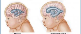
With hydrocephalus, there is a disturbance in the production, outflow and circulation of cerebrospinal fluid in the brain tissues. The disease is characterized by pathological accumulations of cerebrospinal fluid, which increases intracranial pressure. Clinically, hydrocephalus occurs in a severe form, with frequent attacks of the neurological type. A long-term pathological condition leads to the formation of secondary changes in brain tissue, which are usually irreversible. As a result of an uncontrolled course, hydrocephalus becomes the cause of death of the patient. Popularly, the disease has received a second name - dropsy of the brain.
Symptoms of hydrocephalus of the brain
- attacks of intense headache;
- surges in blood pressure against the background of complete well-being;
- attacks of nausea, vomiting;
- episodes of urinary incontinence at night or during the day;
- changes in mental state (development of dementia);
- unsteady, shaky gait.
In children in infancy, there is a rapid increase in head size and bilateral paralysis of the extraocular muscles.
At the beginning of the disease, there are no neurological symptoms, although pathological changes in the brain are already occurring. Observed:
- cerebrovascular disorders;
- reduction of certain parts of the brain;
- organic changes in the cerebral substance.
Hydrocephalus of idiopathic origin is extremely rare.
To find out the causes and nature of the disease, detailed diagnostics using hardware technologies is necessary. Magnetic resonance imaging provides the most complete information about the state of the brain. The diagnosis of hydrocephalus is most often made in children under 3 months of age. Among the adult population, pathology is registered in patients over 60 years of age.
A list of the most common causes of hydrocephalus has been determined:
- tumors of various origins;
- traumatic brain injuries;
- diseases of the vascular system;
- conditions after neurosurgical interventions;
- acute cerebrovascular accidents - strokes;
- hematomas;
- congenital pathologies of the central nervous system.
The treatment program is drawn up by the doctor. It depends on the type of disease and severity. Uncomplicated forms of cerebral hydrocele can be treated with medication. If conservative therapy has shown its failure, and the disease causes a number of severe complications, neurosurgical treatment is performed. As a rule, operations are performed on an emergency basis.
Sinuses
Sinuses are cavity formations, venous sacs that act as containers for venous blood and structures that reabsorb cerebrospinal fluid. These cavities are located between the layers of the dura mater. They receive venous blood from the external and internal veins of the brain.
Anatomy
Sinuses are anatomically similar to the structure of veins. However, the wall of the former, unlike the vessel, is stretched along its length by the wall of the hard shell. Due to the fact that the sinuses are attached to the membranes, their walls do not collapse and ensure a constant outflow of venous blood during various changes in intracranial pressure. This feature ensures uninterrupted functioning of the brain. Also, venous oblong sacs do not have valves.
Venous sinuses
The following venous sinuses of the brain are distinguished:
- Upper. It passes along the falciform process and ends at the level of the occipital protuberance, where it passes into the right sinus.
- Lower. If the previous structure ran along the upper edge of the falciform process, then this one ran along the lower edge. It opens into the straight sinus.
- Straight. Located between the cerebellum and the falx process.
- Transverse sinus of the brain. This cavity is a pair, and was located in the cranial groove of the same name.
- Occipital. Distributed around the foramen magnum. Later it becomes sigmoid.
- Cavernous. Also paired. It is located and surrounds the sella turcica - the place in which the pituitary gland lies. This sinus differs from others in that the internal carotid artery, abducens, oculomotor, ophthalmic and trochlear nerves pass through it.
- There are also intercavernous, wedge-shaped, superior petrosal and inferior petrosal sinuses.
Pathologies and diseases
Venous discirculation is a pathology characterized by impaired outflow of venous blood from the sinuses. The causes of the disease are as follows:
- traumatic brain injuries;
- fractures of the skull bones;
- strokes;
- tumors;
The actions of all these factors come down to one phenomenon - external compression of the walls of the venous sacs. Sooner or later, the patient will begin to be bothered by the following symptoms :
- Constant headaches, especially in the morning.
- Migraine that appears after minor irritants - stress, fatigue, lack of sleep.
- When rising, a person feels darkening in the eyes and dizziness.
- Noise in ears.
- Constant fatigue, asthenia, muscle weakness.
- Insomnia is a sleep disorder.
- Memory deterioration, general inhibition of mental processes.
- Paresthesia on the arms and legs (crawling “goosebumps”, numbness).
Thrombosis of the cerebral sinuses is a serious disease that is manifested by the presence of blood clots (thrombi) in the sinuses. As a result, local blood flow deteriorates. This disease most often appears after:
- past infectious diseases: otitis media, sinusitis, tonsillitis;
- acute bacterial conditions: tuberculosis.
- fungal infections;
- excessive use of hormonal drugs;
- systemic autoimmune diseases: lupus erythematosus, sarcoidosis.
This disease usually develops acutely – within a few days. In a minority of patients, symptoms peak at 30 days. Signs of thrombosis are:
- Severe headache, nausea and vomiting, dizziness, double vision.
- Local seizures.
- Sensory and motor dysfunction. These people may experience sudden numbness or lack of strength in their arm.
In the case when the development of thrombotic disease develops rapidly, septic thrombosis is formed, accompanied by sudden changes in body temperature, extreme sweating and various disturbances of consciousness - from mild delirium to complete loss of consciousness - coma.
Types of hydrocephalus
According to etiological factors, hydrocele of the brain is divided into congenital and acquired.
Based on the type of cerebrospinal fluid accumulation, the disease is divided into types:
- external;
- internal (mono-, bi-, tri-, tetraventricular);
- mixed.
According to the pathogenetic process, hydrocephalus is divided into:
- open - communicating;
- closed - occlusal.
Will an MRI show hydrocephalus?
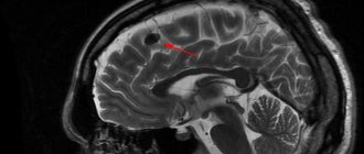
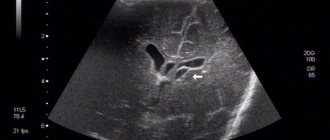
Magnetic resonance imaging shows hydrocephalus in its earliest stages. During the examination, layer-by-layer images of the lobes and parts of the brain are formed. The diagnostician examines all the images obtained, assesses the state of the brain matter, and identifies the most minor deviations from the norm.
MRI is the only diagnostic method that accurately determines the fact of hydrocephalus. The conclusion about the disease is established even in the absence of obvious neurological symptoms. Based on the images obtained, conclusions are drawn about the type of pathology and the presence of associated abnormalities. Signs of hydrocephalus in adults detected on MRI
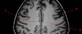
Signs of the disease can be direct or indirect. Direct - occur with hypertrophy of the cerebral ventricles - III, IV and lateral. In the initial stages of hydrocephalus, expansion is observed in areas of the anterior horns and body. Enlargements also appear in the area of the aqueduct and subarachnoid space (convexity, in the area of the basal cisterns, Sylvian fissures).
Indirect signs of hydrocephalus on MRI images include:
- increase in interventricular index more than 0.5;
- with tension hydrocephalus - periventricular edema;
- downward shift of the hypothalamus;
- local protrusion of the roof of the lateral ventricles.
A significant advantage of MRI over other types of diagnostics is the identification of the causes of cerebral hydrops.
Tanks
Brain cisterns are small hollow formations located between the arachnoid and pia mater and containing spinal cerebrospinal fluid. All tanks are connected to each other through various holes. These sacs also communicate with the fourth ventricle of the brain.
Anatomy
The anatomical features of the cisterns are that they completely repeat the surface relief of the telencephalon - the gyri and sulci. These formations are narrow and almost flat oblong passages. In some areas they expand and turn into full-fledged containers of cerebrospinal fluid.
Types of tanks
There are the following types of tanks:
- Cerebellar. This tank is the largest among all the others. It is located between the cerebellum and the medulla oblongata. The posterior wall of this cavity is limited by an arachnoid membrane.
- Basal. Represented in the form of a pentagon.
- Prepontinnaya. It lies in front of the bridge. The basilar artery passes through it, giving its branches to the cerebellum.
- Quadrigeminal cistern. It is located between the cerebellum and the corpus callosum.
- Bypass or enveloping cistern of the brain. This tank looks like a canal running along the sides of the cerebellar peduncles. It is subsequently connected to the previous cavity.