Types of hydrocephalus
There are several variants of the disease depending on the cause of occurrence and localization. By origin, there is a congenital form, which develops as a result of problems suffered by the baby in the prenatal state, and an acquired form, provoked after birth by external factors (injuries, infections).
According to the type of location, they are distinguished:
- external hydrocephalus, when in a child cerebrospinal fluid accumulates mainly in the space under the membranes of the brain;
- internal form, in which cerebrospinal fluid flows into the ventricles of the brain, causing them to expand;
- mixed or general hydrocephalus, which is characterized by a combination of external and internal forms of the disease.
There are also open (communicating) and closed (obstructive) types of pathology: in the first case, the communication between the ventricles and the subarachnoid space is not broken, in the second it is absent. According to the speed of flow, acute, subacute and chronic dropsy of the brain are distinguished.
How it manifests itself
The main function of the ventricles is to secrete cerebrospinal fluid, as well as ensure its normal circulation in the subarachnoid space. If the balance of exchange and production of cerebrospinal fluid is disturbed, then stagnation is formed and, as a result, the walls of the cavities are stretched. The same slight expansion of the lateral segments may be a normal variant, but their asymmetry and enlargement of individual parts (for example, only the horn) will be a sign of the development of pathology.
Enlarged ventricles of the brain in an infant can be diagnosed with a congenital disease such as ventriculomegaly. It varies in severity:
- Slight expansion of the ventricles of the brain up to 11-12 mm, with no significant symptoms. It manifests itself in the child’s behavior: he becomes more excitable and irritable.
- Increasing the depth of the ventricles up to 15 mm. Most often, the pathology is accompanied by asymmetry and impaired blood supply to the affected area, which entails the appearance of seizures, an increase in head size and a lag in mental and physical development.
- Ventricular dilatation up to 20 mm is characterized by irreversible changes in brain structures and is often accompanied by Down syndrome and cerebral palsy in infants.
In adulthood, an increase in ventricular volume is manifested by the following symptoms:
- Gait disturbance, with the child walking “on tiptoes” or vice versa, focusing only on the heels.
- The appearance of visual disorders, such as squint, insufficient focus of the gaze, as well as double images when trying to see small details.
- Tremor of arms and legs.
- Behavioral disorders that manifest themselves in excessive lethargy and drowsiness, while it is difficult to captivate the child with any activity.
- The appearance of headaches due to increased intracranial pressure, sometimes nausea and even vomiting may occur.
- Dizziness.
- Frequent regurgitation, loss of appetite. Some newborns are able to refuse breastfeeding.
Symptoms of hydrocephalus of the brain in a child
Classic signs of hydrocephalus in children over 1–3 years of age are symptoms of increased pressure on the brain, which include:
- intense headache;
- nausea and vomiting;
- spastic paresis, when muscles involuntarily tense and limb movements become sluggish;
- constant fatigue;
- urinary incontinence;
- gait disturbances;
- convulsions;
- strabismus;
- nystagmus;
- developmental delay.
Hydrocephalus, which occurs as a consequence of a brain injury or infection, can cause in an older child a deterioration in memory and attention, decreased visual acuity, irritability, dizziness and head pain that does not go away after taking painkillers.
The following symptoms help to suspect hydrocephalus in infants:
- bulging and pulsation of the fontanel;
- divergence of the sutures of the skull;
- deformation of the head with a predominance of the cerebral region over the facial region;
- rapid increase in head circumference with a significant deviation from the norm;
- increased excitability, moodiness;
- poor appetite;
- frequent volumetric regurgitation, similar to vomiting;
- exotropia;
- Graefe syndrome;
- a distinct pattern of saphenous veins on the head.
In some cases, an additional sign may be a slow heartbeat, tremor (trembling) of the chin and limbs, which persists for a long time.
The size of the ventricles is normal
A healthy newborn normally has 4 ventricles: two lateral, the third is conventionally anterior, and the fourth ventricular component, which is considered posterior. An enlargement of the lateral ventricles entails the production of a large amount of cerebrospinal fluid, which will not be able to circulate normally between the membranes of the brain and, accordingly, perform its functions of regulating metabolic processes. Therefore, when assessing the size of the ventricles of newborns, the following standards are used:
- the lateral anterior horns should fall within the range of 2-4 mm;
- lateral occipital horns - 10-15 mm;
- body of the lateral ventricles - no deeper than 4 mm;
- III ventricle - no more than 5 mm;
- IV - up to 4 mm.
When examining the brain of infants up to a year and older, the use of these standards will be incorrect, since the brain matter and ventricles will grow, so the assessment is carried out using other indicators and corresponding tables.
Reasons for the development of hydrocephalus in children
Factors that can lead to the internal form of the disease are:
- anomalies and malformations of the fetus during the period of intrauterine formation, associated with infections, injuries, and the influence of bad habits of the mother;
- various birth injuries;
- prematurity and premature birth with low body weight.
The causes of acquired hydrocephalus in a child can be infectious diseases of the brain and its structures: meningitis, malignant and benign neoplasms, encephalitis. In some cases, the starting mechanism is traumatic brain injuries and bruises resulting from falls, blows, accidents and other injuries.
Intracranial pressure
It is simply impossible to imagine a woman interested in children's health issues who has not heard of intracranial pressure - ICP.
Phrases like “we have intracranial” or “we treat intracranial pressure” have become so firmly entrenched in the vocabulary of the average visitor to a children's clinic that many have simply stopped thinking about the meaning of these words.
Nevertheless, the frequency of conversations, the frequency of diagnosis and the frequency of treatment do not at all indicate that the very concept of “intracranial pressure” or the diagnosis of “increased intracranial pressure”, in turn, is understandable to the broad masses of workers.
Although at first glance everything seems obvious. And the essence of the problem (from the point of view of the average person) looks something like this. There is a head. There is a brain inside, blood vessels, there is pressure in the blood vessels - everyone knows this - both grandmother and grandfather have pressure. But grandparents have damaged hearts, but for a child everything is different. The heart is healthy, but the pregnancy was unsuccessful, there was not enough oxygen during childbirth, or the umbilical cord got wrapped around, or some other illness happened, or he hit his head, or they gave him the wrong medicine - so the vessels were damaged, now the pressure in the head is high, hence the heap problems: headache, crying, doesn’t listen to mom, doesn’t sleep well, trembles chin, jerks leg, walks on tiptoes, speaks poorly (wrongly), fights in the sandbox, sucks thumb, refuses to eat and dozens, if not hundreds of other consequences of these injuries -promotions. And since the above complaints and symptoms are possible to one degree or another in almost every child, the presence of, in fact, an epidemic of intracranial pressure becomes easily explained, and this epidemic is gaining momentum. Of course, doctors are actively fighting this, and most children recover safely - thanks to medicine, or as the classic used to say: “Glory, glory to Aibolit! Glory to the good doctors!
A doctor’s attempt to approach the problem of intracranial pressure competently, in a modern way, and to treat it like in the best clinics in the world cannot be implemented. Because the epidemic of ICP treatment that has swept the CIS countries is limited to these countries. That is, our overseas friends are somehow disconnected from this topic - either they misunderstand and do not care about the neurological health of children, or they do not diagnose it, or their children are different?
There must be something wrong here: how can there be a disease that pediatric neurologists in our clinics detect in at least 50% of children (this is the most optimistic figure), and at the same time a disease that is completely absent outside the CIS .
No, the phrase ICP exists, its increase is discussed in scientific articles, moreover, tactics to combat this very dangerous phenomenon are being studied, but the list of conditions accompanied by an increase in ICP is very small, and these are increasingly such terrible horror stories that can be easily made conclusion: having increased ICP, you can sooner end up in the intensive care unit than wait in line for an appointment with a pediatric neurologist at the district clinic.
That is, globally, here and there, the approaches to ICP are fundamentally different: there it is a very rare, very dangerous (life and health threatening) condition, usually requiring hospitalization and emergency care, but in our country it is an extremely common disease, easily diagnosed, almost always easily treatable and almost always on an outpatient basis.
No, something is definitely wrong here. And it seems that we need to figure it out: either we don’t understand something, or we are being misled together, or our children are special - not like those in the rest of the world. Since the last statement seems extremely unlikely, and you don’t really want to be lost and misunderstood, let’s consider the topic slowly and in order.
So, what is ICP and where does it come from? What is the pressure on what and how does it all come out?
In the cranial cavity there is a brain, there is blood, there is a special fluid called cerebrospinal fluid (CSF). Liquor is formed from the blood in special choroid plexuses, circulates, washing the brain and spinal cord, after which it is again absorbed into the blood through special venous sinuses. Liquor performs a number of important functions; without these functions, normal brain function is simply impossible.
Liquor does not stand still, but just like blood, it moves all the time. There are vessels for blood movement. For the movement of cerebrospinal fluid, there are special anatomical cavities - the ventricles of the brain and the spinal canal.
This is, so to speak, elementary, or, to be more precise, superficially primitive anatomical and physiological information.
But now we can understand where intracranial pressure comes from. So, a certain liquid is constantly being formed and is constantly being absorbed. You probably already remembered school mathematics with problems about a swimming pool and two pipes - the same is true with liquor. It flows out of one pipe (choroid plexuses), and flows into another pipe (venous sinuses). While it flows, it puts pressure on the walls of the pool (the inner surface of the ventricles of the brain and the spinal canal).
That's all, actually.
Now for some obvious conclusions.
Everyone has intracranial pressure, just like everyone has noses, hands and butts. The phrase “my child has an intracranial condition” is at least ridiculous and certainly does not indicate that this child has something that others do not have.
Another question is that a specific figure indicating the value of ICP in a specific period of time is not a stable concept, which, in fact, follows from the fact that ICP changes all the time. Both the formation of CSF, and the speed of its movement, and the activity of absorption depend on many factors: whether the child is sleeping or awake, lying, sitting or standing, silent or screaming, normal body temperature or elevated, and in general what the temperature around is - comfortable, or hot, or Cold. The connection between the ICP level and all of the above parameters does not seem obvious at first glance, but an elementary illustration: if the room is hot and the child is actively sweating, blood thickening occurs, as a result, the rate at which the choroid plexuses produce cerebrospinal fluid decreases. It is clear that many manifestations of a wide variety of diseases will, in turn, affect the level of ICP - vomiting, coughing, prolonged crying, painful sitting on the potty due to constipation, and much, much more.
And in this aspect, the analogy between blood pressure and intracranial pressure may be quite appropriate.
In an absolutely healthy child who does not suffer from hypertension at all, the level of blood pressure can fluctuate within a fairly wide range. I went for a run, cried, laughed, got scared - it got better; fell asleep, calmed down, caught my breath - it went down. But the concrete and obvious physiological fact of fluctuations in blood pressure does not make anyone want to run after a child with a tonometer and constantly correct this pressure.
The situation with ICP is exactly the same, but logic and common sense do not answer the elementary question: why is so much attention paid to the level of ICP and its fluctuations? Why is talk about ICP so popular, and its supposed treatment so widespread?
We will give the answer a little later, but now let's talk about really increased intracranial pressure (synonym - intracranial hypertension).
From the point of view of modern, civilized, evidence-based medicine, increased intracranial pressure is one of the manifestations of a number of diseases. Rare and very serious diseases. I emphasize once again: intracranial hypertension is not a disease, not an independent disease, but only a symptom of other very specific and definite diseases. In order for ICP to increase significantly, certain preconditions must be met, for example, a sharp increase in the production of cerebrospinal fluid, which occurs with meningitis and encephalitis. Any damage to the brain substance: stroke, tumor, abscess, injury - also affects all three factors that determine the level of ICP - the production of cerebrospinal fluid, its absorption, and its circulation. Excessive production of cerebrospinal fluid can also be observed in some very serious metabolic disorders, for example in very severe forms of diabetes.
Nevertheless, there is a very specific disease when the increase in ICP is quite tangible - hydrocephalus . Hydrocephalus , as a rule, is associated with congenital abnormalities of the brain, when either there is a very active production of CSF, or the reabsorption of cerebrospinal fluid is impaired, or due to certain anatomical defects its circulation is impaired, or when a combination of these factors occurs. Sometimes hydrocephalus is not congenital, but occurs as a complication after very serious diseases (meningoencephalitis, for example) and neurosurgical interventions.
With hydrocephalus, excess or non-exitable CSF puts pressure on the ventricles of the brain, they seriously expand, the consequence of all this is a rapid increase in the size of the head, a corresponding increase in the size of the fontanelles, and separation of the sutures between the bones of the skull. Hydrocephalus comes in varying degrees of severity. Compensated forms, when mental development does not suffer and symptoms appear moderately, are treated conservatively, with special medications that reduce the production of cerebrospinal fluid and activate its outflow, but in severe cases of the disease, rather complex neurosurgical operations are performed.
It is clear that hydrocephalus does not happen suddenly - that is, a normal child in all respects was walking, and suddenly hydrocephalus happened to you - out of the blue. Hydrocephalus is a congenital disease, and its symptoms appear in the first months of life.
Since the main symptom of hydrocephalus is a rapid increase in head size, measuring head circumference is included in the standards of any preventive examination, starting, of course, from the moment of birth. It is very important to emphasize here that it is not the specific size expressed in centimeters that matters, but rather the dynamics of this indicator. That is, stating the fact that the boy Petya at 3 months has a head circumference of as much as 45 cm is not at all a reason to become depressed and urgently save this boy. But the fact that the head circumference has grown by 7 cm over the past month is already alarming and dangerous, and requires both serious attitude and active control. Let me emphasize once again - not immediate treatment, but control. And if the trend continues, then measures should be taken.
Nevertheless, hydrocephalus, to which we devoted four whole paragraphs, is a rather rare disease and occurs with a frequency of 1 case in 2-4 thousand children. And problems with intracranial pressure are detected in almost every second child - a paradoxical situation...
Here another problem looms. When a child’s head quickly increases in size, the increase in ICP is visible to everyone - it’s so pressing... And when everything seems to be normal, and the doctor looks and says - high blood pressure, it needs to be treated, then how did he know about it? Based on what parameters, indicators, symptoms?
When it comes to an increase in blood pressure in a grandmother, everything seems to be clear here - they took out a device (tonometer) and measured it - yes, hypertension - 190 to 120. We treated it, measured it again - we see that it has definitely become better - 160 to 90 - that means they were treated for good reason and with the right medications... Plus, the improvement was not limited to just changing the numbers. Grandma was really unwell - she had a headache, she couldn’t even get up, but now where is she, exactly? I ran to the store to get some potatoes - well, that certainly means it helped...
And what to do with ICP - where can I get a magic device to show - here, mommy, look how high the ICP is. Here are the medicines - save yourself. Come back in a week, we'll try it on again, we'll see.
And here we have to sadly admit: there is no such device! Neither magical, nor real, nor expensive, nor cheap - there is none!
With all the amazing progress of medical science, with all the variety of special equipment, ICP can be reliably measured in only one way: insert a needle either into the spinal canal (lumbar puncture) or into the ventricles of the brain. After the cerebrospinal fluid begins to flow out of the needle, a simple pressure gauge is connected - a graduated glass tube. The measurement is carried out according to the same principle as in a regular home alcohol or mercury thermometer: the level of liquid (CSF) corresponds to a specific line and a specific number on a glass tube. Cerebrospinal fluid pressure is usually measured in millimeters of water. By the way, it should be noted that until now there is no clear opinion among scientists regarding what ICP should be considered normal. Some say that the norm is from 80 to 140 mm of water. Art., others insist that the normal limits are much wider and the pressure may well fluctuate from 60 to 200 mm of water. Art. The given standards are for a horizontal body position. If the patient is sitting, the norms are completely different.
But the main thing for us is not a specific figure, but a statement of the fact that there are no simple, accessible, convenient and at the same time reliable methods for measuring ICP. It is clear that any talk about punctures in a clinic is simply not serious.
However, there are examination methods that allow one to draw a conclusion about the value of ICP based on a number of indirect signs.
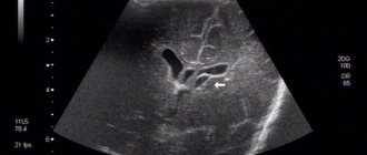
One such method is ultrasound examination of the brain. This method is not used in adults because ultrasound cannot penetrate the skull bones. In children, the situation is completely different, since there is a fontanel - a wonderful window for ultrasound. Neurosonography, which is what ultrasound of the brain is called, is an affordable and absolutely safe method. It allows you to estimate the size of the ventricles of the brain, and an increase in these sizes can well be regarded as an indirect sign of increased ICP. At the same time, as with head circumference, it is not so much the width of the ventricles of the brain that matters, but the dynamics of this indicator.
After the fontanel is closed, the size of the ventricles of the brain can only be seen and assessed using tomography - computed tomography (CT) or magnetic resonance imaging (MRI). At the same time, tomography is a serious, unsafe, expensive method, it is used infrequently - only in cases of real suspicion of serious intracranial pathology.
Another method - outdated, but still widely used - is echoencephalography (EchoEG). Using a special device (echoencephalograph), using the same ultrasound, a number of parameters are assessed, including the pulsation of the blood vessels of the brain. In this case, the amplitude of oscillations of the ultrasonic signal is considered as an indicator capable of assessing ICP.
We emphasize once again: all of the listed methods are not reliable, they do not state, do not confirm, but only admit the possibility, suggest, allow one to suspect an increase in ICP.
This is the result: the examination methods that exist today only give the doctor additional information to think about, but they cannot dot the i’s. This means that you have to rely mainly on specific symptoms. This has its own problems: this is not your grandmother, who, when her blood pressure is high, lies down, and when her blood pressure is normal, runs around shopping. This is a young child, or rather a little month old, who is unreasonable and does not complain about anything in particular.
But the problems are not only due to age and the inability to point with a finger to the place where it hurts. The main problem is that almost all the symptoms that make it possible to suspect an increase in ICP in a child can occur in completely healthy children.
For example, a child’s restlessness, trembling of limbs, and screaming may be manifestations of increased ICP, but may well have nothing to do with ICP at all. And any mother can confirm this, because it is simply impossible to find a child who is always calm and who never shakes anything. Another symptom of increased ICP is strabismus, but it is well known that in children of the first year of life, the extraocular muscles have not yet formed and infant strabismus is often a physiological phenomenon, that is, absolutely normal.
It should, however, be recognized: words such as “anxiety,” “trembling,” “screams,” and “squint” are not capable of seriously frightening the average domestic mother, since everyone hears them and often uses them in everyday life.
It’s a completely different matter when such terrible expressions as “Graefe’s symptom” or “spontaneous Moro reflex” are heard from the doctor’s lips or found in the outpatient card - there is no time for jokes and no time for calm: it is clear that the situation is serious.
Let us try to explain the essence of these wise words.
The essence of Graefe's symptom is the lag of the upper eyelid when the eyeball moves downward. In an additional translation into Russian, this means that when a child looks down and gets scared, several millimeters of the white of the eye are visible above the iris. Looks like a bulging eye. If the child is looking straight, then everything is fine.
The German ophthalmologist Graefe, who lived in the 19th century, described this symptom as typical for patients with goiter (damage to the thyroid gland). In people who do not have goiter, Graefe's symptom can also occur and be a constitutional feature; it can be found in premature infants.
The Moro reflex, or hug reflex, refers to the physiological reflexes of the newborn period. Occurs when hitting the table on which the child is lying, with a sudden loud sound, or when patting the baby on the buttocks or thighs. The reflex consists of two phases. In the first, the child leans back, turns his shoulders, and spreads his arms to the sides. In the second phase, he brings his arms together on his chest. It is clear that the spontaneous Moro reflex is when there were no special external stimuli, and the child throws back his hands... But the absence of “special external stimuli” is a conditional concept. For such a not at all “special”, but quite significant irritant, could well be a doctor’s office - a new environment, an unfamiliar table, a strange aunt-doctor...
It seems that we are completely confused: we promised to explain why the diagnosis of increased ICP and its treatment are so common, but we came to conclusions that were completely opposite. It turns out that additional research methods and examination data in the vast majority of cases do not allow one to confidently diagnose increased ICP. And in situations where such confidence is present, we are almost always talking about extremely dangerous diseases (hydrocephalus, meningitis, tumors, traumatic brain injury) and extremely disturbing symptoms (sharp bulging of the fontanel, disorders of consciousness, vomiting, paralysis).
Let's summarize the main results.
1. Increased ICP is not a disease, but a symptom of certain diseases.
2. Increased ICP is a rare and very dangerous symptom of rare and very dangerous diseases.
3. Treatment of increased ICP has nothing to do with outpatient medicine and almost always requires hospitalization and emergency care.
TTT
To conclude the article, let’s take a break from medicine for a moment and turn to… linguistics. The goal is the curious features of the use of the word “intracranial” itself. The fact is that the phrase “intracranial pressure” in everyday communication among medically savvy mothers is found less and less often. The word “pressure” is omitted as unnecessary, and everyone unanimously “checks the intracranial”, “treats the intracranial” and “complains about the intracranial”.
Specialists in the field of linguistics (linguists) call such things conversion, or the transition of one part of speech to another. This phenomenon is not at all unique. Let us at least recall “ice cream”, “jellied” or closer to medical topics - “laxative”, “ambulance”, “newborn”. Well, who says “sleeping pills” now? Nobody talks, because it’s already clear what they’re talking about. Instead of a children's room - just a children's room, instead of a bakery shop - a bakery that has already become familiar.
The main thing that conversion indicates is the extreme prevalence of a certain word. Conversion in relation to long-suffering intracranial pressure is, alas, a sad phenomenon, because it confirms a rather unpleasant trend: they began to talk about intracranial pressure too often, unreasonably often. This concept is expertly used by the grandmothers on the benches. They will make a diagnosis based on the circumference of the head, more precisely, by the size of the cap, and they also know how to treat. I really wish there were fewer such conversions in our lives. So that the phrase “intracranial pressure” was sometimes uttered by specialists, but grandmothers did not know about intracranial pressure at all, focusing their attention on sleeping pills and aspic.
author Komarovsky E.O. published 06/06/2008 12:08 updated 21/03/2017 — Diseases and treatment
Diagnosis of dropsy of the brain
In infancy, a visual examination is sometimes sufficient to determine the nature of the pathology: the doctor, by examining the baby’s physiological parameters and listening to the parents’ complaints, can make a preliminary diagnosis at the initial appointment.
To clarify the disease in children with an open fontanel, including to identify pathology during intrauterine development, ultrasound is used to assess the degree of enlargement of the ventricles. Additional methods are:
- fluoroscopy;
- computed and magnetic resonance imaging;
- lumbar puncture, necessary for a detailed analysis of the cerebrospinal fluid.
A child with suspected hydrocephalus must be examined by an ophthalmologist or pediatrician; if necessary, concomitant diseases may require consultation with a cardiologist, surgeon, oncologist and other specialists.
Treatment of hydrocephalus in children
Depending on the type and nature of the pathology, treatment for hydrocele can be conservative or surgical. Conservative tactics are used only in cases with a non-progressive open form of the disease, which is caused by external factors. It involves medication to relieve symptoms and eliminate the cause.
In all other situations, it is rational to use surgical treatment methods, which are aimed primarily at eliminating the obstacle that impedes the outflow of cerebrospinal fluid (tumor, abscess, intracranial hematoma, developmental anomalies). In cases where it is not possible to eliminate the cause of the pathology, a special operation is performed - bypass surgery. The technique involves introducing a system of tubes and valves into the brain, which will drain excess cerebrospinal fluid to other parts of the body.
In situations that threaten the child’s life, when immediate assistance is required, external ventricular drainage is performed - an operation during which excess cerebrospinal fluid is quickly pumped out of the ventricles of the brain using the drainage system.
With timely diagnosis and adequate treatment, a child after hydrocephalus grows and develops in accordance with the norms; the disease does not affect the mental or mental state in any way.
Disease prevention
The list of measures that can prevent the development of pathology includes:
- pregnancy planning;
- prevention of birth injuries;
- giving up bad habits before conceiving a baby and during pregnancy;
- vaccine prevention of infectious diseases of the brain (meningitis, encephalitis);
- early perinatal diagnosis;
- regular monitoring of children at risk by a pediatrician and neurologist;
- timely completion of preventive examinations up to a year;
- providing the child with safe living conditions to prevent household traumatic brain injuries.
Parents need to closely monitor the development and condition of young children, promptly contacting doctors in case of infectious diseases, falls and injuries affecting the head and neck area.
Specialists at the SM-Doctor clinic will conduct a detailed diagnosis if hydrocephalus is suspected, plan treatment and monitor the child throughout therapy and rehabilitation.