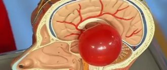
A brain cyst is a benign neoplasm. It looks like a cavity filled with cerebrospinal fluid.
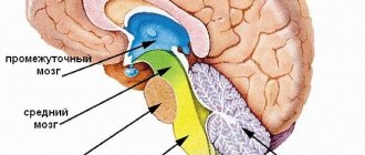
A lacunar cyst of the brain is located between the gray matter of the brain and its membranes; it can also be located in the hemisphere of the cerebellum or in the pons. According to observational data, it occurs in 4% of the population; men are more likely to suffer from the disease than women.
The danger of developing this type of cyst is that this formation can asymptomatically reach significant sizes.
Types of cysts
With lacunar cysts of the brain, the size, norm and location determine their type. But first of all, it is taken into account how they began to develop - in utero, which means they are congenital, or in the process of ordinary life, here the reasons for their appearance are much more diverse.
Moreover, there is a difference between one type of cyst and another based on the location of their appearance in the brain. A retrocerebellar cyst is a neoplasm that arises under the arachnoid membrane of the brain.
If the tumor appears on the outer arachnoid membrane, it is called an arachnoid cyst. Lacunar cerebrospinal fluid cysts occur between the membranes of the brain. A vascular cyst is a tumor that arises in the plexus of blood vessels in the brain. Lacunar cyst of the basal ganglia occurs in the cerebellum, pons, or subcortical ganglion.
You have received only a basic understanding of what it is - a lacunar cyst of the brain. This topic is being studied in medical institutes to this day, which is why quite a lot of types and types of this neoplasm have been discovered, and new tumors and cysts are added to the resulting list every year.
Types of cysts
Lacunar cysts are divided into congenital and acquired. The first type is classified as a primary type of disease, the second - as a secondary one. A congenital cyst develops in a baby in the womb. An acquired lacunar cyst appears due to external factors.
The primary type of the disease, as mentioned above, develops even before birth. This is influenced by the behavior of the mother who abuses bad habits. Alcohol, smoking, and drugs negatively affect the development of the fetus. Such habits cause the child’s genes to mutate and impair fetal nutrition and blood supply. In addition to the appearance of a lacunar cyst, other congenital diseases are possible in a child.
In addition to the bad habits of the mother, the child is affected by diseases suffered by the woman during pregnancy. They affect the further development of the central nervous system of the fetus. Cyst formation occurs at three stages of pregnancy, but often the disease appears in the first trimester.
The secondary type of disease occurs due to acquired diseases. The range of problems is significant: meningitis, head injuries. A lacunar cyst can form due to diabetes, hypertension, or a connective tissue disorder. Its formation is affected by weakened immunity. A mother's weak immune system has a negative impact on the child.
Causes of lacunar cysts
You need to understand that if a patient was diagnosed with a cyst that developed during the period of intrauterine growth, then it is most likely a hereditary disease. It is impossible to insure yourself against this type of tumor. There are cases where a person did not know that he had a congenital cyst until it was accidentally discovered during a routine medical examination. The tumor was 10 cm in diameter and did not interfere with the person’s life at all. What is a cerebral lacunar cyst? This is a formation that creates problems, but not in every case of its occurrence.
If the cyst is diagnosed as an acquired pathology, this means that it was the result of some kind of somatic disease. For example, it appeared as a complication after meningitis or traumatic brain injury. Diabetes mellitus, thrombosis, and hypertension can lead to the appearance of a cyst.
Often, frequent concussions can cause the appearance of a tumor in the brain. This phenomenon occurs in professional sports, where an athlete often receives traumatic brain injuries, for example, boxing or other martial arts.
Post-ischemic lacunar cyst has become widely known in medical circles. From the name it is clear that it was a consequence of ischemic brain disease, which, in fact, leads to chronic hypertension. Postischemic lacunar cysts of the brain are studied and treated simultaneously with stroke. Often they become the cause of craniotomy and brain surgery. But lacunar cerebrospinal fluid cyst is amenable to radiation therapy, and surgery in this case is not always required.
Risk factors
Lacunar infarction can occur at any age, but the likelihood of its occurrence increases with age and reaches its maximum after 85 years. More often, cerebral circulatory disorders occur in men. The most significant risk factors for the occurrence of lacunar cerebral infarctions are:
- hypertonic disease,
- diabetes,
- chronic renal failure,
- post-infarction cardiosclerosis,
- abnormalities in the circulatory system and heart defects,
- rheumatism,
- cardiac arrhythmias,
- disorders of the blood coagulation system, blood diseases.
Signs of a cyst in the brain
Lacunar cysts of the brain may not show symptoms for many years. At the same time, it grows and develops. The first signs that a person has it appear only when the enlarged tumor begins to put pressure on nearby blood vessels, thereby disrupting blood flow or creating pressure on certain parts of the brain. And depending on what lobes these are, characteristic signs appear:
- The patient experiences hallucinations.
- Severe headache, and the location of the pain is located opposite the cyst.
- A person begins to feel sick or vomit for no apparent reason.
- In severe cases, seizures with convulsions and loss of consciousness are possible.
- Coordination of movements is impaired.
- Speech and a person’s ability to write texts are impaired.
- If a cyst appears in the frontal lobe, then a person’s vision may be impaired or completely lost.
- If the tumor is in the temporal lobes, the sense of smell is impaired and the ability to distinguish tastes is lost.
But the neoplasm does not always have at least some manifestation. Often, a lacunar cyst of the brain does not receive any treatment at all, because it has not manifested itself in any way throughout a person’s life. There was no pain or any disturbances in brain function.
The main types of cystic formations
Ovarian
This is a fluid-filled bladder that causes the ovary to enlarge, leading to pain and sometimes infertility. The main reason for its appearance is hormonal imbalance. The disease often occurs without symptoms; when the formation is large, the cycle is disrupted, pain is felt in the lower abdomen, a false urge to urinate occurs, and bleeding occurs outside of menstruation. Methods of surgical treatment: oophorectomy (complete removal of the ovary), laparoscopic cystectomy (excision of the cyst), adenxectomy (surgery to remove the uterine appendages). In most cases, the choice is not classical surgery, but gentle endoscopic removal of the cyst while preserving the patient’s reproductive function.
Read more about ovarian cyst removal
Coccygeal
Most often occurs in men aged 15-30 years. It is an opening in the area of the gluteal fold, approximately 10 cm from the anus. Externally it looks like a fistula. It can be congenital or acquired - due to too much hair in this area. It manifests itself as pain when walking, sitting, redness of the tailbone, a feeling of discomfort and the presence of a foreign body in the area where it is located. At a later stage, pus is released from the hole.
Read more about the treatment of coccygeal cyst
Bartholin gland
Appears due to blockage of the gland duct due to infection or chronic inflammation. The formation has a capsule and is filled with secretion, which gradually gels. If it is large, it interferes with walking and sitting, makes intimate intimacy inaccessible, and can become infected and lead to an abscess. Usually reaches 2 cm, but there are formations up to 9 cm. The main causes are chronic bartholinitis, candidiasis, bacterial vaginosis, decreased local immunity.
Where and how to remove a Bartholin gland cyst
Eye
A hollow formation filled with non-inflammatory fluid - products of the activity of the cornea or conjunctiva. May originate from the cornea, iris, conjunctiva and other eye membranes. Causes: inflammation and trauma, congenital anomalies. The disease is manifested by pain, a feeling of fullness, blurred vision, and the presence of translucent dots in the field of vision.
Read more about surgical treatment of cysts in ophthalmology
Maxillary sinus
A cavity containing fluid and having a membrane attached to the wall (usually the lower) of the maxillary sinus. A cystic formation can be true (its walls consist of mucous membrane) or pseudocyst (the mucous membrane is split and fluid accumulates in it). It manifests itself as headache, difficulty breathing through the nose, a feeling of fullness and heaviness in the eye and cheek area, mucus discharge from the nose and its flow down the wall of the pharynx, discharge of yellow transparent fluid from the nose, frequent sinusitis with suppuration. It is formed due to the peculiarities of the anatomical structure of the nose, blockage of the excretory duct of the glands of the maxillary sinus, inflammation of the teeth, spreading to the roots.
Read more about the treatment of abscesses and cysts of the ENT organs
Mammary gland
A cavity bounded by a capsule of connective tissue and filled with fluid is formed in the ducts and can be single or multiple. It is formed due to an increase in the duct of the mammary gland, the accumulation of secretions in it. The lesion may be round, oval, or irregular in shape. The disease is asymptomatic for a long time; over time, pain and burning appear in the mammary gland, and may be accompanied by suppuration and inflammation. One of the varieties is a fatty cystic formation that occurs when the sebaceous gland of the skin is blocked and filled with secretions. The main provoking factors are mastitis, thyroid disease, inflammation of the genital organs, and ovarian dysfunction.
Learn more about symptoms and treatments
Uterus and cervix
These are dilated and clogged glands, inside of which secretions (mucus) accumulate. The disease occurs against the background of endocervitis, cervitis. Provoking factors: abortion, childbirth, infections, menopause, hormonal imbalance, use of an intrauterine device, infections. Nabothian cystic formations are localized in the vaginal area of the uterus and are not removed until they reach a certain size. Retention occurs due to an excessive amount of secretion in the gland duct. The disease is often asymptomatic; its indirect signs are frequent inflammation.
Read more about surgical treatment of uterine and cervical cysts
Brain
A cystic formation is an accumulation of fluid in the substance or membranes of the brain. When large in size, it entails intracranial hypertension and puts pressure on the surrounding brain structures. Can form at any age. Depending on the location, cerebral (intracerebral) and arachnoid formations are distinguished. The first are found in the internal structures of the brain, in areas of necrosis. The second ones are in the meninges. Provoking factors: inflammatory diseases, trauma, including birth, cerebrovascular accident, parasites, complications after surgery. They manifest themselves as nausea, a feeling of pressure on the eyes, decreased performance, sleep disturbances, pulsation or noise in the head, and visual impairment.
Thyroid gland
Small formations (up to 5 mm) may not have pronounced symptoms. If the thyroid cyst has dense inclusions and a complicated structure, then special studies (for example, ultrasound), tests and biopsy are needed, because such a condition may be a sign of malignant degeneration.
Read more about surgical treatment of thyroid cysts
Larynx
Cysts of the larynx can be located in any part of the larynx. They do not grow into the mucous membrane, but grow towards the lumen of the larynx and thereby narrow it. The cause of retention cysts is blockage of the excretory ducts of the laryngeal glands. There are cysts of the vocal cords that arise due to constant irritation.
More information about surgical treatment of laryngeal cyst (laryngocele)
You can find out the prices for cyst removal endoscopically or by another method, as well as the cost of preoperative diagnostics on our website or by calling honey. +7 (812) 435 55 55.
Diagnosis of the presence of a cyst in the brain
Treatment of lacunar post-ischemic cyst of the brain is prescribed only after a complete comprehensive diagnosis. The same applies to any type of cyst - it is important to know its size and location.
After all, if the cyst does not pose a danger to the functioning of the brain, does not grow, then they simply observe it, without trying to cure it in any way - there is no need. But if it begins to grow and put pressure on the surrounding vessels and brain tissue, then urgent therapy begins.
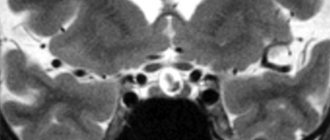
The main method for diagnosing brain tumors is computed tomography. It most clearly and accurately determines the presence, size and location of the cyst.
In order to understand whether its contents are dangerous, that is, whether it can turn into a malignant tumor, a histological analysis of the cells obtained from the cyst as a result of a biopsy is done.
The diagnosis can be clarified by examining the vessels of the head and neck using Doppler sonography. At the same time, the condition of the patient’s blood is studied, the presence of elevated cholesterol levels in it, in order to eliminate the likelihood of cholesterol plaques.
The blood is also examined for the presence of pathogenic microorganisms that can cause inflammation.
Diagnosis may include daily monitoring of blood pressure fluctuations. For this purpose, special sensors are attached to the patient, which accompany all his activities throughout the day.
Drug treatment of lacunar cyst
If the cyst does not bother the patient, does not increase in size and does not pose a danger, then it is not subject to special treatment. In this case, therapy is aimed at eliminating the cause of its occurrence. For example, if a patient has suffered a severe viral disease such as meningitis or encephalitis, then he is treated with antibiotics and immune-strengthening drugs. The specific type of drug, its dosage and regimen of use are prescribed individually depending on the general condition of the patient.
A neurologist may prescribe a drug that breaks up adhesions in connective tissue, blood thinners, and antioxidants. All this allows you to restore impaired blood flow in the vessels of the brain and stabilize pressure.
General information
One of the types of cerebral infarction (ischemic stroke) is lacunar infarction, which is a small (up to 15 mm in diameter) brain damage that occurs when local blood circulation and gas exchange are disrupted. The reasons for this situation are varied and not fully understood, but most often it is blockage of supply vessels as a result of changes in their walls (atherosclerosis, inflammation), emboli (blood clots, fat droplets, bacterial colonies, etc.). Most of them are found in the periventricular region, basal ganglia, and thalamus - the central, deep structures of the brain. Lacunar infarctions account for 20-30% of all strokes.
Surgery
If the cyst poses a danger to the patient’s condition, then surgery is indicated. It can be performed using various methods - bypass surgery, endoscopy and craniotomy.
Each method has both positive and negative sides. Endoscopy can only reach those cysts that are located directly under the skull bone. The instrument, which is a thin tube, does not reach the neoplasms lying deep in the brain matter.
Bypassing a cyst involves suctioning out the fluid filling the cyst using a thin needle. As a result of this procedure, the cyst is reduced until only one shell remains. The negative side of this procedure is the risk of introducing an infection into the brain, which will cause complications.
Craniotomy involves opening the skull to provide access to the cyst at any depth. This type of operation provides a 100% opportunity to completely remove the tumor, but has a long postoperative period.
The type of operation is chosen by the attending physician, based on the patient’s condition, location and size of the cyst, and many other medical indicators. The wishes of the patient in this situation are not taken into account.
Treatment
If the cystic neoplasm is small in volume and does not bother the patient, treatment is not required. You can maintain your condition with trips to a sanatorium and preventive treatment. But you need to monitor the size and condition of the tumor, its position and shape, so as not to develop new symptoms. Scientists have concluded that even a small cyst can accelerate the aging of the body.
Basically, such formations are treated by influencing the causes. They use antibiotics, antiviral drugs, and antihypertensive drugs. All medications are prescribed taking into account the person’s medical history. These drugs are commonly used by doctors:
- antioxidants;
- medications that help restore blood circulation;
- anti-adhesion drugs;
- nootropics;
- immunomodulators.
Treatment with medications is prescribed to improve blood clotting in lacunar tumors to control cholesterol and blood pressure. It is not possible to completely get rid of the disease in this way; medications only support the patient’s life. For a complete cure, surgery is necessary.
Sometimes pills don't do the job. Symptoms become more severe and life becomes worse. In this case, the patient needs to decide on surgery. This method is the only possible treatment option that completely rids a person of the tumor. Treatment methods:
- trepanation;
- endoscopy;
- shunting.
In the latter case, a drainage tube is inserted into the brain to remove fluid from the lacuna. This operation must be repeated for a long time, and the risk is enormous. The method is used only in extreme cases. In the second method, the skull is pierced with an endoscope and the tumor is removed through the puncture. It is the most painful, but safe. In the case of trepanation, there is a risk of damaging the brain, but the operation is the most effective.
Postoperative rehabilitation
The time allocated for postoperative rehabilitation depends on the complexity of the surgical intervention and the severity of the disease that caused the cyst.
For example, after removal of a congenital cyst, which did not complicate the patient’s condition, using bypass surgery, 10-15 days are allotted for rehabilitation.
And if the tumor was caused by an infectious disease such as meningitis, the cyst disrupted some functions in the body, vision, hearing or musculoskeletal function, trepanation was required to remove it, then a complete cure may take 5-6 months.
Special diet
During treatment and in the postoperative period, the patient adheres to a special diet. To reduce cholesterol in the blood, which can cause a blood clot, dishes with fatty, fried meat are removed from his diet. Meat products suitable for consumption in this situation are boiled fish, chicken and veal.
To normalize blood pressure and strengthen the immune system, fresh fruits and vegetables are included in the diet. The patient's diet is adjusted so that he eats 6-7 times a day, but in small portions. This reduces the load on the stomach, but allows all the beneficial substances from food to be fully absorbed into the intestines.
Drinking coffee and alcoholic beverages is strictly prohibited.
Possible complications
If any type of lacunar cyst is left untreated, the patient may develop various complications.
Thus, a pineal cyst leads to encephalitis or hydrocephalus - the accumulation of fluid in the brain. And an arachnoid cyst can lead to epilepsy. In colloidal cysts, complications are even more dangerous - cerebral hernia, hydrocephalus and death.
If an untreated cyst remains in a child's brain, it can delay his intellectual and even physical development. The most dangerous complication is cyst rupture. In this case, a quick and painful death awaits the person.
Consequences and forecasts
In the absence of adequate therapy, the development of a lacunar cyst can have a number of adverse consequences, such as impaired coordination of movements, damage to vision and hearing, inflammatory processes in brain tissue, and death.
Children may develop hydrocephalus - accumulation of cerebrospinal fluid in the ventricles, leading to compression of brain structures.
However, with the correct diagnosis and timely treatment, it is possible to completely get rid of the signs of the disease and prevent the formation of new cysts.
To prevent the disease, you should undergo a preventive examination at least once every 2 years and, if necessary, consult a doctor.
Traditional medicine treatment
Despite the seriousness of the disease, lacunar cysts have several methods of treatment with folk remedies. In general, it, like conservative therapy, is aimed at eliminating the causes of the tumor. Therefore, traditional methods must be used in combination with drug therapy and only with the permission of the attending physician.
Medicinal plants that can positively affect a person’s condition in this situation are hemlock, elecampane, wormwood, chamomile, calendula, yarrow, raspberry, corn silk, and Dioscorea Caucasica.
From these plants you can make decoctions or alcohol tinctures. The decoction is simple to make - 1 tbsp. a spoonful of the plant is poured into a glass of boiling water and simmered over low heat for no more than 15 minutes. After the broth has cooled, it should be strained and taken in a glass half an hour before meals.
The alcohol infusion takes longer to make - the dry, crushed plant is filled with alcohol in a ratio of 1 to 3, that is, 300 ml of alcohol per 100 g of plant, and infused for 2 months in a dark place. The infusion should be shaken once a week. After straining, the product is taken 1 teaspoon per day, half an hour before meals.
Prevention
To prevent the occurrence of a cyst in the brain, it is necessary to take a number of measures aimed at protecting a person from situations in which a cyst begins to develop:
- It is necessary to avoid stress and nervous tension.
- Treat viral diseases in a timely manner, preventing them from becoming chronic.
- Protect your head from injuries at work or in sports. That is, wear a protective helmet or hard hat.
- It is necessary to monitor blood pressure and take measures to reduce it during periods of increase.
- You need to give up bad habits - smoking and drinking alcohol, because they cause many diseases. A person who smokes is several times more likely to get cancer.
Existing diagnostic methods for detecting pathology
Lacunar formation can only be seen on CT, MRI or spiral computed tomography. The images clearly show the location, the affected neighboring areas, the shape and size of the neoplasm. Dopplerography helps in clarifying the clinical picture: it reflects the speed of blood flow, the condition of the arteries, the presence of an aneurysm or atherosclerotic changes in blood vessels. These examination methods are non-invasive. However, when doctors need to clarify the histological structure of the cyst tissue, they resort to invasive methods. The first and most revealing is a biopsy, which accurately determines the nature of the tumor, allowing for a morphological study of the biopsy sample taken.
Laboratory diagnostics give their results:
- cholesterol content and coagulability reflect the possibility of vascular blockage;
- Determination of pathogenic microorganisms in the blood indicates the infectious basis of the neoplasm.
As an additional examination, 24-hour blood pressure monitoring is performed. Pressure drops and extreme blood test values become indirect confirmation of the presence of pathology in the brain.