Formation and functions of the brain convolutions
It has been revealed that the main sections of the contents of the cranium begin to form from the mother's womb. And each of them is responsible for a separate side of the human personality. Thus, the function of the temporal gyri is associated with the perception of written and spoken speech.
Wernicke's center is located here, damage to which leads to the fact that a person ceases to understand what is being said to him. At the same time, you can still pronounce and write down words. The disease is called sensory aphasia.
- Prevention of congenital pathologies
- Temporal lobes, structure
- Frontal lobe of the brain -
- Research on changes in the white matter of the brain
- Brain malformations: polymicrogyria, agyria and pachygyria
- What is this?
- Cerebral cortex
- Embryology
- Frontal cortex and hippocampus age faster
- Furrows and convolutions of the parietal lobe. Postcentral sulcus, sulcus postcentralis.
- Does the number of convolutions affect the level of intelligence?
- Formation of the cortex in embryogenesis
- Structure
- What is this?
- Temporal lobe
In the area of the inferior pubic gyrus there is a formation responsible for the reproduction of words, which is called Broca's speech center. If an MRI reveals damage to this brain region, the patient experiences motor aphasia. This means full understanding of what is happening, but the inability to express your thoughts and feelings in words.
Damage to all departments responsible for speech can cause complete aphasia, in which a person may lose contact with the outside world due to the inability to communicate with others.
The anterior central gyrus is functionally different from the others. As part of the pyramidal system, it is responsible for performing conscious movements. The functioning of the posterior central eminence is inextricably linked with human senses. Thanks to her work, people feel heat, cold, pain or touch.
The angular gyrus is located in the parietal lobe of the brain. Its meaning is associated with visual recognition of the resulting images. It also contains processes that allow sounds to be deciphered. The cingulate gyrus, located above the corpus callosum, is a component of the limbic system.
Memory is of particular importance in human life. She plays an important role in her own education and in the education of new generations. And storing memories would be impossible without the hippocampal gyrus.
Doctors who study neuropathology note that damage to one of the brain regions is more common than disease of the entire organ. In the latter case, the patient is diagnosed with atrophy, in which a large number of irregularities are smoothed out. This disease is closely associated with serious intellectual, psychological and mental disorders.
Brain: synapses, hemispheres, parts, sections and lobes of the brain
The brain is a reliable biological system built from unreliable elements.
John von Neumann
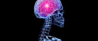
A 1.3-kilogram supercomputer hidden inside your skull simultaneously processes facts and faces, stores memories, and regulates movement and speech. The brain is one of the most mysterious organs of our body. It has been studied by scientists from all over the globe for many years.
EVOLUTION OF THE HUMAN BRAIN
In 1860, the male brain weighed 1370 grams. Now the average male brain weighs 1425 grams, and in old age its weight decreases to 1395 grams. And huge dinosaurs had a tiny brain weighing 70 grams and the size of a walnut.
MAIN PARTS OF THE BRAIN
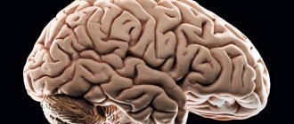
If you look at the image of the brain, you will notice that it consists of several parts, dotted with winding grooves.
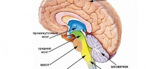
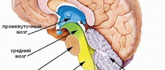
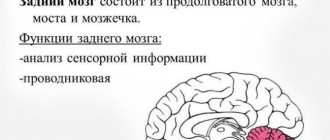
Scientists distinguish three main parts of the human brain: the hindbrain, the midbrain and the forebrain. They are clearly visible already in a four-week embryo in the form of “brain vesicles”. The hindbrain and midbrain are responsible for vital internal functions of the body: maintaining blood flow, breathing. The forebrain is responsible for forms of communication with the outside world.
The brain is physically divided into two hemispheres: left and right. The functions performed by the organ are also divided into hemispheres. Despite their external similarity and active interaction, functional differences are clearly visible in the functioning of the hemispheres. Some functions are better handled by the right hemisphere, while others are handled better by the left hemisphere.
RECORD IQ
Korean child prodigy Kim Un-young has the world's highest IQ of 210. He mastered algebra by the age of eight months and spoke four languages by the age of two. At the age of four, Kim entered university and graduated at 15.
Functions are distributed not only between the hemispheres, but also across different areas of the brain. The cerebral cortex can be divided into four paired lobes: occipital, parietal, temporal and frontal.
- The frontal lobes can be called the command post of the brain. Here are the centers that ensure a person’s independence and initiative, as well as his ability to critically self-assessment. The frontal lobes are also responsible for mastering skills. It is thanks to them that initially complex work becomes automatic and does not require much effort.
- The temporal lobes in the upper regions process auditory sensations, turning them into images. It is in this part of the brain that the words addressed to a person are recognized and filled with meaning, as well as the selection of words to express one’s thoughts. The anterior and middle parts of the temporal lobes are associated with the sense of smell. It is the temporal lobes that store memories. The dominant temporal lobe deals with verbal memory and object names, while the non-dominant temporal lobe deals with visual memory.
- The parietal lobes are responsible for the ability to put parts together into a whole. For example, to read you need to be able to put letters into words and words into phrases. Same with numbers and numbers. This part of the brain is also responsible for sensing your body and distinguishing between the right and left sides. The parietal lobes are the center of the three-dimensional image. It provides a three-dimensional perception of the surrounding world. This side is also involved in spatial orientation and the perception of heat, cold and pain.
- The occipital lobes are responsible for processing visual information. Everything we see, we do not see with our eyes, which only record light and translate it into electrical impulses. We “see” with the occipital lobes, which interpret signals from the eyes.
USEFUL CONNECTIONS
The human brain contains about 100 billion neurons. This value is approximate. After all, it is almost impossible to count all microscopic cells without missing a single one.
The channels or nerve pathways along which signals travel are called synapses. Two cells participate in their formation - transmitting and receiving. Synapses are located in those areas of nerve cells where they contact each other. They are also present where nerve cells connect with muscles or glands.
STORE YOUR MEMORIES IN TERABYTES
A piece of brain the size of a grain of sand contains 100,000 neurons. Each of them is connected to its “neighbors” using 40,000 synapses. It turns out that there are more connections in the brain than there are stars in the Universe. This organ has enough space for 1000 terabytes of information.
The middle part of the synapse is the synaptic cleft—the space between two interacting nerve cells. An electrical impulse passes through this gap.
The final part of the synapse is the postsynaptic terminal. This cell fragment comes into contact with many sensitive receptors.
Prevention of congenital pathologies
Congenital malformations of the structures of the central nervous system account for about 25% of the total mass of all developmental anomalies. In 30% of cases, malformations of the central nervous system lead to the death of the newborn. The prognosis worsens when cerebral pathology is combined with a somatic disease. Measures that prevent the development of structural abnormalities of the cortical structures of the brain include:
- Prevention of infectious diseases and toxic lesions in pregnant women.
- A healthy lifestyle (nutrition, adherence to work and rest, dosed physical activity, walks in the fresh air) during gestation.
- Prevention of anemia and hypovitaminosis during gestation.
- Prevention of fetoplacental insufficiency (impaired placental functions - trophic, respiratory, protective, excretory).
- Prevention of hypoxic and ischemic processes in the fetal brain tissue.
Prenatal diagnosis makes it possible to identify cerebral and somatic pathologies in the early stages. The main causes of developmental abnormalities are associated with genetic mutations and impaired embryogenesis.
Gyri and sulci are structural formations of the cortical layer of the brain that play a decisive role in the formation of intelligence, motor activity and behavior. Damage to these areas of brain tissue leads to disruption of higher mental functions.
Signs of defeat
Disturbances in the structure of the sulci and gyri in humans are more often associated with malformations at the stage of embryogenesis, less often with TBI - if such changes are present, the likelihood of developing epilepsy and mental retardation increases. The clinical picture in the presence of malformations of the cortical relief includes convulsive and epileptic seizures, dementia, paresis and spastic paralysis. Common malformations:
- Pachygyria. Thickening, flattening, expansion of cortical convolutions.
- Lissencephaly. Underdevelopment or absence of convolutions (effect of a smooth surface of the brain).
- Polymicrogyria. Abnormal folding of the cortical layers. Lack of clear relief - in some cases the convolutions merge.
Damage to the cortical layers of the brain is accompanied by symptoms: agnosia (impaired perception - visual, auditory, tactile), amnesia (partial or complete loss of the ability to remember past experiences), aphasia (impaired formed speech), apraxia (impaired complex voluntary movements while maintaining basic motor skills) .
Damage to individual areas of the cortex is accompanied by specific symptoms. Analysis of symptoms allows us to identify the localization of the pathological focus. Damage to the gyrus recta, which lies in the cerebrum, is associated with a slowdown in the formation of a cognitive response, a disturbance in the emotional background and behavioral skills.
Research shows a connection between damage to this area of the cortex and disturbances in social perception (social role, social status, interpersonal relationships). Damage to the angular gyrus, which lies in the cerebral cortex, manifests itself as difficulties in developing writing, speaking, and reading skills.
A patient with damage to brain structures of such localization experiences difficulties in performing mathematical operations, is unable to distinguish between fingers, and does not distinguish between the right and left half of the body. Damage to the supramarginal gyrus leads to loss of the ability to make complex voluntary movements leading to a specific goal.
Damage to the fusiform gyrus leads to impaired recognition of faces and symbols. When the lingual gyrus is damaged, the processes of perception and analysis of visual stimuli are disrupted. With the help of vision, a person receives about 90% of information coming from outside. Damage to the medulla in this area is accompanied by hemianopsia - partial loss of visual fields (vision is usually preserved in the central field).
Damage to the central gyrus is accompanied by impaired motor activity and the appearance of convulsive syndrome. Damage to the cingulate cortex is associated with the development of schizophrenia and other psychopathologies. The function of social cognition and the formation of emotions is impaired in patients.
Temporal lobes, structure
The temporal lobes are separated from the frontal and parietal lobes by a groove called the lateral. In a direction almost parallel to the lateral one, two more grooves are located on the lateral portion of the temporal lobes:
The anterior portion of the temporal lobe is called the temporal pole, and its edge, under which the insula is located, is called the temporal operculum. The convolutions are located parallel to the main grooves.
The superior temporal gyrus is located between the lateral and superior temporal sulcus. It contains the so-called Heschl convolutions, which are transverse formations of short length. The middle temporal gyrus is located between the superior and inferior temporal sulci. The inferior temporal gyrus is located on the inferior portion of the temporal lobes.
On the left side, in the posterior part of the temporal gyrus, there is an auditory analyzer (in left-handed people it is located not on the left, but on the right). Its nucleus is located on the surface of the temporal lobe that faces the insular lobe.
The parahippocampal gyrus is located almost in the center of the temporal lobe. The hippocampus itself, a small section of the temporal lobes located on their inner surface, is responsible for long-term memory. All human memories are stored in the temporal lobes.
This gyrus contains the center responsible for smell and taste, and in general, the anterior and medial sections of the temporal lobes are responsible for the functioning of the sense of smell. The curved anterior edge of the gyrus is called the parahippocampal uncus.
The left and right temporal lobes have a similar structure, but functionally they are asymmetrical. The functions they perform are determined primarily by which hemisphere is dominant in a person. A right-handed person has a left-dominant hemisphere, and a left-handed person, accordingly, has a right-dominant one.
Functions performed by the temporal lobe of the hemisphere that is dominant:
ability to understand spoken words;
memory: long-term;
the opportunity to learn by listening to information;
the connection of streams of auditory and visual information, the awareness of what a person saw, together with what he heard: the understanding that visible objects can sound.
responsibility for the synthesis of senses: tactile, auditory and visual images.
harmonization of the emotional background.
For the non-dominant hemisphere:
recognition of music and rhythm;
recognition of voice intonations;
recognition of faces, recognition of their expressions;
ability to learn using visual sources.
Main convolutions
Although the shape and size of some of the sulci and convolutions of the cerebral hemispheres differ from individual to individual, their number is normally unchanged. Every person, regardless of age and gender, has the following grooves:
- Sylvian fissure - separates the frontal lobe from the temporal lobe;
- lateral sulcus - separates the temporal, parietal and frontal lobes, and is also one of the deepest in the brain;
- Roland's fissure - separates the frontal lobe of the brain from the parietal lobe;
- parieto-occipital sulcus - separates the occipital region from the parietal;
- cingulate sulcus - located on the medial surface of the brain;
- circular - is the boundary for the insular part on the basal surface of the cerebral hemispheres;
- The hippocampal sulcus is a continuation of the cingulate sulcus.
The relief of the cerebral cortex is very complex. It consists of numerous convolutions of different shapes and sizes. But we can highlight the most important of them, which perform the most important functions. The main convolutions of the brain are presented below:
- angular gyrus - located in the parietal lobe, involved in recognizing objects through vision and hearing;
- Broca's center - the posterior part of the inferior frontal gyrus on the left (in right-handers) or on the right (in left-handers), which is necessary for correct speech reproduction;
- Wernicke's center - located in the posterior part of the superior temporal gyrus on the left or right (similar to Broca's area), is involved in the understanding of oral and written speech;
- cingulate gyrus - located on the medial part of the brain, takes part in the formation of emotions;
- hippocampal gyrus - located in the temporal region of the brain, on its inner surface, necessary for normal memorization;
- fusiform gyrus - located in the temporal and occipital regions of the cerebral cortex, is involved in face recognition;
- lingual gyrus - located in the occipital lobe, plays an important role in processing information coming from the retina;
- precentral gyrus - located in the frontal lobe in front of the central sulcus, necessary for processing sensitive information entering the brain;
- postcentral gyrus - located in the parietal lobe behind the central sulcus, necessary for voluntary movements.
Frontal lobe of the brain -
In the posterior part of the outer surface of this lobe the sulcus precentralis runs almost parallel to the direction of the sulcus centralis. Two grooves extend from it in the longitudinal direction: sulcus frontalis superior et sulcus frontalis inferior.
Due to this, the frontal lobe is divided into four convolutions - one vertical and three horizontal. The vertical gyrus, gyrus precentralis, is located between the sulcus centralis and sulcus precentralis. The horizontal gyri of the frontal lobe are as follows:
- superior frontal, gyrus frontalis superior, which runs above the sulcus frontalis superior, parallel to the upper edge of the hemisphere, reaching its medial surface;
- the middle frontal gyrus, gyrus frontalis medius, stretches between the superior and inferior frontal sulci and
- The inferior frontal gyrus, gyrus frontalis inferior, is located between the sulcus frontalis inferior and the lateral sulcus.
The branches of the lateral sulcus, extending into the inferior frontal gyrus, divide the latter into three parts: pars opercularis, lying between the lower end of the sulcus precentralis and ramus ascendens sulci lateralis, pars triangularis, located between both branches of the lateral sulcus, and, finally, pars orbitalis, located in front from ramus anterior sulci lateralis.
Neurologist
Neurosurgeon
What tests and diagnostics do you need to undergo for the Frontal Lobe of the brain:
MRI of the brain
CT scan of the brain
Is something bothering you? Do you want to know more detailed information about the Frontal Lobe of the brain or do you need an examination? You can make an appointment with a doctor - the Eurolab clinic is always at your service! The best doctors will examine you, advise you, provide the necessary assistance and make a diagnosis. You can also call a doctor at home. The Eurolab clinic is open for you around the clock.
How to contact the clinic: Phone number of our clinic in Kyiv: (+38 (multi-channel). The clinic secretary will select a convenient day and time for you to visit the doctor. Our coordinates and directions are listed here. Look in more detail about all the clinic’s services on its personal page.
Important Registration of a disability group after a stroke
| (+38 |
If you have previously performed any tests, be sure to take their results to a consultation with your doctor. If the studies have not been performed, we will do everything necessary in our clinic or with our colleagues in other clinics.
It is necessary to take a very careful approach to your overall health. There are many diseases that at first do not manifest themselves in our body, but in the end it turns out that, unfortunately, it is too late to treat them. To do this, you simply need to be examined by a doctor several times a year in order not only to prevent a terrible disease, but also to maintain a healthy spirit in the body and the body as a whole.
If you want to see a doctor, use the online consultation section, perhaps you will find answers to your questions there and read tips on caring for yourself. If you are interested in reviews about clinics and doctors, try to find the information you need on the forum. Also register on the Eurolab medical portal to be constantly aware of the latest news and updates about the Frontal Lobe of the Brain on the site, which will be automatically sent to you by email.
Other anatomical terms starting with the letter “L”:
| Forehead |
| Face |
| Elbow |
| Lungs |
| Lymph |
| The lymph nodes |
| Leukocytes |
| Elbow joint |
| Wrist joint |
| Spatula |
| Radius |
| Elbow bone |
| pubic bone |
| Ankle |
| Lymphocytes |
| Bulb of the vestibule of the vagina |
| Left atrium |
| Left ventricle |
| Pulmonary trunk |
| Pulmonary veins |
| Radial artery |
| Ulnar artery |
| Lymphatic vessels |
| Lymphatic vessels of the lower extremities |
| Lymphatic vessels of the pelvis |
| Lymphatic vessels of the abdomen |
| Lymphatic vessels of the chest |
| Lymphatic vessels of the upper extremities |
| Lymphatic vessels of the head and neck |
| Limbic region of the brain |
| Ulnar nerve |
| Radial nerve |
| Facial nerve (VII) |
| Frontal bone |
| Elbow muscle |
Research on changes in the white matter of the brain
Other studies have only looked at changes in the white matter of the brain that occur with aging. Their overall conclusion is that small changes in white matter may be noticeable around age 50.
On the tomogram these changes are visible as small bright white spots. The older the person, the more damage is noticeable. They are not necessarily serious, but are still related to declining cognitive abilities.
This was demonstrated in an MRI study conducted at the University of Edinburgh with a group of people with an average age of 78 years. An unusual feature of the experiment was that the scientists had access to the results of tests of mental abilities of the same subjects, but at the age of 11 years.
In 1932, they already took part in a large-scale study that included tests on logical thinking, information processing speed and memory. After brain scans, the same people answered a series of questions measuring cognitive function.
The main goal of the study was to determine which is a better predictor of subjects' current mental abilities: their childhood test scores or visible changes in white matter?
The finding was remarkable: both were equally good predictors of outcome. About 14% of the current results were explained by childhood tests, and the other 14% by changes in white matter. In other words, the better you did on tests as a child, the higher the chance you had of performing well in old age.
But changes in white matter affect the overall picture. If they are observed, you will perform worse on tests of memory and processing speed
Individual differences were of interest. If Gareth had a better memory than Mary in 1932, this may have changed sixty-seven years later if he had more changes in his white matter
Brain malformations: polymicrogyria, agyria and pachygyria
These anomalies arise as a result of a disrupted process of formation of the brain or its individual structures during the prenatal period, and represent abnormal changes in the structure of brain structures.
There are no exact data on the prevalence of congenital brain defects, but polymicrogyria is considered the most common.
More details about each malformation:
- Polymicrogyria is a rare pathology of the development of the cerebral cortex, in which a significant number of small and underdeveloped convolutions are formed on the surface of the cerebral hemispheres. It is often combined with other genetic pathologies and abnormalities in the development of the cerebral cortex. Most often, pathology develops in the area of the Sylvian fissure (60% of cases), but can occur in any part of the cerebral cortex. Pathology can be diffuse, multifocal and focal. It can also be divided into unilateral (40% of cases) and bilateral (60% of cases), symmetrical and asymmetrical.
- Agyria (lissencephaly) is an anomaly in the formation of the cerebral cortex, in which the gyri are underdeveloped, weakly expressed, or absent, and the architecture of the cortex is disrupted. The appearance of the child's brain is identical to the appearance of the fetal brain at 3-4 months of the prenatal period. Agyria is a severe form of lissencephaly. It can be either an idiopathic disease or accompany other pathologies (Miller-Diecker syndrome, Norman-Roberts syndrome, Walker-Warburg syndrome, Fukuyama congenital muscular dystrophy).
- Pachygyria is a rare developmental anomaly of the central nervous system, in which a small number of wide and flat convolutions are formed in the cerebral cortex. With pachygyria, the main gyri are enlarged, and the secondary and tertiary gyri are absent, the sulci are shortened and straightened, and the architecture of the cerebral cortex is disrupted. This pathology is considered an “incomplete”, milder form of lissencephaly.
Is there a connection between a person's gender and the number of convolutions?
The long-known fact that the male brain weighs more than the female brain has given rise to many ridiculous jokes and stereotypes. However, a worthy answer to the jokers was given by scientists who found that the female brain, as opposed to the male brain, has a more complex structure with a significantly larger number of convolutions, which compensates for less weight.
The human brain is the largest element of the central nervous system, which determines the complexity of its structure. It is he who makes a person himself, gives him the miracle of consciousness. Naturally, scientists have long been interested in whether there is a connection between the appearance of the brain and the kind of personality it makes its owner.
For now, we can say for sure: neither its mass, nor how many convolutions a person has in the brain, defines an individual as smart or stupid. The grooves in the gray matter are just folds of a huge organ squeezed into the human skull. Attempts to calculate their average number are pointless, because for each person this number is individual, and in structure and appearance they can be both deep and barely visible to the eye, which makes the counting process impossible.
What is this?
The topography of the contents of the cranium showed that the surface of the organ responsible for the functioning of the human body is a series of elevations and depressions, which become more pronounced with age. This is how the area of the brain expands while maintaining its volume.
Gyri are the folds that characterize an organ in the final stage of development. Scientists associate their formation with different levels of tension in the brain in childhood.
The grooves are the canals that separate the convolutions. They divide the hemispheres into main sections. According to the time of formation there are primary, secondary and tertiary types. One of them is formed during the prenatal period of human development.
Others are acquired at a more mature age, remaining unchanged. The tertiary sulci of the brain have the ability to transform. Differences may relate to shape, direction and size.
Brain convolutions: what are they and why are they formed
The human brain is the most complex organ. It consists of more than one hundred billion neurons. This is not surprising, because this organ is the main control center that controls all processes in our body; it gives self-awareness, which makes a person a person, an individual.
Retaining all this number of elements in a limited space, the surface of the brain, called the cerebral cortex, is naturally covered with countless grooves. Such anatomy is a consequence of the body’s adaptation to “crowding,” that is, the limited space of the skull.
The mechanism of formation of convolutions can be easily illustrated as follows: it is easier to push a square leaf into a small round box by crumpling it. In this case, the lump into which the once square sheet has turned becomes a set of grooves similar to those found in the brain when the organ is compactly located in the cranium.
Contrary to popular belief, the number of grooves on a person’s gray matter can neither increase nor decrease, regardless of what activity he engages in throughout his life. The structure of the brain, externally similar to the kernels of a walnut, is formed in humans while still in the embryonic state. Thus, the smooth surface of the gray matter begins to be streaked with grooves at the twentieth week of pregnancy, and they cease to appear in the child at the age of one and a half years.
Cerebral cortex
The cerebral cortex forms a thin layer of gray matter responsible for higher mental function. On the superficial part of the cortex you can visually see grooves, which is why all parts of the brain have a folded surface. The central organ of each person has a different shape of grooves, depth and length, thus creating an individual pattern.
Important Paranoid and paranoid schizophrenia: features and differences of diseases
Studies of brain structures have made it possible to determine the most ancient cortical layer and the evolutionary development of the organ through histological analysis. The bark is divided into several types:
- Archipallium - the oldest part of the cortex, regulates emotions and instincts;
- Paleopallium is the younger part of the cortex, responsible for vegetative regulation and maintains the physiological balance of the whole organism;
- Neocortex is a new area of the cortex that forms the upper layer of the cerebral hemispheres;
- Mesocortex - consists of an intermediate old and new cortex.
All areas of the cortex are in close interaction with each other, as well as with subcortical structures. The subcortex includes the following structures:
- The thalamus (visual thalamus) is a collection of large masses of gray matter. The thalamus contains sensory and motor nuclei, and nerve fibers allow it to be connected to many parts of the cortex. The visual thalamus is connected to the limbic system (hippocampus) and is involved in the formation of emotions and spatial memory;
- The basal ganglia (nuclei) are a collection of white matter in the thickness of gray matter. The layer is located on the side of the thalamus, near the base of the hemispheres. The basal ganglia carry out higher processes of nervous activity, the active phase of work occurs during the daytime, and stops during sleep. Neurons in the nuclei are activated during mental work of the organ (concentration), and produce electrochemical impulses;
- Brainstem nuclei - regulate the mechanisms of redistribution of muscle tone, and are responsible for maintaining balance;
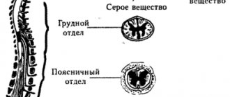
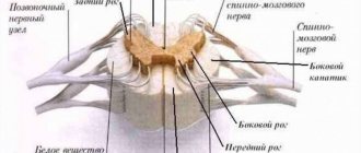
- The spinal cord is located in the spinal canal and has a cavity filled with cerebrospinal fluid. It is presented in the form of a long cord and provides communication between the cerebrum and the periphery. The spinal cord is divided into segments and performs reflex activities. Information flows through the spinal canal to the brain.
The hierarchy of these structures in relation to the cortex is lower, but each performs important functions and, in case of violations, independent self-government is triggered. The subcortical region is represented by a complex of various formations that are involved in the regulation of behavioral reactions.
Embryology
First at 3 months. During embryonic development, the lateral (Sylvian) fossa appears. Its bottom is formed by a slowly growing bark, which later forms an island. Rapidly growing neighboring areas of the cortex cover it and form folds—tires. The line of their contact forms the lateral (Sylvian) fissure. At 5-6 months. During embryonic development, the central, parieto-occipital and calcarine grooves appear. Following them, in the subsequent months of development, the remaining grooves and convolutions are formed. Based on the timing of the appearance of grooves and convolutions in the process of development, their depth and constancy, D. N. Zernov identified 3 types of grooves: primary grooves - constant, deep, appear early in the process of ontogenesis; secondary furrows, also permanent, but more variable in configuration, appear later in the process of ontogenesis; tertiary grooves, inconsistent, may be absent, very variable in shape, length and direction. By deep primary grooves, each hemisphere is divided into lobes: frontal (lobus frontalis), parietal (lobus parietalis), temporal (lobus temporalis), occipital (lobus occipitalis) and insula (insula); some authors also identify the limbic lobe or region.
In the cerebral hemispheres, there is a superficial (cortical) mantle part with grooves and convolutions located on it. Based on phylogenetic development, the brain cloak is divided into ancient (paleopallium), old (archipallium) and new (neopallium). So-called primitive grooves belonging to paleopallium and archipallium, generally very few in number, are already visible in reptiles. In mammals, grooves are also present in the neopallium.
Frontal cortex and hippocampus age faster
Areas such as the frontal cortex and hippocampus are more susceptible to aging than other parts of the brain. This may manifest as a decrease in the volume of gray and white matter and the level of activity in these areas.
The frontal cortex is used in planning and predicting the outcome of actions, it is responsible for controlling our behavior, while the hippocampus is extremely important for long-term memory and storing information (in turn, the frontal cortex is responsible for using this stored information).
So one answer to why older people become more forgetful may be that their hippocampus shrinks, making it harder to find and retrieve information.
Some of these studies compared a group of older adults (aged 60 to 80 years) with a younger group (aged 20 to 40 years). This is not an ideal method, since within-group differences independent of age also play a role. Older people lead different lifestyles, have different social contacts or eating habits, which influence the results. From a scientific perspective, more convincing results can be obtained by following one group of people using MRI scans every 10 years or so to track changes in their brains.
Location of the hippocampus in the brain
Large-scale studies using MRI to measure brain volume have shown that the frontal regions shrink more in size compared to areas at the back.
Furrows and convolutions of the parietal lobe. Postcentral sulcus, sulcus postcentralis.
Parietal lobe
. On it, approximately parallel to the central groove, there is a sulcus postcentralis, usually merging with the sulcus intraparietalis, running in a horizontal direction.
Depending on the location of these grooves, the parietal lobe is divided into three gyri, of which one is vertical and the other two are horizontal.
The vertical gyrus, gyrus postcentralis, runs behind the sulcus centralis in the same direction as the gyrus precentralis, separated from it by the central sulcus. The superior parietal gyrus is located above the sulcus intraparietalis
, or slice,
lobulus parietalis superior, which extends to the medial surface of the hemisphere.
Below the sulcus intraparietalis lies the lobulus parietalis inferior, which, going backwards, bends around the ends of the lateral groove and sulcus temporalis superior and is lost in the region of the occipital lobe. The part of the lobulus parietalis inferior that goes around the lateral sulcus is called gyrus supramarginalis; the other part, which goes around the sulcus temporalis superior, is called gyrus annularis.
Occipital lobe
The boundaries of the occipital region of the brain are outlined by the following formations: it is separated from the parietal lobe by the parieto-occipital recess, from below the occipital part smoothly flows into the basal surface of the brain.
It is in this area of the brain that the most unstable structures are located. But the posterior occipital gyrus of the brain is present in almost all individuals. Moving closer to the parietal region, transitional gyri are formed from it.
On the inner surface of this area there is a calcarine groove. It separates three convolutions from each other:
- wedge;
- lingular gyrus;
- occipitotemporal gyrus.
There are also polar grooves that have a vertical direction.
The function of the most posterior lobe of the brain is the perception and processing of visual information. It is noteworthy that the projection of the upper half of the retina of the eyeball is in the wedge, but it perceives the lower part of the visual field. And the lower half of the retina, which receives light from the upper visual field, is projected in the region of the lingual gyrus.
Does the number of convolutions affect the level of intelligence?
Today it has been scientifically proven that the number of convolutions, as well as the mass of the brain, cannot in any way affect the mental development of a person. And even if you read the works of ancient Greek philosophers from morning to night, you will not gain as many gyri as you do grams of weight. This is logical, because the human convolutions in the form in which they remain throughout life are formed during the period of intrauterine development, and the weight of the brain depends on the body composition.
Some scientists and ordinary citizens who donate their bodies to science after death have allowed repeated studies to be conducted that have established that physiological differences between the brains of ordinary people and scientists do not correlate with the intelligence demonstrated during life.
Formation of the cortex in embryogenesis
The grooves and gyri in neuroanatomy that give the brain its wrinkled appearance serve two critical functions. They help increase the surface area of the cortex, which allows more neurons to pack into it and enhance the brain's ability to process information. The sulci and convolutions of the brain form divisions, creating boundaries between the lobes of the brain, dividing it into two hemispheres.
Important What does an MRI of the pituitary gland show and when is it prescribed?
Main grooves:
- The interhemispheric fissure is a deep groove in the center of the brain that contains the corpus callosum.
- The Sylvian fissure (lateral sulcus) separates the parietal and frontal lobes.
- Roland's fissure (central sulcus), separating the fusiform gyrus and the hippocampal gyrus on the inferior surface of the temporal lobes.
- Parieto-occipital - separates the parietal and occipital lobes.
- The calcarine fissure (spur-like groove or prominent fissure) is located in the occipital lobes and divides the visual cortex.
The main convolutions of the brain:
- The angular gyrus of the parietal lobe helps in processing auditory and visual recognition.
- Broca's gyrus (Broca's center) is an area of the brain located in the left frontal lobe in most people that controls functions related to speech production.
- The cingulate gyrus, an arched fold located above the corpus callosum, is a component of the limbic system and processes sensory input regarding emotions and regulates aggressive behavior.
- The fusiform gyrus is located in the temporal and occipital lobes and consists of lateral and medial parts. It is thought to play a role in word and face recognition.
- The hippocampal gyrus lies on the inner surface of the temporal lobe, which borders the hippocampus. Plays an important role for memory.
- The lingual gyrus in the occipital lobe is involved in visual processing. It is limited by the collateral groove and calcarine fissure. In front it is in contact with the pararpopampal gyrus, and together they form the medial part of the fusiform gyrus.
It is followed by the central one, separating the motor cortex (precentral gyrus) from the somatosensory cortex (postcentral gyrus). Most of the cortical sulci and gyri of the brain, the anatomy of which begins to take shape between 24 and 38 weeks of pregnancy, continue to grow and develop after the newborn is born.
The early state of the brain has a strong influence on the final level of gyrification. In particular, there is an inverse relationship between cortical thickness and gyrification. Brain regions with low thickness values have a higher level of gyrification. The opposite is also true: brain regions with a high thickness value (for example, thickening of the hippocampal gyrus cortex) have a low level of gyrification.
Embryogenesis is the intrauterine development of the fetus from the moment of conception to birth. First, uneven depressions form on the cerebral cortex, which give rise to furrows. The primary grooves are formed first. This occurs around the 10th week of intrauterine development. After this, secondary and tertiary depressions are formed.
The deepest groove is the lateral one; it is one of the first to form. It is followed in depth by the central one, which separates the motor (motor) and sensory (sensitive) zones of the cerebral cortex.
Bottom surface
The lower, or basal, surface of the brain is formed by parts of the frontal, temporal and occipital lobes. However, in addition to these structures, the so-called olfactory brain is also located on the basal surface. It consists of the olfactory sulcus, surrounded by the straight gyrus and orbital sulci.
The temporal lobe, based on the brain, contains the inferior temporal and occipitotemporal sulci, between which the gyrus of the same name is located. The lingular gyrus is also detailed nearby.
Structure
The pattern of convolutions and sulci of the brain is best seen in schematic images. The depressions dividing the cortex into two parts (hemispheres) are called primary. In addition, there are other fundamental limitations of the cortex, namely:
- Sylvian fissure (lateral, lateral): separates the temporal and frontal cortex.
- Roland's fossa (central): separates the parietal from the frontal.
- Parieto-occipital fossa: separates the occipital and parietal lobes of the brain.
- The cingulate recess, which passes into the hippocampal recess: separates the surface of the olfactory brain from other parts.
These structures also have another name: first-order sulci of the brain.
Each part of the telencephalon contains several convolutions, separated by secondary cavities. Tertiary depressions develop purely individually: their presence depends on the personal characteristics of a person and his mental abilities. The third type of notches gives individual relief to the folds.
Superolateral part of the hemisphere
This area of the telencephalon is limited by three sulci: the lateral, part of the occipital and central. The lateral cavity originates from the lateral fossa. Developing slightly upward and backward, the formation ends on the superolateral surface.
The central sulcus begins at the upper edge of one of the hemispheres. From its middle it goes backward and partially forward. In front of this notch is the frontal lobe of the brain, and behind it is the parietal cortex.
The end of the occipital region serves as the edge of the parietal region. This groove does not have a clear boundary, so the separation is carried out artificially.
Medial surface of the brain
This part of the hemispheres has permanent deep grooves. When talking about the formations of the medial surface, first of all, as a rule, one thinks of the groove of the corpus callosum (1). Above this groove there is a belt cavity (2), forming a knee and subsequently a branch. Also in this area is the hippocampal sulcus (3) or seahorse sulcus. Closer to the occipital lobe is the collateral groove (4). On the territory of the posterior part of the median surface there is a calcarine groove (5).
Between the first two formations is the encircling gyrus. And the hippocampal and collateral groove limits the gyrus belonging to the temporal cortex of the hemisphere.
Furrows and convolutions of the lower surface of the cortex
This part of the brain is distributed in different parts of the cortex - temporal, occipital and frontal. The lower surface includes the following grooves:
- Olfactory (1)
- Orbital (2)
- Straight (3)
- Inferior temporal (4)
This area of the hemisphere does not have prominent gyri, however, one should still be noted - this is the lingular gyrus (5).
Medial surface
The sulcus of the corpus callosum is located most medially, which then passes into the sulcus of the hippocampus, which borders the hippocampus itself. Next to the callosal sulcus are the subparietal and callosal-marginal sulci. The rhinal sulcus runs parallel to the hippocampus.
The recesses of the brain listed above limit a specific system, which is called limbic. It, in turn, consists of the cingulate and hippocampal gyri.
In addition to the limbic system itself, on the inner surface of the brain there are also structures that continue their course from the outer part of the cerebral cortex. In this way, the parieto-occipital groove extends, behind which the precuneus is located (a gyrus resembling a trapezoid in shape). Next to this depression there is also a calcarine groove, which extends from the back of the head and forward all the way to the corpus callosum. Between the two recesses mentioned above is the sphenoid gyrus.
What is this?
The topography of the contents of the cranium showed that the surface of the organ responsible for the functioning of the human body is a series of elevations and depressions, which become more pronounced with age. This is how the area of the brain expands while maintaining its volume.
Gyri are the folds that characterize an organ in the final stage of development. Scientists associate their formation with different levels of tension in the brain in childhood.
The grooves are the canals that separate the convolutions. They divide the hemispheres into main sections. According to the time of formation there are primary, secondary and tertiary types. One of them is formed during the prenatal period of human development.
Others are acquired at a more mature age, remaining unchanged. The tertiary sulci of the brain have the ability to transform. Differences may relate to shape, direction and size.
Types of furrows
Fissures are, roughly speaking, gaps in the brain that form more convex parts - convolutions. The following main grooves of the brain can be distinguished:
- primary formed - the deepest, divide the cortex into separate lobes (frontal, occipital, temporal, insular, parietal);
- secondary - less deep, they divide the brain into small convoluted parts - convolutions;
- additional (tertiary) - the most superficial, designed to give a specific shape to the gyri and to increase the surface of the cortex.
Temporal lobe
This structure of the brain is limited by the following grooves: the lateral one from above, a conventional line between the lateral and posterior occipital grooves at the back.
The temporal lobe, by analogy with the frontal lobe, consists of three large convolutions:
- superior temporal;
- average;
- lower
The name of the depressions corresponds to the convolutions.
On the lower surface of the temporal region of the brain, the hippocampal gyrus and the lateral occipitotemporal gyrus are also distinguished.
Wernicke's speech center is located in the temporal lobe, which was already mentioned earlier in the article. In addition, this area of the brain performs the functions of perception of taste and olfactory sensations. It provides hearing, memory, and synthesis of sounds. Specifically, the superior temporal gyrus, as well as the inner surface of the temporal region, is responsible for hearing.
Thus, the lobes and convolutions of the brain are a complex and multifaceted topic to understand. In addition to the parts discussed in the article, there is also the limbic cortex with its own relief, a structure called the insula. There is a cerebellum, which also has a cortex with its own characteristics. But the anatomy of the brain should be studied gradually, so this article provides only basic information.
Structure of the cortex
Thanks to the presence of the cortex, a person is able to experience emotions, navigate himself and the surrounding space. What is noteworthy is that the structure of the bark is unique. The grooves and convolutions of the cerebral cortex of one person have a different shape and size than that of another. But the general plan of the building is the same.
What is the difference between the sulci and convolutions of the brain? Fissures are depressions in the cerebral cortex that look like slits. They are the ones who divide the bark into shares. There are four lobes of the cerebral hemispheres:
- frontal;
- parietal;
- temporal;
- occipital
Gyri are convex areas of the cortex that are located between the furrows.