The median nerve is formed by fibers of a number of spinal nerves and has mixed functions. Its main purpose is to innervate some muscles of the forearm and hand. With the development of median nerve neuritis, deterioration in the functions of the affected limb, severe pain and a number of other symptoms appear. If treatment is not provided in a timely manner due to poor quality, neuropathy of the median nerve can result in a lifelong loss of the ability to fully use the arm, up to and including disability.
Contacting specialist neurologists from the Health Workshop network of clinics will help you quickly carry out a diagnosis, draw up a list of necessary health measures and create conditions for complete relief from the manifestations of this disease. Trust your health to our doctors!
Causes
Inflammatory disease of peripheral nerves can be triggered by various factors:
- hypothermia;
- infectious diseases - influenza, measles, herpes, brucellosis, etc.;
- injuries accompanied by damage to nerve fibers;
- vascular diseases;
- lack of vitamins in the body;
- intoxication of exogenous and endogenous nature.
Often neuritis develops in the musculoskeletal canals. Their natural narrowness, due to anatomical features, may predispose them to the formation of inflammatory processes.
Another common cause of neuritis is compression of the peripheral nerve by surrounding tissues. This condition can be provoked by sleeping in an uncomfortable position, prolonged maintenance of an unnatural body position, which creates a load on individual organs, or mechanical pressure.
Neurolysis of peripheral nerves of the extremities
Causes of damage to the integrity or conduction of peripheral nerves
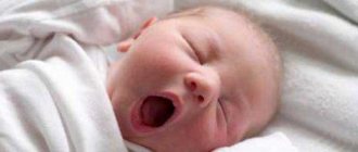
Violation of the integrity and conductivity of peripheral nerves can occur as a result of domestic or industrial trauma, injury or sports training. Damage to the nerves of the limbs occurs in newborns in cases of birth trauma. The appearance of hematomas, tumors, aneurysms, scarring after injuries, and the formation of bone calluses also leads to impaired sensitivity. Nerves are compressed, torn, thinned, which leads to loss of integrity and conductivity.
Lack of treatment provokes the replacement of the nerve with connective tissue, which aggravates the process, nerve cells are eliminated, the limbs completely lose sensitivity, functionality decreases, and chronic pain appears. Surgical intervention “neurolysis” is aimed at eliminating the causes of nerve compression, restoring the sensitivity of the tissue and limb.
Methods for diagnosing injury
The initial examination can detect damage to peripheral nerves in patients of any age. In the process of providing first aid, it is necessary to identify possible disorders of the skeletal system, muscles and exclude injuries to nerve endings. Signs of peripheral nerve damage:
- motor dysfunction;
- loss of sensation;
- weakness or loss of motor reflex.
The initial examination involves palpation and analysis of the complexity of the damage.
Neurolysis of peripheral nerves of the extremities
Neurolysis is the release of nerve fiber with the elimination of factors that interfere with its conduction.
Surgical intervention is indicated at different stages of the disease.
There are three periods: primary, delayed, late - an operation can be performed in any of them. Neurolysis is an effective new generation technique that restores damaged nerve endings and returns sensitivity and full functionality to the limbs.
In some cases, neurolysis surgery requires nerve transposition. The procedure for displacing or transferring a nerve fragment from another part of the body to the damaged area for implantation at the site of the rupture has been successfully used in practice. The operation is performed to remove factors such as scars, bone calluses - causes that cause not only impaired sensitivity, but also interfere with normal performance, causing pain due to constant irritation of nerve endings.
Surgical intervention is performed with a significant tissue incision. This is required to isolate the nerve from surrounding tissues. The surgeon determines the need for projection, non-projection roundabout or angular approach on the bends, paying special attention to maintaining the muscle connection with the nerve fibers.
The operation involves removing areas of the nerve that cannot be restored. After the procedure, the ends of the fibers are sutured, and the fragment is transposed. Carrying out neurolysis requires experience, special surgical techniques, special suture materials and micro-instruments.
Postoperative period and rehabilitation
Recovery after neurolysis includes:
- fixation of the limb with a splint to limit movement;
- wearing postoperative dressings;
- paraffin therapy courses;
- electrical stimulation treatment;
- completing a rehabilitation course of physical therapy;
- taking prescribed vitamins and nootropic drugs.
Diagnostics
The presence of the median nerve is indicated by the following results of functional tests:
- with the palm pressed to the table, the patient cannot make scratching movements with the 2nd finger;
- when the affected hand is clenched into a fist, bending the first three fingers is most difficult;
- the patient is unable to place the little finger and thumb on the same plane.
Additionally, various electrophysiological procedures may be prescribed. In each individual case, the neurologist working with you at the Health Workshop clinic will draw up an individual list of required examinations.
Diagnosis of median nerve neuropathy
When the C7-spinal nerve or the middle trunk of the brachial plexus is damaged, the function of the median nerve is partially affected, as a result there is a weakening of the flexion of the hand and its inward rotation in combination with damage to the radial nerve.
Almost the same loss of function of the median nerve occurs with damage to the external bundle of the brachial plexus, into which the fibers of the upper peduncle of the nerve pass from the middle trunk, but in combination with damage to the musculocutaneous nerve. When the spinal nerves C8–Th1 , the lower trunk and the internal bundle of the brachial plexus are damaged (Dejerine-Klumpke palsy), those fibers of the median nerve that make up its lower leg suffer in combination with damage to the ulnar nerve (weakening of the finger flexors and thenar muscles).
The motor function of the median nerve mainly consists of inward rotation of the hand, palmar flexion of the hand due to contraction of the corresponding muscles, flexion of fingers, mainly I, II and III, extension of the middle and terminal phalanges of II and III fingers.
The place of compression of the median nerve in carpal tunnel syndrome is at the level of the carpal tunnel.
Sensitive fibers of the median nerve innervate the skin of the palmar surface of the I, II, III and radial half of the IV fingers, the corresponding part of the palm, as well as the skin of the rear of the terminal phalanges of these fingers.
When the median nerve is damaged (median nerve neuritis), inward rotation of the hand suffers, palmar flexion of the hand is weakened, flexion of the 1st, 2nd and 3rd fingers and extension of the middle phalanges of the 2nd and 3rd fingers are impaired.
Superficial sensitivity in case of median nerve neuritis is impaired on the hand in an area free from the innervation of the ulnar and radial nerves. Articular-muscular sensation with neuritis of the median nerve is always impaired in the terminal phalanx of the index, and often in the second finger.
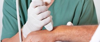
Diagnosis of the level of damage to the median nerve during neuritis is made using electroneurography (ENG).
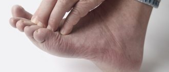
Muscle atrophy with damage to the median nerve is most pronounced in the thenar region. The resulting flattening of the palm and bringing the thumb close and in one plane to the index finger create a peculiar position of the hand, which is called “monkey”. Pain when the median nerve is damaged, especially partial, is quite intense and often takes on a causal nature. In the latter case, the position of the hand may become bizarre.
Vasomotor-secretory-trophic disorders are also common and characteristic of lesions of the median nerve: the skin, especially the 1st, 2nd and 3rd fingers, becomes bluish or pale in color; nails become “dull”, brittle and streaked; skin atrophy, thinning of fingers (especially II and III), sweating disorders, hyperkeratosis, hypertrichosis, ulcerations, etc. are observed. These disorders, as well as pain, are more pronounced with partial rather than complete damage to the median nerve.
The median nerve, like the ulnar nerve, gives off its first branches only to the forearm, so the clinical picture with high damage along the entire length from the axillary fossa to the upper parts of the forearm is the same. When the median nerve is damaged in the middle third of the forearm, the functions of internal rotation of the hand, palmar flexion of the hand and flexion of the middle phalanges are not affected.
The use of acupuncture is very effective in the treatment of median nerve neuritis.
With lower lesions of the nerve, the function of flexion of the terminal phalanges of the I, II and III fingers may be preserved, and then all symptoms of the lesion are limited to damage to the thenar and lumbrical muscles and sensory disturbances in the typical zone.
The main tests to determine movement disorders that occur with damage to the median nerve (median nerve neuritis) are the following:
- When the hand is clenched into a fist, fingers I, II and partly III do not bend
- Bending the terminal phalanges of the thumb and index fingers is impossible, as is scratching the index finger on the table with the hand tightly adjacent to it.
- During the thumb test, the patient cannot hold a strip of paper with a bent thumb and will hold it by adducting the straightened thumb with the adductor muscles from the preserved ulnar nerve
Treatment of tunnel neuropathies
The treatment of tunnel syndromes is based on conservative therapy, the goal of which is to decompress the nerve trunk and restore its functions. Physiotherapeutic methods (ultrasound therapy, UHF), acupuncture, and physical therapy are also used.
Pharmacotherapy of tunnel neuropathies includes the use of the following groups of drugs:
- decongestants (diuretics, L-lysine aescinate);
- non-steroidal anti-inflammatory drugs;
- analgesics (gabapentin);
- venotonics (phlebodia, venosmil);
- neurometabolic agents (alpha-lipoic acid preparations: dialipon, berlition);
- B vitamins;
- antihypoxants, antioxidants (actovegin).
Blockades with Xefocam are carried out locally into the tissue surrounding the nerve or the corresponding channel. Muscle massage is also performed in the innervation zone and directly at the point of compression.
A radical method of treating tunnel neuropathies is surgical intervention (the tissue surrounding the nerve is dissected, canal plastic surgery is performed), which is carried out when conservative methods are ineffective, there are severe neurological disorders, rapid progression of symptoms, and frequent relapses.
Symptoms of tunnel neuropathies
Signs of tunnel neuropathies depend on the location of the lesion; the main symptoms are severe pain in the area of innervation of the affected nerve, paresthesia, impaired sensitivity and motor function. Despite the topographical typicality of the localization of sensitivity disorders and pain, the signs of tunnel syndromes can vary significantly, which is due to areas of overlap of adjacent nerves, asymmetry of compression, as well as psychogenic characteristics.
Carpal tunnel syndrome
Carpal tunnel neuropathy, or carpal tunnel syndrome, occurs when the median nerve is compressed in the carpal tunnel, which is formed by osteoarticular structures and ligaments. It is the most common type of tunnel neuropathy and is thought to affect 1% of the population to varying degrees. Frequent, prolonged work with a computer mouse and keyboard plays an important role in the development of this pathology. Most often, carpal syndrome has a chronic course with periods of exacerbations.
The first symptoms of carpal tunnel syndrome are numbness of the hands, paresthesia and pain at night; As the pain progresses, it also occurs during the day, and there is a decrease in sensitivity in the area of 1-4 fingers and thenar muscles. When raising the arm and holding it above the head, the symptoms intensify (pain, paresthesia), which is associated with insufficient blood supply to the affected nerve. The pain is also provoked by strong flexion of the hand, as well as compression of the arm in the shoulder area by the cuff of the blood pressure measuring device until the pulse disappears. When movement disorders occur, the patient experiences difficulty fastening buttons and cannot lift an object with his index finger and thumb.
Ulnar nerve tunnel syndrome
Compression of the ulnar nerve can develop in the area of the elbow joint (in the cubital canal or in the area of its exit, where the nerve is surrounded by a fibrous arch), as well as in Guyon's bed (in the area of the wrist joint). Most often, ulnar nerve neuropathy occurs due to prolonged compression in patients who are unconscious (the ulnar nerve is compressed between the arm and the bed), and during long telephone conversations (the nerve trunk is compressed when holding the arm in a bent state). The main symptoms are paresthesia, pain, itching in the area of the ulnar edge of the hand (on the side of the 5th finger), in the fifth and half of the fourth finger. Symptoms are aggravated by tapping the nerve in the elbow joint. In the case of Guyon's neuropathy, there is a violation of abduction of the 4th and 5th fingers.
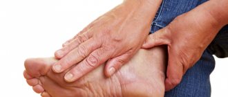
Tunnel neuropathy of the external cutaneous nerve of the thigh
This type of tunnel neuropathy develops when the nerve is compressed under the inguinal ligament or in the area of the anterior superior iliac spine. The cause is most often obesity, as well as wearing tight belts. Paresthesia and pain are localized along the anterolateral surface of the thigh, possibly impaired hair growth and decreased sweating.