Peroneal nerve neuropathy
Neuropathy of the peroneal nerve, or peroneal neuropathy, occupies a special position among peripheral mononeuropathies, which also include: neuropathy of the tibial nerve, neuropathy of the femoral nerve, neuropathy of the sciatic nerve, etc. Since the peroneal nerve consists of thick nerve fibers that have a larger layer of myelin sheath, then it is more susceptible to damage due to metabolic disorders and anoxia. This point is probably responsible for the fairly wide prevalence of peroneal neuropathy. According to some data, neuropathy of the peroneal nerve is observed in 60% of patients in traumatology departments who have undergone surgery and are treated with splints or plaster casts. Only in 30% of cases, neuropathy in such patients is associated with primary nerve damage.
It should also be noted that often specialists in the field of neurology have to deal with patients who have a certain history of peroneal neuropathy, including the postoperative period or time of immobilization. This complicates treatment, increases its duration and worsens the result, since the earlier therapy is started, the more effective it is.
Anatomy of the peroneal nerve
The peroneal nerve (n. peroneus) arises from the sciatic nerve at the level of the lower 1/3 of the thigh. It consists predominantly of fibers of the LIV-LV and SI-SII spinal nerves. After passing through the popliteal fossa, the peroneal nerve exits to the head of the bone of the same name, where its common trunk is divided into deep and superficial branches. The deep peroneal nerve passes into the anterior part of the leg, descends, passes to the dorsum of the foot and divides into internal and external branches. It innervates the muscles responsible for extension (dorsal flexion) of the foot and toes, pronation (raising the outer edge) of the foot.
The superficial peroneal nerve runs along the anterolateral surface of the leg, where it gives off a motor branch to the peroneal muscles, which are responsible for pronation of the foot with its simultaneous plantar flexion. In the area of the medial 1/3 of the tibia, the superficial branch of n. peroneus passes under the skin and is divided into 2 dorsal cutaneous nerves - the intermediate and medial. The first innervates the skin of the lower 1/3 of the leg, the dorsum of the foot and the III-IV, IV-V interdigital spaces. The second is responsible for the sensitivity of the medial edge of the foot, the back of the first toe and the II-III interdigital space.
Anatomically determined areas of greatest vulnerability of the peroneal nerve are: the place where it passes in the area of the head of the fibula and the place where the nerve exits the foot.
Causes
Most often, neuritis (neuralgia) of the peroneal nerve is caused by its compression by anatomical structures according to the principle of tunnel neuropathy, mainly near the head of the bone. Thickening of the walls of anatomical tunnels can occur as a result of a general disease (gout, polymyositis, rheumatoid arthritis, diabetes, osteochondrosis, etc.).
Promote the development of neuritis (neuralgia) of the peroneal nerve:
- injuries;
- prolonged squatting or kneeling;
- acute and chronic infectious diseases;
- intoxication with heavy metals, alcohol, etc.;
- wearing tight shoes;
- impaired blood supply to the legs due to thrombophlebitis and other diseases of the arteries and/or veins;
- tumor formations, etc.
Treatment methods and duration
To eliminate pathological changes at the initial stage, conservative treatment methods are used. After examination by a specialist, the patient undergoes a therapeutic course at home. Ointments with anti-inflammatory non-steroidal properties with an analgesic effect, compresses with dimexide, diuretics, and vitamin-mineral complexes are indicated for use. The affected areas are fixed with bandages, splints or orthoses. Physical activity is limited. After the symptoms have weakened, simple gymnastic exercises are recommended. If the disease progresses, the patient is hospitalized. Treatment includes intramuscular and intravenous administration of non-steroidal drugs.
An effective remedy in the fight against painful symptoms is considered to be novocaine blockade with the simultaneous administration of hydrocortisone.
Elimination of exacerbations and complications by surgical method
If all the steps taken do not give a lasting result, the disease periodically worsens, and surgical intervention will be required. A drastic measure will prevent a decrease and complete loss of motor functions. The operation is also indicated for the development of syndrome after a bone fracture with displacement of bone fragments. The operation is performed by orthopedic surgeons, neurosurgeons, and general surgeons. Having numbed the surgical field, the specialist makes a dissection of the skin, subcutaneous tissue and ligament. Carefully examines the carpal tunnel, eliminates compression of the nerve fibers by hypertrophied ligaments, after which the incision is sutured. In addition to the open incision technique, a new technique is used - endoscopic surgery. A small device with a camera equipped with a magnifying lens is inserted into a narrow depression made in the wrist. Special surgical instruments are inserted into the incision in the palm. Medical equipment helps the doctor navigate the anatomical structure of the hand and carry out the necessary manipulations with the least complications for the patient. The consequences of surgical treatment are long-term rehabilitation. It may take up to 2 months to restore functional abilities.
Recommendations for daily lifestyle
A person's health depends on his lifestyle. Daily activity, balanced nutrition, proper rest, emotional stability are the handrails that keep the body in good condition. If the biological system fails, you should change your behavior tactics. If possible, reorient your type of work activity. As a last resort, choose a more comfortable position and ergonomic tools. When working at a computer, adjust the height of your chair in relation to the table. Choose a work chair with comfortable armrests. Use a wrist rest. You can do it yourself. Choose a mouse and keyboard based on your individual needs. Wear a brace or a regular elastic bandage on your hand. Non-monotypic movements are the best prevention of carpal tunnel syndrome. Take short breaks from work. A person’s left hand can easily perform all the functions of the right hand and vice versa. Improve your abilities - this will allow you to change hands while working. Try to be in the fresh air. Oxygen improves blood circulation and activates metabolic processes. Watch your body weight. Eliminate foods high in cholesterol and carcinogens from your diet. Control the duration of your workload. Workaholism is a painful psychological addiction.
Treatment
Treatment of neuritis (neuralgia) of the peroneal nerve is aimed at identifying and eliminating the main cause of the disease. At the First Medical Clinic, only non-surgical methods are used to achieve this goal.
Relief of the inflammatory process is successfully achieved through local injection therapy - the introduction of anti-inflammatory, analgesic, antibacterial, vitamin and other drugs into the area of the affected nerve. therapeutic blockades are indicated - injections of anesthetic into the points of maximum pain.
Pathological changes in tissue that cause compression and neuritis of the peroneal nerve are well eliminated by injections of platelet autoplasma - plasma obtained from the patient’s blood using a special technology. This method is called autoplasma therapy and is widely used in the treatment of various diseases. Autoplasma therapy relieves swelling, inflammation and pain, and also stimulates the self-regeneration of damaged tissues.
In the most common variant of the pathology, when pinching and subsequent inflammation of the nerve is caused by overload of the peroneal muscles due to problems with the spine, the use of muscle relaxants, physiotherapy, therapeutic massage, reflexology, manual therapy, shock wave therapy and osteopathy is effective.
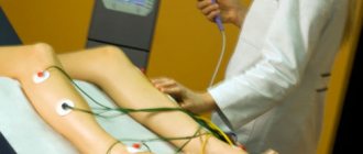
To stimulate muscle function, transcutaneous electromyoneurostimulation - delivering weak impulses of electric current to the area of nerve endings.
The required amount of therapeutic assistance can only be determined by a specialist after diagnostic procedures. If symptoms of neuritis (neuralgia) of the peroneal nerve appear, we recommend that you contact the First Medical Clinic in a timely manner.
You can make an appointment for a consultation and appointment by phone
Reasons for the development of pathology
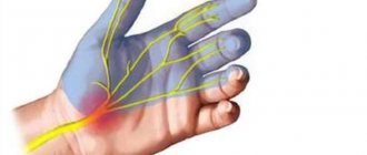
Carpal tunnel syndrome is a little-known diagnosis, but quite common. The focal point of the pathology is the joints. Due to increased motor activity, they are most often subject to degenerative, traumatic, and inflammatory changes. Human activity—professional, sports, and everyday life—leads to frequent macrotraumatization of the nerve trunk. Therapeutic fasting and long-term diets provoke pathology. Subcutaneous fat tissue acts as a shock absorber in the body. The loss of the soft elastic pad leads to a malfunction of the biological system. Tunnel syndrome is included in the status of occupational diseases of freelancers, programmers, drivers, cashiers, musicians, and hairdressers. Persons whose activities involve monotonous movements are at risk. Gamers who spend days and nights playing computer games, in the heat of passion, forget that their hobby can have serious consequences. They are susceptible to carpal tunnel syndrome, the most common type of tunnel neuropathies.
Factors that can give impetus to the development of a specific disease are diabetes, gout, arthritis, kidney failure, blood diseases, alcohol and drug addiction. Medical intervention leads to damage to the ulnar and median nerves during prolonged intravenous invasions. The syndrome is associated with endocrine disorders: pregnancy, lactation, menopause, thyroid dysfunction, long-term use of contraceptives. In some cases, a genetic predisposition is a prerequisite for the occurrence of pathology: hereditary narrowness of the canals and increased vulnerability of nerve fibers.
Diagnostic measures
A neuropathologist is responsible for the diagnostic determination of pathology. During the examination, the specialist assesses the sensitivity of the fingers. Then, using a dynamometer, he measures the strength of the arm muscles.
- Phalen test - increased pain when raising the arms up indicates a pinched nerve in the carparal canal;
- opposition test - the inability to connect the thumb with the little finger;
- Tinel test - pain and tingling in the fingers when tapping on the wrist;
- Durkan test - a shooting sensation when clenching your hand into a fist.
- electromyography - a diagnostic method that allows you to evaluate the bioelectrical activity of muscles and nerve endings at rest and in motion; The procedure identifies areas of muscle damage. Nerve conduction analysis, a test that determines the speed and strength of electrical signals in nerves; slowing of the impulse indicates pinching of the median nerve.
To exclude pathologies similar in their symptoms to carpal tunnel syndrome, radiography is prescribed.
1. Reasons for violation of the integrity or conductivity of peripheral nerves
Impairment of the integrity or conduction of peripheral nerves
occurs as a result of domestic or work injuries, wounds (stabbing, gunshot) or sports training (sprains, fractures, dislocations).
There are frequent cases of birth trauma in newborns, aggravated by damage to the nerves of the limbs. Also complicated by impaired nerve conduction can be tumor diseases, hematomas, aneurysms, calluses and scars after suppuration. The nerve can be completely dissected, torn, compressed, or thinned. In the absence of proper surgical treatment at the site of injury, nerve cells are replaced by connective tissue. These scars that form impede nerve conduction. The limb loses sensitivity, loses functionality, this may be accompanied by chronic pain
.
Surgical treatment “neurolysis”
is used precisely in such situations. It is aimed at releasing the nerve from compression and restoring its conductivity.
A must read! Help with treatment and hospitalization!
Symptoms
Manifestations of neuritis (neuralgia) of the peroneal nerve depend on which part of it is affected.
When deep branches are affected, flexion/extension on the back of the fingers and feet is impaired, and sensitivity in the space between the first two fingers is reduced.
With the development of pathology in the upper part of the nerve, the function of the muscles in the anterior region of the leg is disrupted.
Inflammation of the superficial branch is accompanied by paralysis of the peroneal muscles.
If the common peroneal nerve is damaged, all or some of the symptoms described above may develop, and the following may also be observed:
- foot drop with tucking inward and curling of toes;
- “cock” gait (steppage, peroneal gait) - when walking, the leg is raised high so as not to touch the surface with the toe, and when lowered, first the toe of the foot touches it, then the outer edge and only then the heel;
- inability to move on your heels;
- pain (neuralgia of the peroneal nerve), which is usually unexpressed, has a burning sensation, and is localized along the outer posterior surface of the leg and foot.
