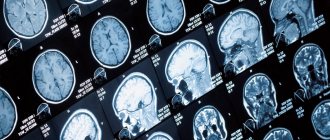
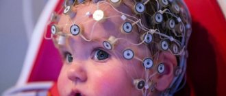
Image of the brain on MRI results Magnetic resonance imaging is one of the best methods for diagnosing the brain. It is painless and has a small list of contraindications.
Neurosurgeons prescribe a study for an accurate diagnosis after injuries and when pathologies are suspected, manifested by complaints of acute or persistent headache, tinnitus, confusion, weakness, memory impairment, etc.
Along with computed tomography, MRI plays an important role in recognizing life-threatening diseases. When a doctor refers patients for a magnetic wave scan, many people wonder if they need to prepare for an MRI of the brain. The answers are in this article.
The essence of the brain MRI procedure
To know how to prepare for an MRI of the brain, you need to understand the features of the procedure. During the examination, the magnetic field of the device affects the hydrogen atoms present in the body. The tomograph's sensors record pulses, which are modeled into images of areas of interest using software.
MRI allows you to identify brain diseases in the early stages, detect neoplasms and inflammatory processes, visualize blood vessels, etc. Using a contrast agent, you can measure a tumor, determine the exact location of the lesion, and the presence of metastases. Magnetic resonance imaging is absolutely painless and safe, does not have a negative effect on the body, and therefore can be performed repeatedly, even for pregnant women and children.
How preparation for research affects the result
It is important to remain still during the examination, otherwise the image quality may be somewhat distorted. Throughout the examination, the patient is under the supervision of a specialist. Inside, there is a button at hand to contact staff in case of an emergency. When examining children in the examination room, in exceptional cases (depending on age), an accompanying person may be present.
If you have a pacemaker, there are serious contraindications for MRI examination. You should consult your cardiologist as MRI may affect the implants and their function. When receiving a referral for diagnostics, you should have the implant passport with you.
Metal parts that are in the body (surgical screws, replaced joints, artificial heart valves, tattoos with metal particles) must be told to the doctor before the examination. Magnets can interfere with the normal functioning of pacemakers and can move pieces of metal that are present in the body.
A strong magnetic field can affect magnetic cards, electronic devices, and any magnetized metal body located inside or outside the body. Therefore, before entering the examination room, you must remove jewelry and watches. Personal belongings may be handed over to accompanying persons or placed in storage rooms located outside the examination room.
Rules for undergoing MRI of the brain
Before the procedure, many are interested in how to prepare for an MRI of the brain. The upcoming diagnosis requires compliance with the following recommendations:
- Choose clothes without metal inserts, buttons, buttons, locks, etc. Inspect the pockets for small change, keys, and magnetic cards.
- You should come to the examination without jewelry (earrings, rings, chains), otherwise you will have to take it off in the locker room.
- Enter the room where diagnostic equipment is located without a phone, belt, watch or other accessories containing metal.
Magnetic resonance imaging
Before an MRI of the brain, the attending physician clarifies the nuances that may be a reason to refuse the procedure:
- Scanning using a magnetic field cannot be done if there are metal objects in the body in the form of:
- plates;
- clips on vessels installed during the treatment of an aneurysm;
- implants;
- pins, etc.
The above structures may become displaced during the procedure, which can lead to unwanted complications. Among the metals that do not react to the magnetic field of the device are titanium and gold. If the patient has undergone surgery for joint replacement, installation of a shunt, etc., it is necessary to clarify before the study what alloy the product is made of.
- Magnetic resonance scanning is dangerous if there are electronic devices in the body (pacemaker, etc.). The induction field negatively affects the operation of devices.
- MRI has limitations during pregnancy. Scans of the brain and other organs are done no earlier than 13 weeks due to the potential negative impact of magnetic radiation on the embryo.
- MRI can be used from early childhood, but up to five years of age, children are usually examined in a hospital under anesthesia.
- The procedure in a tunnel tomograph is contraindicated for people with a fear of closed spaces. To exclude panic attacks in such patients, the study is performed on an open-type device or in a state of medicated sleep.
How to Prepare for a Magnetic Resonance Imaging Scan
The principle of MRI is the use of a magnetic field that affects water dipoles in cells. The saturation of tissues with liquid is assessed by the intensity of the reverse reaction. The procedure lasts approximately half an hour, if contrast is used - 45 minutes.
The patient is asked to lie down on a mobile couch, after which his head is secured with straps to ensure immobility. This is all part of the preparation - the final manipulations.
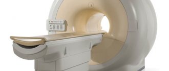
A magnetic pulse generator with indicators that read data is installed in a wide part of the tomograph, which has a pipe configuration. The platform with the patient slides into the scanning apparatus section, where the brain is examined in various projections.
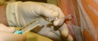
It is possible to increase the information content of the examination through the use of a contrast agent, the main component of which is gadolinium. The contrast is injected into a vein and is non-toxic. Its removal from the body takes 1-2 days. A contrast agent is used to illuminate brain vessels and visualize the slightest deformations in soft tissues and blood vessels.
During the examination, the tomograph makes noise; headphones or earplugs help reduce discomfort. During the consultation, the doctor explains to the patient that he will monitor the process from the next room and that if necessary, he can be contacted through a two-way voice communication system.
To perform an MRI examination of the brain, no complex preparation is required. The patient should consult a physician to exclude the presence of factors that are considered a contraindication for magnetic resonance examination. These include:
- The presence of metal products in the patient’s body – these can be pins with dental crowns or implants.
- Electromagnetic devices integrated into the body - pacemaker, insulin pump.
- Numerous tattoos on the skin, made using inks with a high concentration of metal.
What to take for an MRI of the brain?
In the medical department, MRIs are performed upon the direction of a doctor and upon self-referral. To obtain informative photos, it is recommended to take to the clinic previous examination results, an extract from the outpatient card, papers from the hospital and other documents related to the disease.
Before an MRI of the brain, tell your doctor if you have a fixed denture, an orthodontic structure to correct your bite, or tattoos that contain metal in their ink.
By adhering to the recommendations, the patient has a better chance of receiving comprehensive information about the pathologies of cerebral structures and the dynamics of changes during treatment.
Basic guidelines that people should follow when preparing for an MRI
When using a contrast agent, you should inform the specialist performing the procedure about the presence of allergies, problems with the heart, liver or kidneys, or other previously identified diseases. The patient must tell the doctor what medications he took in anticipation of the procedure.
If a person experiences fear of closed spaces, this should also be reported to the doctor. In such cases, doctors often prescribe sedatives to help prevent panic attacks during the examination and complete it successfully.
It is not advisable for women in the first trimester of pregnancy to undergo an MRI. This is explained by the fact that the effect of a magnetic field on an embryo during organ formation has not been fully studied. Patients during lactation should avoid breastfeeding for two days after the procedure. This will prevent the penetration of gadolinium salts into the baby’s body during MRI with contrast.
Immediately before the examination, the patient is asked to remove his clothes and change into a more comfortable medical gown. He should not enter the diagnostic room with bank cards, telephone, jewelry or costume jewelry made of metal. You must also remove your watch if you have one.
Having completed all these instructions, the patient lies down on a retractable table and follows all the doctor’s recommendations during the procedure.
Is it possible to drink alcohol before an MRI of the head?
Alcoholic drinks are strictly prohibited before the study. Drinking alcohol is permissible no later than 48 hours before the MRI. If contrast is required, a patient in a state of intoxication will not be able to undergo the examination. The administration of the drug is highly undesirable because:
- the indicator is sensitive to alcohol;
- alcohol dilates and then constricts blood vessels - a violation of the blood supply to the brain gives a corresponding signal on MRI.
A ban on alcohol before an MRI.
Going through a native procedure in an inadequate state is also problematic: a person will not be able to calmly endure the session, which will affect the quality of the images. Experts are unanimous in their opinion that alcohol should be excluded in the process of preparing for an MRI of the brain with and without contrast.
Smoking recommendations allow nicotine consumption no later than 120 minutes before the procedure.
Possible contraindications
Even the safest procedures must be done as prescribed by a specialist. The following situations require special attention:
- the presence of metal elements inside the body, implanted prostheses that cannot be removed even for a short time;
- severe diabetes mellitus;
- failure of the liver, kidneys, heart;
- neurological diseases with tics, uncontrolled movements;
- psychical deviations;
- intolerance to certain medications.
The listed restrictions may become conditional if you consult a doctor in advance. He will select an MRI examination program that will not cause harm and will overcome contraindications.
Is a diet indicated before an MRI of the head?
The procedure for preparing for MRI of the brain does not provide any restrictions in this regard. The patient can adhere to his usual diet and diet. During the procedure it is necessary to remain still, so before the session it is important to eliminate discomfort in the abdomen. You should not eat exotic dishes, diuretic foods, or foods that cause excess gas. Avoid excessive consumption of coffee, energy drinks and other stimulating drinks.
Visualization of pathologies on MRI with and without contrast
How often can I scan with a tomograph?
During scanning of the vascular system of the brain, the body is not exposed to radiation, so there are no restrictions on the frequency of such examinations. The decision on the appropriateness of such a diagnosis is made by the attending physician. For multiple sclerosis, for example, the procedure is performed 1 to 2 times a year. It is prescribed after strokes; in such cases, preventive examinations can be done up to 4 times a year. In Alzheimer's disease, a one-time diagnosis is sufficient to confirm the presence of pathology.
Why is virtual angiography needed?
With the help of such an examination it is possible to establish:
- Structural anomalies;
- Aneurysms;
- Dissection of vascular walls;
- Atherosclerosis;
- Neoplasia of various origins;
- Secondary foci of cancer;
- Degree of stenosis;
- Ischemic and hemorrhagic stroke;
- Hemorrhage;
- Pathological vascularization;
- Hemodynamic disturbances leading to decreased cerebral circulation;
- Vertebrobasilar insufficiency;
- Thrombosis of the cerebral sinuses.
Registration for examination
To book the type of study you need, contact the service advisory service at the number at the top of the page. Operators will select the required options, answer remaining questions, provide information on addresses and costs, and reserve an MRI or CT session with special discounts for portal users. Consultation and other services of the recording service are absolutely free.
Sources used:
- Belova, L.A. Venous cerebral discirculation in chronic cerebral ischemia: clinical picture, diagnosis, treatment / L.A. Belova // Neurological Bulletin named after. V.M. Bekhtereva – .2010. – T. 42, No. 2. – P. 62–67.
- Bozhko, O.V. The role of neuroimaging methods in the early detection of Alzheimer's disease / O.V. Bozhko, S.I. Gavrilova, Ya.B. Fedorova // “Psychiatry”. – 2006. – No. 2 – P.54–59.
- Alzheimer's disease: textbook. allowance / A.Yu. Emelin et al. – St. Petersburg, 2016. – 76 p.
- Varakin, Yu.Ya. Prevention of cerebrovascular accidents / in the book. Essays on angioneurology // Yu.Ya. Varakin; edited by BEHIND. Suslina. – M., 2005 – 368 p.
- Vasenina, E.E. Primary progressive aphasia / E.E. Vasenina, O.S. Levin // Journal of Neurology and Psychiatry. – 2014. –T. 6, No. 2. – P. 3–12.
- Venous dysgemia and cognitive deficit in patients with dyscirculatory encephalopathy / S.V. Lobzin et al. // Vestn. Northwestern State Medical. University named after I.I. Mechnikov – .2013. – T.5, No. 2. – pp. 12–18.
List of references on the topic:
- Modern methods of diagnosis, treatment and prevention of diseases: collection. instructional method. doc.: in 7 volumes / Ministry of Health Rep. Belarus. - Vol. 9. - Minsk: RNMB. – 2008.T. 7: Oncology. Radiology and medical radiology. — 160 s.
- Gusev E.I., Nikiforov A.S., Konovalov A.N. Nervous diseases, neurosurgery. M., 2001.
- Shtulman D.R., Levin O.S. handbook of a practical physician on neurology. - M., 1999.
- Diseases of the nervous system. Guide for doctors / Ed. N.N. Yakhno.
The text was checked by the doctor Mikhail Mikhailovich Motov
Therapist
Advantages of recording through our service
We do not advertise individual organizations, as we cooperate with all clinics in St. Petersburg on equal terms. By accessing the service, you:
- save time on searching and comparing offers;
- receive up-to-date information about prices and available dates for the procedure;
- in a few clicks find centers for pediatric, contrast, expert, night diagnostics;
- you can select equipment according to technical characteristics;
- Receive timely assistance from the support service.
Special conditions and discounts are provided for portal users. Operators of the consulting service will tell you more about them. Call and sign up for the study at a convenient time.
General urine analysis
For a general clinical urine test, you should use “morning” urine, which is collected in the bladder during the night. It is first necessary to thoroughly clean the genitals.
You need to collect an average portion of urine:
- Start urinating into the toilet;
- After 2–3 seconds, place the container for collecting the analysis;
- After filling the container to 2/3–3/4 volume, continue urinating into the toilet;
- Close the container tightly with a lid, and legibly write your surname and initials, the date and time the analysis was collected.
The day before, it is not recommended to consume foods and medications (for example, beets, vitamins, and other medications) that cause urine coloration.