The organ of the endocrine system, the size of which does not normally exceed 15 mm, controls many physiological processes in the body, including regulating metabolism, responsible for the formation of the skeleton, muscular system, functionality of the reproductive system, and also provides resistance to stress. Pituitary adenoma accounts for about 10% of all intracranial neoplasms diagnosed in neurosurgery. The age at risk when the tumor develops most often is between 25 and 50 years, but in recent years there has been a trend toward younger age of the disease. Pituitary adenoma is becoming increasingly common in childhood and adolescence.
Classification
Adenomas are classified according to tumor size as follows:
- 16–25 mm – small formations;
- 26–35 mm – medium-sized tumors;
- 36–59 mm – major pathologies;
- more than 50 mm – giant lesions.
Microadenomas are formations up to 10 mm in size that do not cause changes in the size of the sella turcica. Tumors are divided into hormonally inactive forms (not producing hormones) and hormonally active forms, that is, producing hormones in excessive amounts. Depending on what hormone is produced by the adenoma, the following formations are distinguished:
- prolactinomas (synthesize the hormone prolactin);
- thyrotropinomas (thyroid-stimulating hormone is produced in excess, changing the normal functioning of the thyroid gland, resulting in the development of the disease thyrotoxicosis);
- gnadotropinomas (stimulate the synthesis of human chorionic gonadotropin);
- somatotropinomas (excess somatropic hormone provokes the disease acromegaly);
- adenocorticotropinomas (excessive amounts of adrenocorticotropic hormone cause improper functioning of the adrenal glands, resulting in the development of Cushing's disease).
In addition to the listed types of adenomas, there are formations in the pituitary gland that stimulate the production of several hormones at once. Hormonally inactive tumors and adenomas that synthesize the hormone prolactin account for about 40% of the total number of diagnosed neoplasms in this category.
According to the nature of their spread, hormonally active adenomas can be:
- endosellar (do not extend beyond the pituitary fossa);
- endoextrasellar (extend the contours of the sella turcica) and spread
- in one of the following ways:
- into the cranial cavity;
- under the dura mater;
- towards the sinuses;
- into the area of the ethmoid labyrinth or orbit;
- in the region of the posterior cranial fossa.
According to histological characteristics, neoplasms can be chromophobic, basophilic, acidophilic, or adenohypophyseal.
Sources
- Guyton A.K. Medical physiology / A.K. Guyton, J.E. Hall / Per. from English; Ed. IN AND. Kobrina. - M.: Logosphere, 2008. - 1296 p.: ill.
- Physiology of the hypothalamic-pituitary system: textbook / N.V. Vorokhobina, V.R. Slobodskoy, S.N. Vogt; Northwestern State honey. University named after I.I. Mechnikov. – St. Petersburg, 2012. – 50 p.
- Eremenko T.V., Mathesius I.Yu., Matsievsky N.A. Prolactinoma: textbook. - St. Petersburg: Publishing house of North-Western State Medical University named after. I. I. Mechnikova, 2021. - 40 p.
- Vorokhobina N.V., Baranov V.L., Mathesius I.Yu. Acromegaly: etiopathogenesis, clinical picture, diagnosis, treatment: educational manual. - St. Petersburg: Publishing house of North-Western State Medical University named after. I. I. Mechnikova, 2021. - 52 p.
- Itsenko–Cushing syndrome: classification, etiopathogenesis, clinical picture, diagnosis, treatment: educational manual / I.P. Serebryakova, Z.R. Shafigullina. - St. Petersburg: Publishing house of North-Western State Medical University named after. I.I. Mechnikova, 2021.— 36 p.
- Igisheva Lyudmila Nikolaevna Information systems in a comprehensive assessment and forecast of the health status of school-age children // Ministry of Foreign Affairs. 2008.
Signs of development of pituitary adenoma
The clinical manifestations of pituitary adenoma directly depend on the type of tumor. Significant disturbances in the functioning of the body are observed with adenomas that synthesize hormones.
The consequences of the disease are pathologies such as acromegaly and gigantism, manifested by a disproportionate increase in the size of the limbs, disproportions of the skeleton and skull. Hyperprolactinemia is characterized by decreased sexual function, infertility in women, and impotence in men.
Itsenko-Cushing's disease, which is also included in the list of common pathologies that develop as a result of pituitary adenoma, leads to atrophy of muscle tissue, uneven obesity, and weakening of the walls of blood vessels. The pathology is easy to identify by external signs: moon-shaped face, rosy cheeks, saggy belly, changes in the appearance of the skin.
In case of dysfunction of the thyroid gland caused by a pituitary adenoma, there is increased nervousness, causeless anxiety, increased appetite, tearfulness, mood swings, heart rhythm disturbances, and increased sweating.
In hormonally inactive forms of pituitary adenoma (PAA), the most common signs of the development of the disease are decreased visual acuity (up to complete blindness), attacks of severe headache that cannot be eliminated after taking analgesics, and short-term disturbances of consciousness. These symptoms become more severe as the tumor grows and puts more pressure on nearby brain tissue, most often affecting the optic nerve.
Adenomas differ in the degree of aggressiveness: there are tumors that quickly increase in size and compress tissue, while other neoplasms are not prone to growth. Rare cases include malignant transformation of neoplasms with the formation of metastatic foci in the brain.
All hormonally active pituitary adenomas affect certain target organs, disrupting their functioning due to pathological changes in hormonal levels. Secondary pathologies are similar in symptoms to other endocrine diseases, so differential diagnosis is necessary to establish the correct diagnosis.
Symptoms
At the onset of the disease, there are no symptoms, and the tumor, due to its very small size, is not even visible on X-ray images. In this state, the body can still eliminate it on its own, and self-healing often occurs, which the patient is not even aware of. As the tumor grows, symptoms arise, primarily associated with compression of surrounding tissues. Such manifestations of the disease include:
- migraines that are not relieved by taking medications;
- dizziness;
- double vision;
- decreased visual acuity.
Further, as the disease progresses, significant impairments in memory and speech, as well as changes in motor coordination, may occur. It is not uncommon to detect congestion of the fundus and some drooping of the eyelids. If treatment is not started in a timely manner or the tumor develops particularly rapidly, the risk of death is especially high, since the skull limits the space for growth, causing the tumor to compress the brain, injuring it in such a way that the damage becomes incompatible with life.
The patient also exhibits general symptoms, which are inherent in all malignant tumors. Such manifestations of pathology include:
- weakness;
- weight loss;
- slight drop in temperature;
- burning sensation in the area of the tumor;
- a feeling of pins and needles in the area of the tumor;
- fainting is characteristic only of cancerous formations located inside the skull.
It is important that the patient seeks medical help in a timely manner, without waiting for severe manifestations of the disease, when treatment is practically impossible.
Causes of development of pituitary adenoma
Medical science has not established exactly what causes lead to the development of pituitary adenoma. This area continues to be under the radar of researchers. The list of factors that can most likely lead to the onset of the disease includes:
- head injuries;
- infections affecting the brain;
- intrauterine infections;
- frequent cases of pregnancy;
- heredity;
- long-term use of hormonal drugs;
- exhaustion of the nervous system, frequent stress.
Pituitary adenoma in women occurs 1.8–2.3 times more often than in men.
Diagnostics
Comprehensive diagnostics is the basis for correct diagnosis. To determine the hormonal status, a blood test is prescribed with an assessment using the RIA method (radioimmune assessment of hormones). The analysis allows you to determine the level of pituitary tropic hormones.
MRI is the main diagnostic method used to detect pituitary adenomas of various sizes. Computed tomography is used less frequently and mainly in cases where it is necessary to visualize hard tissues, the walls of the sella turcica and formations that have spread beyond its boundaries.
CT angiography is one of the most important diagnostic methods performed before planned operations for the removal of large pituitary adenomas. The surgeon must know how the formation is located in relation to the cerebral arteries and optic nerve. Imaging protocols are used directly during surgery.
Dynamic contrast-enhanced magnetic resonance imaging is considered the most informative method for assessing small formations in the pituitary gland. Using a tomograph, you can determine the boundaries and size of the tumor, while ensuring high resolution resolution in the image.
Treatment of hormonally active tumors
Treatment of pituitary adenoma is developed on an individual basis and only after receiving accurate diagnostic findings. The main methods are surgery, drug therapy and radiation therapy.
For example, prolactinoma is treated conservatively, with the use of dopamine antagonists. If therapy does not produce results, surgery is prescribed. Radiation is indicated in cases where the above methods of therapy did not achieve the planned result or the operation was not performed effectively enough (tumor fragments remained). Radiation therapy continues until the tumor begins to shrink in size and its tissue disintegrates, which is accompanied by the restoration of physiologically normal hormonal balance.
In the treatment of pituitary adenoma, stereotactic methods are used, when the radio beam is directed directly to the area where the tumor is localized with precise focusing. This approach allows you to keep the surrounding tissues healthy.
Radiation therapy is divided into several methods:
- radiological surgery (radiation exposure is applied during one session);
- radiotherapy (a set of procedures is prescribed at a certain interval).
Radiosurgical treatment of pituitary adenoma is carried out, depending on the indications and capabilities of the clinic, using Gamma Knife medical equipment, remote gamma therapy, a proton beam, and a linear accelerator.
Somatotropinoma, complications of which are acromegaly, gigantism and obesity, is treated mainly surgically. The main surgical method is endoscopy with endonasal access (through the sinuses). Conservative treatment is prescribed as part of the preparation program for surgery, as well as in cases where the operation was not radical enough. Six months after surgery, re-diagnosis using MRI is indicated to detect a possible relapse.
Corticotropinoma, which causes excessive accumulation of the hormone cortisol, which is synthesized by the adrenal glands, occurs most often in women aged 20–45 years. In the absence of adequate treatment, complications such as diabetes mellitus, heart failure, obesity, osteoporosis and other dangerous pathologies develop. Surgical removal of a tumor up to 1 cm in size leads to complete restoration of health.
If there are contraindications, radiation therapy is performed or medication is prescribed.
Contraindications to surgical treatment
Surgical treatment for pituitary adenoma is in most cases the most effective method. Based on the diagnosis, a tumor removal scheme is developed (open or endoscopic surgery with the choice of optimal access).
Contraindications to radical treatment are:
- advanced age;
- heart and kidney failure in the stage of decompensation;
- diseases of the hematopoietic organs;
- untreated infections of any location;
- post-infarction and post-stroke conditions;
- systemic diseases that occur in severe form.
If surgery is not possible, other methods are prescribed to stop the progression of the pathological process and mitigate the symptoms of the disease.
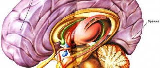
Location of the gland
The organ is localized in the lower part of the brain and belongs to its processes. The pituitary gland is located in the center of the skull, above it is the hypothalamus. The appendage is located in the recess of the sphenoid bone, in most cases it completely fills it, sometimes only half of the volume. With hypertrophic changes, the organ may extend beyond the bone pocket.
The size of the iron is small:
- in height – 3-8 mm;
- width – 10-16 mm;
- weight – does not exceed 1 g.
On the posterior, inferior and anterior sides, the pituitary gland is protected by bone structures and is covered on top by a diaphragm with an opening. A leg passes through it, connecting the gland to the hypothalamus.
Treatment of hormonally inactive forms of pituitary adenoma
Hormonally inactive pituitary adenomas account for 25% of all adenomas (prevalence rate: 6 cases per 1 million population per year). The rarest disease in adults is craniopharygioma. Other types of neoplasms without hormonal activity (hemangioma, dysgerminoma, harmathroma) are detected somewhat more often.
Symptoms indicating the development of tumors in this group are menstrual irregularities in women and decreased erectile function in men. Other signs of pituitary adenoma are weakness, hypotension, and increased fatigue. If the tumor appears in childhood, growth and sexual development are delayed. Impaired water metabolism and neurological symptoms occur in 75% of patients. With active tumor growth, nausea and vomiting intensify, and the most serious consequence is cranial nerve palsy.
The gold standard of treatment is surgical removal of the tumor. Radiation therapy is prescribed when diagnosing remnants of adenoma or in cases of relapse. After surgery, the patient is observed by a doctor, and six months later an MRI examination is performed. The prognosis depends on the location and size of the pituitary adenoma.
With successfully chosen treatment tactics and correctly performed surgery, complete recovery and restoration of working capacity occur. If, as a result of the progression of the pituitary adenoma, visual functions are lost and persistent neurological disorders appear, then the patient receives permanent disability.
Forecast
With minor changes, the prognosis is favorable. Most pathologies of the pituitary gland are treatable, and replacement therapy allows you to avoid side diseases.
Poor prognosis for large malignant tumors with elements of ischemia and local bleeding.
But when assessing the prognosis, it is also important to take into account age, concomitant diseases and the degree of their compensation. Do not forget that the course of the disease depends on the psycho-emotional background of the patient and his desires.
When should you see a doctor?
Pituitary adenoma is accompanied by endocrine disorders, which always cause negative symptoms. Be sure to make an appointment with your doctor if you experience weight loss or gain without changing your diet, blurred vision, changes in the appearance of the skin, headaches, or sexual dysfunction. You will be prescribed a series of tests, based on which a specialist will make an accurate diagnosis. The sooner you contact a medical clinic, the sooner you will receive a conclusion, the shorter and more effective the treatment will be.
First appointment with a therapist. Next, based on complaints and examination results, the specialist will write a referral to a specialized doctor. Women are examined by a gynecologist-endocrinologist, men by an andrologist. Consultations with doctors of other specializations may be required.
The MedCom clinic in Ryazan has everything necessary for the diagnosis and treatment of pituitary adenoma. Experienced doctors will conduct an emergency examination and develop the most effective and safe treatment regimen. Make an appointment by calling +7 (4912) 77–92–02 or using the online contact form. Do not delay your visit to the doctor - pituitary adenoma can be successfully treated in the early stages!