Heterogeneity in the structure of the pituitary gland can be either a normal variant or a condition preceding adenoma. To clarify the diagnosis, it is necessary to take into account the clinical signs of impaired hormone formation, as well as the results of their analysis in the blood. In doubtful cases, it may be necessary to administer a contrast agent during MRI of the pituitary gland, examine a neurologist and an ophthalmologist.
In the absence of other disorders, diffuse tissue heterogeneity does not require treatment. Patients need to be periodically examined by an endocrinologist and follow recommendations for lifestyle and nutrition correction.
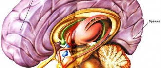
Morphology and localization
Pituitary adenomas, depending on their size, are divided into micro- (up to 1 cm) and macro-adenomas (more than 1 cm in diameter). Microadenoma is a lesion located entirely within the pituitary gland.
Direct signs of the presence of microadenoma:
- volumetric education,
- often hypointense on T1,
- T2 hyperintense or hypointense.
Indirect signs:
- funnel displacement,
- dredging,
- increase in the size of the pituitary gland,
- bulging of the superior contour of the pituitary gland.
In the absence of signs of microadenoma, its presence cannot be rejected without resorting to a study with dynamic contrast.
Using CT to diagnose microadenomas is not objective: bone artifacts and low tissue contrast. MRI is used exclusively. In cases with macroadenomas, CT is not replaceable for differential diagnosis.
Fig. 1549-1541
Microadenoma in the right half of the pituitary gland (arrowheads in Fig. 1549, 1550), which has relatively clear and even contours, is well differentiated by T2 (Fig. 1549) and a weakly hypointense MR signal by T1 (Fig. 1550). Bulging of the upper contour of the pituitary gland (arrow in Fig. 1551) is also a sign of pituitary microadenoma.
Fig.1552-1554
Also, signs of the presence of a microadenoma are a deepening of the bottom of the sella turcica (arrowhead in Fig. 1552), a clearly heterogeneous structure of the gland (arrows in Fig. 1552, 1553) and displacement of the funnel in the direction opposite to the microadenoma (arrow in Fig. 1554).
Assessing cavernous sinus invasion can be difficult. The most convenient way is to assess the extent of coverage of the cavernous part of the internal carotid artery. Less than 90 degrees makes sinus involvement very unlikely, while coverage greater than 270 degrees almost certainly indicates cavernous sinus involvement [45].
Invasion of the dural sinuses often leads to a direct release of the hormone from the functioning adenoma into the blood, which is manifested by an increase in the level of the hormone in the blood, for example, a convincing sign of prolactinoma of the pituitary gland invading the cavernous sinus is a prolactin level over 1000 ng/ml [2].
Invasion of adjacent structures occurs in 35% of cases and is not evidence of malignancy [48].
Fig. 1555-1557
Pituitary adenoma with invasion into the right cavernous sinus (arrow heads in Fig. 1555, 1556), which is well differentiated on contrast-enhanced MRI (arrowhead in Fig. 1557).
Macroadenoma is a large formation of the chiasmal-sellar region with an expansive type of growth and expansion of the sella turcica of the skull (the change is noticeable on the craniogram).
Extrasellar spread can occur in any direction, accordingly, indicated by the type of growth:
- suprasellar,
- antesellar,
- retrosellar,
- infrasellar,
- laterosellar.
It may spread in several directions at once.
Adenomas with a suprasellar growth pattern have a typical “dumbbell” or “8” appearance, which is explained by the tumor passing through the diaphragm of the sella turcica [48]. On MRI they are usually isointense to gray matter on T1 and hyperintense on T2 [72]. On CT, macroadenomas have a density of 30–40 HU [114].
Fig.1558-1560
Pituitary macroadenoma with a suprasellar type of growth (asterisk in Fig. 1558) in the form of a large formation above the sella turcica, compressing the chiasm and the third ventricle. Adenoma with a suprasellar type of growth (asterisk in Fig. 1559) and fouling of the siphons of the carotid arteries (arrowheads in Fig.). Macroadenoma with a retrosellar type of growth (arrowheads in Fig. 1560).
As it grows, it spreads to the sides, the tumor causes destruction of the bone structures of the sella turcica, and can also be complicated by liquorrhea.
If it reaches a large size, macroadenoma can even cause indirect compression of the foramina of Monroe and lead to occlusive hydrocephalus. Large lesions are often heterogeneous and vary in signal due to areas of cystic change, necrosis, and bleeding [115].
Fig.1561-1563
Macroadenoma with a laterosellar type of growth to the left from the area of the sella turcica (asterisk in Fig. 1561). Laterosellar spread to the right, and coverage of the cavernous segment of the right internal carotid artery (arrows in Fig. 1562). Destruction of the apex of the pyramid of the right temporal bone and the sphenoid bone, in the process of spreading of the pituitary adenoma, into the right middle cranial fossa (arrows in Fig. 1563).
Fig.1564-1566
Pituitary adenoma with growth into the sinus of the main bone (asterisk in Fig. 1564). Deepening of the bottom of the sella turcica due to bone atrophy and restructuring caused by the growth of adenoma (arrows in Fig. 1565). A giant macroadenoma with extensive distribution in several directions (arrowheads in Fig. 1566).
Bone destruction
In the process of expansive growth, macroadenoma leads to atrophic changes in the bone structures of the sella turcica, which at first looks like its expansion, and then like the absence of the back, inclined processes and the upper wall of the sphenoid sinus.
Fig. 1567-1569
Cystic adenoma
Cyst-shaped adenoma with expansion of the sella turcica (asterisk in Fig. 1567, 1568). The heterogeneous structure of the pituitary adenoma on CT is revealed due to the difference in density characteristics and, with contrast enhancement, objectively demonstrating the heterogeneity of the tumor (Fig. 1569).
Fig.1570-1572
A pituitary macroadenoma can be suspected by the expansion of the sella turcica during tumor growth (asterisk in Fig. 1570). CT gives a complete picture of the state of the bone structures of the base of the skull, which reflects a significant expansion of the pituitary fossa (asterisk in Fig. 1571), thinning of nearby bone structures (arrow in Fig. 1581) and disappearance of the dorsum sella (dotted line in Fig. 1571 and Fig. .1572).
Cystic degeneration occurs in 18% of cases, calcification is not typical [48].
Fig. 1573-1575
Cysts are found in the tumor stroma, which are limited areas with homogeneous liquid content, which is a reflection of dysmetabolic degenerative changes (arrows in Fig. 1573-1575).
Hemorrhagic adenoma
Macroadenomas are diagnosed as cysts containing blood, which is divided into phases depending on the specific gravity (separation of the contents into phases with different densities, in this case, plasma and formed elements - the effect of sedimentation). The presence of sedimentation effect is a specific sign of cystic degeneration [2].
Fig.1576-1578
Hemorrhage in the cavity of a cystic macroadenoma, with visualization of the level of sedimentation of formed elements and the liquid part of the plasma (arrows in Fig. 1576, 1577, 1579). Macroadenoma (asterisk in Fig. 1578) has a supra- and infrasellar distribution, compressing the chiasma and deepening the sella turcica (arrows in Fig. 1578, 1580, 1581), as well as laterosellar (arrowhead in Fig. 1578).
Fig. 1579-1581
Fig. 1582-1584
Hematoma in the left half of the adenoma (arrows in Fig. 1582, 1584), having an MRI signal in T1 (arrow head in Fig. 1583) and T2 (arrow in Fig. 1583).
There may be hemorrhage into the adenoma (apoplexy of the adenoma), reflecting convincing tumor invasion of the cavernous sinus, but this is not a hemorrhagic adenoma.
How to eat with a heterogeneous structure of the pituitary gland
If the structure of the pituitary gland is heterogeneous, patients are prescribed a complete balanced diet, the basic rules for creating a menu:
- eating 4-5 times a day;
- drinking water 1.5-2 liters, salt – 8-10 g;
- the source of protein should be lean meat products, fish (at least 3 times a week), seafood, milk, cottage cheese, freshly prepared fermented milk drinks without additives, moderate-fat cheese;
- carbohydrates come mainly from cereals, whole grain bread, fresh and boiled vegetables, fruits and berries, and juices from them;
- fats should predominate of plant origin, nuts and seeds are healthy;
- you need to limit fatty, fried meat, sausage, canned food, processed foods, fast food, hot sauces, sugar and white flour, mustard and pepper, sweet soda, chips.
We recommend reading the article about pituitary gland enlargement. From it you will learn about the signs and symptoms of changes in the size of the gland, the consequences of vertical enlargement of the pituitary gland, as well as methods for diagnosing and treating pituitary enlargement.
And here is more information about Rathke's cyst of the pituitary gland.
A heterogeneous structure of the pituitary gland can also occur in absolutely healthy people and become a signal of the onset of adenoma formation. With minor changes, you can only get by with lifestyle and nutrition adjustments. When identifying diseases, the help of an endocrinologist, ophthalmologist and neurologist may be required.
Contrast enhancement
In a native (conventional, without contrast) study, microadenoma is most often isointense in T1 and T2 tissues of unchanged areas of the adenohypophysis. Thus, in the absence of structural signs, the presence of a tumor cannot be excluded. The use of contrast enhancement using the standard method of intravenous administration, with further scanning, in most cases, gives false negative results, i.e. The pituitary gland uniformly accumulates the contrast agent and homogeneously increases the MR signal on T1. Rarely, microadenoma is distinguished by the absence of contrast accumulation with standard administration.
Fig. 1585-1587
Microadenoma in the left half of the pituitary gland (arrowhead in Fig. 1585), having a weak T1 MR signal on a conventional (non-contrast) study. After standard intravenous contrast, a reduced accumulation of the contrast agent in the adenoma is determined (arrowheads in Fig. 1586, 1587), compared to the unchanged pituitary tissue that intensively accumulates contrast.
To verify the presence of a microadenoma, dynamic contrast is used, during which it is possible to evaluate the phase-by-phase accumulation of contrast in the adenohypophysis.
Dynamic sequences demonstrate a rounded area of delayed (or, less commonly, advanced) enhancement at the microadenoma site compared to the rest of the gland [131]. In the late phases of dynamic contrast enhancement, the accumulation of contrast by the adenoma and pituitary gland is aligned with T1. Dynamic contrast-enhanced MRI has a sensitivity of 90% [140].
Fig.1588-1590
On native scans and scans with conventional contrast, there is no convincing evidence for the presence of a microadenoma (Fig. 1588), however, using a bolus injection of a contrast agent, in the first 20 seconds, there is a lack of contrast in the pituitary gland zone (Fig. 1598), where the microadenoma is located, followed by equalization of the intensity of contrast adsorption in the pituitary gland and adenoma (Fig. 1590) and the absence of tumor differentiation.
Macroadenomas always accumulate contrast agent intensely and homogeneously, depending on their structure (ie, with the exception of cysts and areas of hemorrhage).
Fig. 1591-1593
Macroadenomas accumulate the contrast agent intensively and relatively homogeneously (Fig. 1591-1593).
Fig. 1594-1596
Macroadenomas on CT (arrow in Fig. 1594) have a density slightly higher than the density of brain matter (Fig. 1595). After intravenous contrast, an increase in the density of the adenoma is determined (Fig. 1596, 1598), and a clearer differentiation of the tumor (arrow in Fig. 1597) with the ability to visualize it when constructing volumetric reconstructions (Fig. 1599).
Fig. 1597-1599
Symptoms
With small focal changes in the tissue, symptoms may be absent or have minor manifestations. But such foci can degenerate, acquiring a malignant course, unite into larger foci (merge) or disappear completely.
Symptoms indicating a malfunction of the pituitary gland:
- -type headaches : debilitating diffuse headache of a pulsating nature, taking antispasmodics does not help, and painkillers bring temporary improvement;
- deterioration of vision: changes in visual fields up to the formation of a tunnel, decreased ability to focus the gaze;
- the appearance of isolated episodes of minor epileptic seizures;
- changes in hormonal levels - laboratory parameters of prolactin, thyroid-stimulating hormone (TSH) and adrenocorticotropic hormone (ACTH) are increased several times.
It is also possible to have a disturbance in circadian rhythms, episodes of insomnia, and a disturbance in spatial orientation. In women, hair growth may increase and change to a male type.
Biological behavior and follow-up
There are functioning (3/4 of all adenomas) and non-functioning (1/4 of all adenomas) adenomas. Microadenoma, in the overwhelming majority, is a functioning tumor and, clinically, is manifested by signs of hormonal disorders and associated symptoms. Macroadenomas are more often non-functioning (but may also be hormone-producing), as a result of which they grow latently (clinically, without manifesting themselves in any way), and reach large sizes, discovered during an examination for focal and cerebral symptoms (visual disorders, increased ICP and etc.).
Nonfunctioning adenomas are usually present in patients with delayed puberty, short stature, or primary amenorrhea (in girls) [137].
The morphological structure of the tumor does not correlate with the type of hormone produced. However, prolactinomas and somatotropinomas are more often located in the lateral parts of the adenohypophysis, and adenomas secreting ACTH, TSH and FSH (LH) in the middle [2].
There are also some indirect signs associated with secondary endocrine changes. For example, a secondarily elevated level of GH leads to acromegaly (thickening of the skull bones, widening of the lower jaw on images), an increased level of ACTH leads to pathological compression fractures and “buffalo hump” back deformities [48].
Statistically, prolactinomas are most often found - 1/2 of endocrine active adenomas and 1/3 of all adenomas.
Among active adenomas, in descending order of frequency of occurrence, there are:
- prolactinomas (48%),
- somatotropinomas (10%),
- corticotropinomas (6%),
- thyroprolactinoma (1%),
- adenocarcinoma,
- mixed and non-secreting (35%) [175].
Large and nonfunctioning tumors can also disrupt hormonal balance. Hypopituitarism or moderately elevated prolactin, in both cases, is visible due to the so-called “crossing the stem” effect; Prolactin (unlike other pituitary hormones) is suppressed by a prolactin-inhibitory hormone (such as dopamine) and, if the pituitary infundibulum is compressed, can lead to elevated systemic prolactin levels due to interruption of normal inhibition of secretion [45].
Incidentaloma is an adenoma without any clinical manifestations; more often, these are non-functioning microadenomas (2-30% of adenomas), which attract attention, can be misinterpreted, and may not be justifiably subjected to unreasonable treatment.
An increased level of prolactin over 100-150 nm/ml is a sign of prolactinoma; with lower values of plasma prolactin, there is probably another reason: a tumor of the hypothalamus, drugs (phenothiazines), hypothyroidism and extraglandular hyperprolactinemia (liver, APUD system), physical activity, breastfeeding, nipple stimulation and recent sexual intercourse. ACTH may be ectopically produced in small cell lung cancer [193].
Malignant prolactinomas are extremely rare and cannot be determined either histologically or by imaging, but only retrospectively, starting from the presence of distant metastases [46]. Anaplastic adenoma and pituitary cancer are rare and develop mainly after long periods of relapse and repeated courses of complex treatment [193].
Do I need treatment?
Even if a tumor is detected in the pituitary gland, treatment is prescribed only if:
- intensive growth;
- large sizes with compression of adjacent tissues;
- high hormonal activity of the tumor;
- deficiency of one or more hormones with clinical manifestations.
All other cases are an indication for observation by an endocrinologist. Patients should undergo consultation and testing at least once a year. Sometimes repeated MRIs, examination by a neurologist and an ophthalmologist are required.
Examination by an ophthalmologist
If there is only diffuse heterogeneity of the pituitary gland, then this condition does not require the use of medications or surgical intervention. Such people do not need restrictions in physical activity or professional activities, but they should avoid risk factors for the development of adenoma:
- smoking;
- alcohol abuse;
- uncontrolled use of medications, hormonal contraceptives;
- self-treatment with herbs and dietary supplements.
If changes in the structure of the pituitary gland are detected, it is necessary:
- consult a doctor and undergo a full course of treatment for infectious diseases;
- be under the supervision of a gynecologist throughout pregnancy and the postpartum period;
- When prescribing hormonal therapy, strictly follow the recommendations on drug dosages and duration of use, and monitor the results using blood tests.
Differential diagnosis
There is a rough differential diagnostic rule: in the presence of a sellar formation - in childhood - craniopharyngioma, at 20-40 years - an adenoma, and at an older age - meningioma; if the size of the sella turcica remains unchanged, one should think about an aneurysm of the circle of Willis or meningioma [193]. In this case, there are a lot of nuances that should be taken into account.
Meningioma
Meningioma has bone hyperostosis, is accompanied by sclerosis of the bone, is located on a broad base, spreading to the carotid artery - leading to its narrowing [2]. Macroadenoma causes bone destruction (atrophy from pressure of the dorsum sellae, inclined processes of the sphenoid bone and the upper wall of the sphenoid sinus), if it covers the carotid artery, it does not cause a narrowing of its diameter.
Fig.1600-1602
Meningioma is located on a broad base, spreading along the dura mater (arrows in Fig. 1600), uniformly and intensively accumulates a contrast agent (asterisks in Fig. 1600, 1601), and when the internal carotid artery is involved, it covers it, causing a narrowing of the diameter of the vessel ( arrow head in Fig. 1600). In addition, meningioma is accompanied by hyperostosis and sclerosis of the underlying bone (arrow head in Fig. 1601). Adenoma expands the area of the sella turcica, not due to bone destruction, but due to bone restructuring due to an increase in the volume of formation (asterisk in Fig. 1602), and when covering the carotid arteries, it does not lead to a narrowing of their diameter (arrows in Fig. 1602).
Rathke's pouch cyst
The leading role in the diagnosis of pituitary adenomas is given to hormone analysis, and MRI is used to exclude or confirm the presence of a sellar formation (in the absence of a sellar formation, a decision is made about extraglandular hormone production). The cyst does not accumulate contrast, but microadenomas, in the overwhelming majority, do [2].
Fig. 1603-1605
Rathke's pouch cyst is a developmental anomaly. It arises from Rathke's pouch, from which the adenohypophysis also develops, so it has a structure similar to adenomas, and rarely occurs at the same time. A cyst is a cavity located in the sellar region, filled with serous (the MR signal of the arrow is isointense to the cerebrospinal fluid in Fig. 1603) or mucinous fluid (the MR signal is characteristic of a colloid, arrows in Fig. 1606, 1607), easily visible and differentiated from “ empty” sella turcica on MRI using thin-section cisternography (arrowheads in Fig. 1604). If the cyst is large in size, it puts pressure on the adenohypophysis and leads to displacement of the infundibulum (arrow in Fig. 1605). In addition, the cyst does not accumulate the contrast agent (arrow in Fig. 1608).
Fig. 1606-1608
Pituitary hyperplasia
Pituitary hyperplasia occurs mainly during puberty, is not accompanied by pathological peaks of hormonal activity and has a homogeneous contrast of the adenohypophysis.
Fig. 1609-1611
In a state of physiological hyperplasia (puberty, pregnancy), the pituitary gland increases in size (arrowheads in Fig. 1609, 1610), simulating the presence of a microadenoma. On T1 in the coronal projection, the slice may pass through the neurohypophysis, creating a false impression of the presence of a microadenoma (arrowhead in Fig. 1611). Contrast enhancement does not detect areas of altered contrast material accumulation.
Craniopharyngioma
Macroadenoma more often has a solid structure, occurs in the middle age range, does not contain petrification, and craniopharyngioma, more often, has a heterogeneous structure with the presence of cysts and petrification, and is found in childhood and old age.
Fig.1612-1614
Craniopharyngioma, as a formation of the chiasmatic-sellar region, can simulate an adenoma, but is characterized by greater heterogeneity of structure, the frequent presence of cystic components (asterisks in Fig. 1612, 1613, 1616), which sometimes reach large sizes. Craniopharyngiomas cysts are often filled with various types of contents (blood, colloid), which can occur in different chambers of the same tumor (arrowheads in Fig. 1614). With contrast enhancement in craniopharyngiomas, accumulation occurs in solid areas of the tumor and the walls of cysts containing tumor elements (arrow head in Fig. 1613).
Craniopharyngiomas are distinguished by the heterogeneity of their structure, due to the content of cholesterol crystals and shell-like petrificates in the walls (arrow heads in Fig. 1615), which are convincingly detected on CT (arrow head in Fig. 1617).
Fig. 1615-1617
Aneurysm
A functioning aneurysm accumulates contrast in the arterial phase, is visible on MRA (on PC and TOF) as a sac-like protrusion from the artery, and also causes the formation of artifacts from pulsation in the direction of the phase-encoding signal. A thrombosed aneurysm does not accumulate contrast, always ^T1 and vT2, also visible on time-of-flight MRA (TOF only), but not visible on phase contrast (PC). There may be petrification in the wall of the aneurysm. An aneurysm often does not lead to bone destruction, but a macroadenoma destroys bone barriers during its growth.
Fig.1618-1620
A thrombosed aneurysm has a layered structure and an MR signal in T2 (arrowheads in Fig. 1618, 1619), ^ in T1 (arrow in Fig. 1618). On TOF, a thrombosed aneurysm has an MR signal (arrow head in Fig. 1620). The aneurysm accompanied a disturbance of blood flow in the right middle cerebral artery, which led to an infarction in the area of its blood supply, with the formation of consequences in the form of cystic-glial changes (arrows in Fig. 1619) and weakening of blood flow in the right middle cerebral artery (arrows in Fig. 1620) .
Fig. 1621-1623
A functioning (not thrombosed) aneurysm is filled with a contrast agent during the passage of the bolus, increasing the density on CT, becoming similar to the lumen of the main cerebral artery (arrowheads in Fig. 1621-1623). After a decrease in the concentration of contrast in the arterial blood, a weakening and, ultimately, absence of contrast in the aneurysm is determined in unison with a decrease in the contrast in the cerebral arteries. The study was performed for meningioma of the greater wing of the sphenoid bone (arrows in Fig. 1621, 1622).
Astrocytoma
Astrocytomas of the chiasmatic-hypothalamic region have an age peak of 2-4 years, they are often associated with neurofibromatosis type 1. In MR spectroscopic measurements of astrocytoma, peaks of Cho, NAA, Cr appear, and in measurements of adenoma there are peaks of Cho and Lac/Lip complex.
Fig. 1624-1626
Astrocytoma arises from the chiasma (arrowheads in Fig. 1624, 1625), can spread in any direction, in this case infrasellar into the cavity of the sinus of the main bone (arrows in Fig. 1624, 1625). Astrocytomas have a cystic-solid structure. The contrast enhancement is intense, heterogeneous due to the presence of cysts in the tumor (Fig. 1626), while the affected areas of the optic nerves and tracts can be contrasted (arrows in Fig. 1626).
Chordoma
Statistically, adenoma is much more common, and chordoma has a predominantly retrosellar growth direction. An adenoma grows from the pituitary gland, and a chordoma displaces it. An adenoma expands the sellar region during growth, and a chordoma destroys the clivus. For chordomas, the age peak is 40-60 years, for adenomas a little earlier - 20-40 years.
Fig. 1627-1629
Chordoma of the sellar region (asterisks in Fig. 1627, 1628) leads to destruction of the dorsum sellae (arrow head in Fig. 1630, 1631) and spreads retrosellar, compressing the pons of the brain (arrows in Fig. 1627, 1630). With pituitary adenoma, expansion of the sella also occurs and there may be atrophy of the dorsum sellae (Fig. 1629). On CT, chordoma > orv in relation to the brain substance.
Fig.1630-1632
Pituitary apoplexy
Pituitary apoplexy is an infarction or hemorrhage of the pituitary gland. Pituitary apoplexy is manifested by a set of clinical symptoms - Sheehan syndrome (panhypopituitarism), and is also accompanied by acute headache and loss of vision, which occurs during childbirth or after childbirth, due to high blood pressure or massive blood loss. Approximately 5% of pituitary tumors present with symptoms of “pituitary apoplexy” due to bleeding or infarction of the adenoma. This is usually an indication for emergency surgery [48].
Fig.1633-1635
Hemorrhage into the pituitary gland is determined by the presence of a hematoma in the sellar region or in the pituitary gland, which has a T2 and T1 MRI signal (arrowheads in Fig. 1633-1635). The pituitary gland increases in size and has a heterogeneous structure. Apoplexy is distinguished from a thrombosed aneurysm by its acute onset and severe clinical condition, as well as the absence of layered structure on MRI.
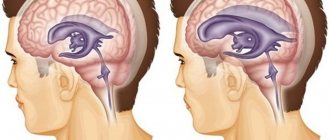
Picture of the heterogeneous structure of the pituitary gland on MRI
Normal pituitary tissue is homogeneous, and its density on MRI is equal to the white matter of the brain, but it is important to take into account that in a completely healthy person there is heterogeneity of the structure in the form of transparent inclusions up to 3 mm - they are located in the center of the anterior lobe, closer to the pedicle and represent clusters proteins.
In addition, small elements can be observed when:
- the special structure of the bones of the sella turcica (pituitary gland bed);
- during the period of active growth of adolescents;
- in pregnant women;
- due to excessive formation of prolactin.
All these conditions are not associated with tumors and do not require additional research.
If widespread heterogeneity is combined with hormonal imbalances and/or increased organ size, the doctor may recommend the administration of a gadolinium-based contrast agent. In this case, the uniformity of its accumulation in tissues is assessed. If a tumor is present, its cells intensively absorb contrast, which helps identify even small formations - microadenomas.
Complications
Diffuse heterogeneous changes in the pituitary gland can lead to complications such as:
- hormonal imbalance ;
- when enlarges, compression of nearby structures occurs;
- cerebral ischemia
- development of epilepsy;
- progression of visual up to blindness;
- development of diabetes mellitus;
- changes in the structure of other endocrine organs.
Treatment methods used
Standard treatment for pituitary adenoma consists of drug, radiation and neurosurgical therapies. The optimal method of exposure is determined based on the diagnostic results: it depends on the patient’s condition, the characteristics of the tumor itself, and the presence of concomitant diseases.
Drug therapy
Drug therapy is prescribed in 90% of cases. The drugs help get rid of the symptoms of the disease and improve the patient’s well-being. Initially, vitamin and restorative complexes are prescribed. Next, you need to determine the type of tumor:
- for prolactinomas, dopamine agonists or ergoline drugs are prescribed,
- for somatotropinomas - somatostatin agonists,
- for corticotropinoma - steroidogenesis blockers.
If necessary, hormone replacement therapy is performed. If the chosen treatment regimen does not bring results for a long time, then a more radical method of treatment is selected: radiation therapy or surgery.
Radiation therapy
For microadenomas, proton, gamma and remote therapy are used. The essence of the tactic is the introduction of a radioactive substance directly into the pathological area. Thanks to this effect, it is possible to stop the growth of the tumor and achieve regression. Typically, radiation therapy is performed when surgery is not possible and the patient refuses surgery. After such exposure, the pituitary adenoma gradually decreases in size and can be completely destroyed.
Surgical intervention
If the tumor is large and associated complications develop (hemorrhages, cyst formation, visual impairment), its removal using the transcranial method is indicated. The operation involves craniotomy. In some cases, intervention may be performed using endoscopic techniques.
The choice of specific treatment tactics mainly depends on the type of tumor. For prolactinoma, radiation exposure is practically useless, while for corticotropinoma it is highly effective. If the tumor does not cause any disturbances in the body and the patient does not complain of deterioration in health, doctors choose a wait-and-see approach.