Subdural hematoma is a pathological condition characterized by a limited accumulation of blood between the dura and arachnoid membranes of the brain. It accounts for up to 40% of all intracranial hematomas. Most often it occurs as a result of traumatic brain injuries in male patients over the age of forty.
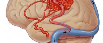
The danger of a subdural hematoma is that it negatively affects the brain structures located around it through pressure on them, their displacement and even damage. So she gives them some serious time. The reasons for its appearance are hemorrhages and traumatic injuries to the blood vessels of the brain.
The Neurosurgery Department of CELT invites you to undergo diagnostics and treatment of hematomas in Moscow. Our multidisciplinary clinic is equipped with the latest technology and has high-precision diagnostic equipment that allows you to make a correct diagnosis. Our specialists use gentle techniques to provide treatment in accordance with international standards.
Etiology of subdural hematomas
This type of hematoma is most often a consequence of traumatic brain injuries and ruptures of blood veins that develop as a result of them, passing in the subdural space. However, the reasons do not necessarily lie in injuries; sometimes hematomas can be provoked by pathological conditions characterized by the destruction of the walls of blood vessels and a decrease in their plasticity. Other reasons are as follows:
- Blood diseases that change its properties;
- Diseases of the blood vessels of the brain;
- Increased intravascular and blood pressure;
A hematoma formed on the side of the injury appears with a small contact area and even without direct contact. This can happen when stopping too suddenly or changing the direction of movement, as well as when falling on the lower limbs or legs. In the process, a sharp shaking of the head occurs, provoking a displacement of the cerebral hemispheres inside the skull, leading to rupture of the blood veins.
As for a hematoma formed on the opposite side of the injury, it can form when the head hits a large, inactive object, causing a large contact area. Much less often, hematomas are formed due to injuries directly to the veins and arteries when the integrity of the dura cerebral membrane is violated.
Recommendations
Consultations with a neurosurgeon and computed tomography of the brain are recommended.
| • | Leading specialists and institutions for the treatment of this disease in Russia: |
| Director of the Research Institute of Neurosurgery named after. Burdenko N.N. Konovalov A.N. | |
| • | Leading specialists and institutions for the treatment of this disease in the world: |
| Professor Duke Samson, USA. |
Clinic of subdural hematomas
Clinical manifestations of subdural hematomas are presented in two large groups, which can be found in our table below:
| Group of symptoms | Clinical manifestations |
| General cerebral symptoms of hematomas |
|
| Focal symptoms of hematomas |
|
Clinical picture
Symptoms appear simultaneously while you are awake. Simultaneously with the disturbance of consciousness, the symptoms of the disease increase over several hours and days. Subdural hemorrhage is characterized by sudden intense headache, vomiting, and weakness/numbness in one side of the body. Drowsiness or agitation, cramps in the limbs, decreased memory, and increased blood pressure may be expressed.
A neurological examination of the patient reveals psychomotor agitation (50%), impaired consciousness (80%), cerebral syndrome (90%), focal/generalized seizures (35%), homolateral mydriasis (40%), nystagmus (35%), ophthalmoplegia ( 15%), contralateral hemiparesis/hemihypesthesia (40%), amnesia (10%). Bradycardia is observed in 40% of cases, respiratory rhythm disturbances - in 20%.
Diagnosis of subdural hematomas
Recognition of a subdural hematoma can be seriously complicated by the variety of its clinical manifestations. In order to correctly make a diagnosis, the neurologist takes into account the following factors:
- Nature and characteristics of traumatic injury;
- Development of disturbances of consciousness;
- Presence/absence of a light gap;
- Neurological status of the patient.
Instrumental diagnostic studies that are prescribed to the patient are presented as follows:
- X-ray of the skull;
- Detailed examination of the fundus (ophthalmoscopy);
- Cerebral angiography (study of cerebral vessels);
- Computer and magnetic resonance imaging, incl. and with contrast.
Life forecast
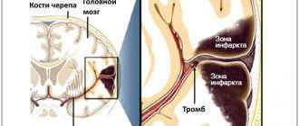
The prognosis of life with chronic subdural hematoma depends on the timeliness of seeking medical help. Severe consequences are observed mainly in elderly patients. Lethal cases occur not only due to the presence of the hematoma itself, but also against the background of traumatic effects on brain tissue. Edema, cerebral ischemic processes, and dislocation of brain structures may occur.
After surgical treatment, it is necessary to establish strict monitoring of the patient's condition. In the first hours after surgery, cerebral edema may occur.
Surgery for chronic hematoma is one of the most effective treatment methods. Conservative therapy is used as an additional way to help the patient. The Medsi Clinic successfully performs neurosurgical operations for such conditions.
Treatment of subdural hematomas
The treatment tactics for subdural hematomas are based on the results of diagnostic studies and the individual indications of the patient. It may involve the use of conservative or surgical techniques.
| Hematoma treatment methods | What are they? |
| Conservative | Indicated for patients who have not lost clarity of consciousness with hematomas up to 10 mm thick, accompanied by a displacement of structures by no more than 3 mm. They are also used if the patient is in a deeply depressed state of consciousness or in a coma, provided that the volume of the hematoma does not exceed forty milliliters and the intracranial pressure is not lower than 25 mm. rt. Art. Conservative treatment involves the use of drugs to prevent cerebral edema, relieve pain, vomiting and convulsions. |
| Neurosurgical | Surgical intervention is practiced for hematomas with symptoms of brain compression, focal manifestations, or in case of increased intracarnial pressure syndrome. Removal can be carried out using endoscopic techniques through a specially created opening. In addition, craniotomy is used, aimed at removing not only the hematoma, but also the areas of crush injury. |
The Department of Neurology at CELT sees neurologists and neurosurgeons with many years of experience in scientific and practical work. You can make an appointment with them online on our website or by contacting our operators: +7 (495) 788 33 88.
Make an appointment through the application or by calling +7 +7 We work every day:
- Monday—Friday: 8.00—20.00
- Saturday: 8.00–18.00
- Sunday is a day off
The nearest metro and MCC stations to the clinic:
- Highway of Enthusiasts or Perovo
- Partisan
- Enthusiast Highway
Driving directions
4. Treatment of the disease
Depending on the patient’s condition and the volume of the hematoma formed, treatment can be either conservative or surgical. Both treatment methods have their own advantages and disadvantages. Modern medicine has new methods for treating this pathology, which combine higher efficiency compared to conservative ones and are less invasive compared to surgical ones (craniotomy). This method is called closed external drainage of the hematoma.
To eliminate the hematoma, the skull is perforated through the skin with an intraosseous needle. Then drainage
with a catheter diameter of no more than 3 mm with a fluid receiver located below the level of the head.
Practice has confirmed the high efficiency of the new method and shown a number of advantages of hematoma drainage:
- surgical removal of compression (squeezing) of the brain;
- reducing the risk of postoperative infection;
- maintaining the integrity of the skull;
- minimizing the likelihood of relapse.
Incidence (per 100,000 people)
| Men | Women | |||||||||||||
| Age, years | 0-1 | 1-3 | 3-14 | 14-25 | 25-40 | 40-60 | 60 + | 0-1 | 1-3 | 3-14 | 14-25 | 25-40 | 40-60 | 60 + |
| Number of sick people | 0 | 0 | 0.01 | 6 | 6 | 13.8 | 17.4 | 0 | 0 | 0.01 | 3 | 3 | 9.6 | 15.3 |
Prevention
The main preventative measure is to prevent injury. Preventive steps:
- Always wear a helmet when riding a bicycle.
- Reduce adrenaline sports.
- Do not take risks when playing sports, respect the instinct of self-preservation.
- Drink alcohol in moderation.
- Do not smoke.
- When treating hypertension, take medications regularly and undergo examinations.
- Try to lose weight.
Recommendations related to hematoma:
- If you have hemophilia diagnosed at birth or use anticoagulants to thin your blood, reduce contact and adrenaline sports, which have an increased risk of injury.
- In case of a head injury with short-term loss of consciousness, nausea or other suspicious symptoms, consult a doctor for a CT scan.
- Try to stay in good physical shape. This will prevent the risk of falling.
- If necessary, use appropriate shoes and rehabilitation aids such as walkers, French sticks.
- Wear glasses to correct your vision.
- Drink enough fluids, especially in summer.
- Reduce the use of sleeping pills and blood pressure-lowering medications if they are not urgently needed. This will prevent possible collapse with subsequent falls and injury.
- If blood circulation in the extremities is impaired, use special medications and ointments.
Where is the danger hiding?
As a rule, with a head injury, a concussion occurs, but in the case of rupture of venous or arterial vessels, subdural and epidural hematomas can develop, differing from each other in the nature of compression of the brain and localization.
What is the difference between a subdural and epidural hematoma
The occurrence of a bruise is almost always associated with a head injury, and the nature of its occurrence can be different, for example, a subdural or epidural hematoma occurs due to:
- A blow with a blunt object to the skull area leads to indentation or fracture of the bones of the skull; as a result, venous or arterial vessels may rupture, leading to hemorrhage.
- A blow to the head while falling from a moving vehicle, falling out of a window, etc., which is also accompanied by indentation of the skull bones, leads to similar consequences.
Subdural
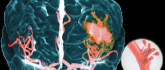
The most common form. Subdural hematoma of the brain is characterized by the ability to affect several areas of the brain, as it is correctly localized between the arachnoid membrane of the brain and the dura mater. In most percent of cases, it occurs as a result of rupture of the venous vessels of the head, in particular when the bridging veins rupture.
Classification:
- acute;
- subacute;
- chronic.
Acute – occurs in the first few hours after injury.
Subacute – manifests itself within a few days (no later than 2 weeks).
Chronic – makes itself felt after a few weeks.
If in the case of the acute type the main cause is primary hemorrhage, then subacute subdural hematoma or chronic can occur with secondary hemorrhage.
Subdural hematoma in the picture
In addition, there is a danger of developing reverse hemorrhage, which is a bruise formed on the side opposite to the point of impact.
Epidural
An epidural hematoma occurs as a result of a head injury, but in this case the bones of the skull are depressed, which explains the location of the bruise. Unlike the subdural, it almost always forms at the site of the impact, and not on the opposite side.
In addition to local, epidural hematoma of the head can be general - that is, affect several parts of the brain.
Classification:
- acute;
- subacute
No chronic course of this disease was observed.
From the editor: What is the difference between thinking and intelligence
As a rule, a hematoma is formed due to bleeding from the middle meningeal artery (in most cases) or from the anterior ethmoidal artery, so localization is noted in the temporal and frontal lobes of the brain. Blood accumulates between the skull and the dura mater.
Adults are more often susceptible to this hematoma, since young children have structural features of the skull that physically do not allow such hemorrhage to occur.