Liquor is the cerebrospinal fluid that fills and washes the anatomical spaces of the brain and spinal cord. It carries the most important functions of protecting these organs from damage and shock, helps maintain the constancy of liquid media, and maintains intracranial pressure. In addition, it is involved in the release of brain metabolites and the transport of nutrients. Analysis of cerebrospinal fluid helps to identify pathologies of the central nervous system, their degree and development, since cerebrospinal fluid immediately reacts by changing parameters to any changes in the brain.
Characteristics of cerebrospinal fluid
The difficulty of understanding cerebrospinal fluid, what it is and for what purpose it is present in the human body, experts draw an analogy with tissue. After all, it contains cells and vitamins, organic as well as inorganic compounds and salts. This composition and structure allows it to perform basic functional duties:
- absorb - in fact, the brain is practically not attached to the bone structures, therefore, in the process of moving a person, it is subject to load and friction, which is leveled by the cerebrospinal fluid;
- participation in metabolic processes - since nervous tissues are independently unable to extract and deliver nutritional components, as well as oxygen molecules, the cerebrospinal fluid performs this function for them.
The circulation of cerebrospinal fluid occurs constantly and continuously - this ensures support for the internal environment. In the event of chemical or functional failures, a person immediately feels a deterioration in health in the form of pain, difficulty moving, and general intoxication. Based on the nature of the unpleasant symptoms, doctors judge the possible causes and prescribe laboratory tests of the brain fluid.
Composition of cerebrospinal fluid
Cerebrospinal substance is produced, on average, at a rate of about 0.40-0.45 ml per minute (in an adult). The volume, rate of production, and most importantly, the component composition of CSF directly depends on the metabolic activity and age of the body. Typically, tests show that the older a person is, the more reduced production is.
This substance is synthesized from the plasma part of the blood, however, both the substrate and the producer differ significantly in ionic and cellular content. Main components:
- Protein.
- Glucose.
- Cations: sodium, potassium, calcium and magnesium ions.
- Anions: chlorine ions.
- Cytosis (presence of cells in the cerebrospinal fluid).
An increased content of protein and cell aggregates indicates a deviation from the norm, which means it is a condition that requires further tests and mandatory consultation with the attending physician.
Comprehensive study of cerebrospinal fluid in bacterial purulent meningitis
Bacterial purulent meningitis (BPM) takes a leading place in the structure of neuroinfections. Despite significant advances in the treatment of HBM, mortality over the past 40 years has remained at a stable level of 6–24%, depending on the etiology of HBM and the quality of treatment [1–5]. Research conducted in 2008–2014 improved the quality of medical care for patients with HD due to, among other things, the development and implementation of new methods for studying cerebrospinal fluid (CSF): polymerase chain reaction (PCR), lactate level, D-fibrin dimer (D-dimer). DF), lactate dehydrogenase (LDH) and its isoforms, protein fractions, pH, pO2 and pCO2, study of indicators of local humoral and cellular immunity.
Materials and research methods
The CSF was studied in 1806 patients with HD of various etiologies; the comparison group consisted of 25 patients with serous viral meningitis and 10 patients with non-inflammatory lesions of the central nervous system (CNS). Spinal puncture was performed upon admission to the hospital (acute period of illness), on days 3–5 (complicated course) and on days 8–18 of treatment.
Research results
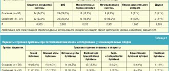
Bacteriological examination of the CSF, being the “gold standard” of diagnosis, provides the etiological decoding of meningitis in no more than 30–40% of cases [1, 4, 6], and immunological methods, in particular, the latex agglutination reaction (LAR), in 60 % of cases [7, 8]. The use of PCR makes it possible to decipher the etiology of meningitis in the late stages of the disease and against the background of ongoing antibacterial therapy, when the bacteriological method does not give a positive result and the amount of capsular polysaccharide antigens in the CSF is not sufficient for their detection by the X-ray method [8–10]. The use of PCR also helps to differentiate viral and bacterial meningitis in doubtful cases, determining the treatment strategy for the patient. The use of PCR increased the efficiency of decoding the etiology of meningitis by an average of 40% compared to the results of bacteriological and immunological studies (N. meningitidis, H. influenzae and Str. pneumoniae), and in combination with other methods of etiological diagnosis of meningitis, it allowed to increase the deciphering of meningitis from 46% up to 88%, and with early admission and no treatment in 100% (Table 1).
According to the results of various diagnostic methods over the past 7 years, significant changes in the structure of the brainstem have been identified. As before, the leading pathogens of BGM (64%) are meningococcus and pneumococcus, and there is a decrease in the number of patients with meningococcal meningitis by 1.25 times (50.1–41%). Hemophilic meningitis is registered in children under 5 years of age, and due to the use of vaccination, the number of patients decreased by 2.5 times (from 9.9% to 4.0%). Significant is the increase over the past 5 years in the role of staphylococcal meningitis to 22.3%. The incidence of pneumococcal meningitis remains at a consistently high level (23%). In some cases, the causative agents of BGM were Listeria, Klebsiella, various types of streptococci, gram-negative bacteria - no more than 9% (Fig. 1).
The disadvantage of etiological diagnosis is the length of time it takes to obtain the result (up to three days) and often a negative result if previously taken antibacterial drugs. Therefore, to carry out empirical antibacterial therapy, it is necessary to use express diagnostic methods that allow, within 2–3 hours, to differentiate BGM from viral meningitis and other diseases of the central nervous system, and to identify criteria for the severity of the disease. In domestic and foreign literature, there are works on the study of lactate levels in the CSF [11, 12], D-DP [12–15], changes in acute phase proteins [12, 16–18], lactate dehydrogenase (LDH) and its isoforms simultaneously in the CSF and blood [12, 19, 20], indicators of acid-base status and electrolytes in the blood and CSF [21, 22]. However, these reports are descriptive in nature, regardless of the etiology, severity, treatment, and prognosis of HBM. The lactate level in the CSF can be considered as an integral indicator of the metabolic activity of the pathogen, which is inversely correlated with glucose. CSF lactate does not depend on its level in the blood, unlike glucose, since it is formed directly in the subarachnoid space and is a product of the metabolism of bacteria and leukocytes [23, 24]. With HD of various etiologies, the lactate level increases to 5.5–25.0 mmol/l (average 11.6 ± 0.7 mmol/l, normal 1.1–2.2 mmol/l). With viral serous meningitis and non-inflammatory lesions of the central nervous system, this figure remains in the range of 0.9–3.9 mmol/l (1.9 ± 0.5 mmol/l). A lactate level above 4.0–4.5 mmol/L is a reliable criterion for HDM, which allows it to be used as a reliable differential diagnostic test. Parallel determination of CSF and blood lactate is essential, since bacterial meningitis always develops against the background of bacteremia or the presence of a purulent-septic focus. The level of blood lactate, especially in secondary HMG (pneumogenic, otogenic, rhinogenic, sepsis), was determined from 3.1 to 4.8 mmol/l (normal 1–2.1 mmol/l) in the acute period. Changes in the dynamics of CSF lactate levels make it possible to evaluate the effectiveness of treatment (Table 2).
With effective antibacterial therapy, after 2–3 days the lactate concentration decreases by 1.5–2.0 times or more; in the absence of effect or unfavorable outcome of the disease, there was no positive dynamics, which indicates that the pathogen retains biological activity in the subarachnoid space. Thus, CSF lactate can be used as a diagnostic, differential diagnostic and prognostic biochemical marker in HBM. We have established the presence of components of the hemostasis system and fibrinolytic activity in the CSF during meningitis. Of greatest interest is the determination of D-DP, which is the main product of fibrinolysis [13–15]. Normally, the level of D-DF in the CSF does not exceed 0.5 µg/ml (500 mg/ml). In the acute period of the disease (upon admission), its amount in the subarachnoid space increases and is above 1.0 µg/ml in 90% of those examined. During fibrinolysis of the inflammatory purulent exudate, on days 3–7 of treatment, the amount of D-DP in the CSF continued to increase by an average of 1.7 times (especially with pneumococcal etiology of meningitis) and varied in individual patients from 3 to 21 µg/ml (on average 13.5 ± 1.41 µg/ml), which was significantly higher (p < 0.005) than at admission. In viral meningitis and non-inflammatory diseases of the central nervous system, the level of D-DP did not exceed 3.0 µg/ml (1.05 ± 0.28 µg/ml) (Fig. 2).
The content of D-DP in the CSF also correlates with other indicators characterizing the severity of the inflammatory process in the subarachnoid space. The correlation coefficient with cytosis was 0.48, with protein content - 0.65, with glucose level - 0.65, with lactate level - 0.73. At the same time, there was no fibrinogen in the CSF of the examined patients, which indicates a high coagulation activity of the CSF, leading to the formation of fibrinous clots in the membranes and substance of the brain. Thus, D-DF can be used as a diagnostic marker of HD, and the dynamics of D-DF can be used to judge the effectiveness of the therapy and, in the absence of its reduction, to predict an unfavorable outcome of the disease. Indicators characterizing BGM are also changes in CSF acidity [21, 24, 25]. The CSF pH is normally slightly alkaline, corresponding to the blood plasma due to an increase in pCO2 to 50–60 mmHg. Art. (norm 45 mm Hg). With the development of HBM, the cerebrospinal fluid of 84.5% became more acidic (pH 7.0–7.3). There is also a decrease in pO2 in the CSF in more than 50% of patients to 40–45 mm Hg. Art. (norm 60 mm Hg), these indicators had a direct correlation with the severity of the patient’s condition (Table 3).
With effective therapy over time, by the 3rd day of treatment for BGM, the pH of the cerebrospinal fluid increases by an average of 0.2 and is 7.25–7.35, pO2 increases (more than 60 mm Hg), which indicates an improvement in blood flow in brain, and pCO2 decreases, which normalize in the complicated course of the disease by 5–8 days of treatment. Thus, the CSF pH level can serve as a diagnostic criterion for HD, an objective indicator of the severity of the disease, and a criterion for assessing the effectiveness of therapy. Almost all enzymes involved in metabolism in the brain are found in the CSF [12, 24–27]. However, due to the low content in the CSF, the determination of their activity is associated with a number of difficulties. Of the detected enzymes, LDH is of greater diagnostic value - the most sensitive indicator of brain hypoxia and lactatrachia. LDH activity in the CSF increases with various brain lesions (normal range is 5.0–40.0 U/l) [19, 20]. Moreover, the study of the isoenzyme spectrum of LDH is more informative than the general activity. In healthy people, LDH activity in the CSF is lower than in the blood serum, and the LDH4 and LDH5 isoenzymes are most often not detected at all. Thus, it has been established that the activity of LDH1–2 in the CSF has a positive correlation with the severity of traumatic brain injury [18]. Inflammatory changes in purulent meningitis are caused by an increase in the proportion of LDH4–5. A study of LDH isoforms (LDH1, LDH2, LDH3, LDH4, LDH5) in the CSF in the acute period and in the dynamics of the disease of HD of various etiologies and severity showed that with HD in the CSF there is an increase in the activity of LDH4 and LDH5 by 2.5 times in the acute period diseases. There was a significant increase in the fractions of LDH4 and LDH5 in the CSF in all BGM, and with meningococcal meningitis their level reached a maximum already on the 1st day, with pneumococcal meningitis - by the 3rd–5th day of illness (p < 0.05) and averaged for LDH4 - 17 ± 9.5% (in the comparison group - 8.8 ± 5.7%), LDH5 - 33.2 ± 12.4 (3.3 ± 2.1). With a fatal outcome of the disease, the increase in LDH fractions is more significant than with moderate-severe HD, which reflects the involvement of the brain substance in the process in the complicated course of HD (Fig. 3).
The dependence of LDH activity in the CSF on protein content and pH has been established [21, 23, 24]. In viral meningitis, in contrast to BHM, the activity of the LDH1 and LDH3 isoenzymes in the CSF increased, which correlated with the content of lymphocytes. Thus, the appearance of LDH4–5 in the CSF can serve as a diagnostic criterion for HD, and the normalization of these indicators indicates the effectiveness of the therapy. An increase in protein levels in the CSF during inflammatory processes is due to an increase in the permeability of the vascular walls [24, 28]. In this case, the ratio of albumin to globulins ranges from 2–3. Albumins and globulins in the CSF are an important indicator of the permeability of the blood-CSF barrier (HLB), and are also markers of acute inflammation (acute phase proteins), since their function in HBM is the recognition of foreign agents, the formation of an antigen-antibody complex with them and their neutralization [12, 28]. The CSF proteinogram is significantly more informative compared to the total protein value, since in some cases, with a normal content of total protein in the CSF, significant fractional changes are detected during electrophoresis [12, 24, 25]. Using electrophoresis, CSF proteins can be divided into albumins, α-1-globulins, α-2-globulins, β-globulins and γ-globulins; we conducted these studies depending on the severity of the patient’s condition in the acute period and the dynamics of treatment. In all groups of patients with HD in the acute period of the disease, an increase was noted? -1-globulins in the CSF, which is presented in table. 4.
A significant direct correlation was revealed between the same indicators: albumin (R = 0.59) and α-2-globulins (R = 0.53) in blood serum and CSF, which confirms the increased permeability of HLB in patients with breast cancer. It has been established that during purulent inflammatory processes in the CSF there is an increase in the content of α-1-, α-2- and γ-globulins, which characterizes an acute inflammatory process in the subarachnoid space, and the percentage of albumin decreases. Upon admission to the hospital (days 1–3 of illness), with BGM, α-1-globulins in the CSF increase by 1.8 times and α-2-globulins by 1.5 times, in contrast to viral meningitis, in the absence of such abnormalities in the blood, which indicates the possible production of specific acute phase proteins in the acute period of the disease directly in the choroid plexuses of the brain. From the 5th day, γ-globulins increased 2 times compared to the control group (p < 0.05). In the following days (days 3–8 of treatment), CSF γ-globulins increased by 1.7–2.1 times compared to the control group (p < 0.05) in all examined patients with HD. Thus, an increase in α-1- and α-2-globulins in the CSF may have a differential significance between meningitis of HBM and meningitis of viral etiology (serous) and is a marker of acute inflammation in HBM, as opposed to viral meningitis. The cerebrospinal fluid electrophoresis method makes it possible to evaluate the effectiveness of therapy and has clinical and prognostic significance when comparing protein fractions of CSF and blood serum in patients with HD. As for γ-globulins, they are of both plasma and brain origin [27, 28]. An increase in γ-globulins in the CSF is associated with increasing processes of local sanogenesis (IgA and IgM). Changes in the proteinogram are not strictly specific, and they are used for diagnosis, differential diagnosis and prognosis of the disease along with other routine and specific indicators. Immunological methods occupy a significant place in liquor studies [29, 30]. The immune system of the central nervous system functions autonomously and is a factor of sanogenesis in HD; there is no significant correlation (p > 0.05) for all parameters between immunity indicators in the blood and CSF. We have found that there are significant differences between the indicators of cellular and humoral immunity in the blood and CSF. In the CSF, there was a significant activation of immune parameters that was large and qualitatively different from the blood. Thus, IRI in the CSF was 3.0 ± 0.4, in the blood 1.9 ± 0.3 (p < 0.05), which was associated with an increase in CD4+ to 62.7 ± 5.4% (in the blood 45 .1 ± 4.8%, p < 0.05). This indicates a high phagocytic activity of CSF leukocytes. It should also be noted that in two patients with IRI in the CSF less than one, late sanitation of the cerebrospinal fluid was observed (19 and 27 for treatment). Long-term pleocytosis during clinical recovery is due to a local immunocellular reaction with the accumulation of T-helper cells and an increase in IRI to 4.5–5.0. Cytotoxic activity in the CSF was carried out mainly due to CD8+, and in the blood - NKT. At the same time, the B-cell system is activated and Ig of various classes accumulates in the CSF. Despite the fact that the content of Ig in the CSF was many times lower than in the blood, their concentration per 1 g of protein was significantly higher, which suggests that Ig is produced directly in the subarachnoid space. Ig levels in g% protein also varied significantly. The IgA content per gram of protein in the CSF was 0.150 g%, in the blood - 0.043 g% (p < 0.001), i.e. 3.5 times higher, IgM, respectively, 0.239 g% and 0.030 g/m (p < 0.001) , i.e. 7.6 times higher. It is difficult, especially in severe cases, to assess the effectiveness of antibacterial therapy in the early stages, especially in patients with severe HD, which often leads to unjustified replacement of the antibacterial drug. For example, with Hib meningitis, replacement was performed in 68%, but was justified in only 28%. It is not always possible to clinically evaluate the effectiveness of treatment, because with ineffective etiotropic therapy, mistakes are made when a temporary therapeutic effect is achieved through detoxification and dehydration therapy. With effective antibacterial therapy, the lack of positive dynamics or negative dynamics in the patient’s condition can be caused by the progression of the inflammatory process as a result of bacteriolysis under the influence of bactericidal drugs, intracranial complications (progression of cerebral edema-swelling, subdural and subarachnoid hematoma, blockage of the cerebrospinal fluid tract), extracranial complications ( pneumonia, etc.), activation of herpes infection, which is confirmed by the detection by PCR of replication of herpes viruses of types 1, 2, 5, 6, and febrile reactions to the administration of antimicrobial drugs. Our studies have shown that the effectiveness of etiotropic therapy in complicated disease can be assessed only on the basis of liquorological studies. They make it possible to evaluate the effectiveness of etiotropic therapy within 3 days in 91% of patients with meningococcal, in 82% with pneumococcal, in 88% with hemophilic influenzae and in 89% with meningitis of unknown etiology. It has been established that sometimes, with ineffective antibacterial therapy, the criteria for routine examination are not always informative; for example, there is a decrease in CSF pleocytosis by 1.5–2.0 times, and the percentage of neutrophils decreases. Therefore, the main criterion for assessing the effectiveness of antibacterial therapy is additional liquorological indicators: a decrease in protein levels by 2 times, an increase in glucose concentration by 1 mmol/l or more, a decrease in lactate and D-DP levels by 1.5–2.0 times within 2 –3 days, increase in pH by 0.2 and pO2 by 1.5 times, decrease in pCO2, decrease in LDH4 and 5 fractions, decrease in α-1- and α-2-globulins (Table 5).
The absence of positive dynamics or worsening of these indicators is prognostically unfavorable. Despite the existing criteria for discontinuation of antibiotics, in particular, a decrease in cytosis to 100 cells in 1 μl, in 5–10% of patients lymphocytic pleocytosis of up to 200–300 in 1 μl remains in the CSF for 1.0–1.5 months in good general condition . We have established that pleocytosis in these cases is caused by a local immunocellular reaction with the accumulation of T-helper cells and an increase in IRI to 4.5–5.0. At the same time, the B-cell system is activated and Ig of various classes accumulates in the CSF. Thus, the totality of this data in most cases allows, within 2–3 days, to realistically assess the effectiveness of treatment and make an informed decision on the duration of treatment with the same drug or on its replacement. Evaluation of the effectiveness of antibacterial therapy is objectively possible no earlier than after 2–3 days of treatment, and for pneumococcal meningitis - after 3–5 days.
Conclusion
The study of CSF allows one to obtain a huge amount of information about the nature of the pathological process in the membranes and substance of the brain, and in case of BGM it is the main method of diagnosis, differential diagnosis and assessment of the effectiveness of antibacterial therapy and prognosis of the disease. To increase the diagnostic value of CSF examination, it is advisable to determine the level of lactate, D-DP, and pH both in the acute period of the disease and during treatment.
Literature
- Lobzin Yu. V., Pilipenko V. V., Gromyko Yu. N. Meningitis and encephalitis. St. Petersburg: Foliot, 2006. 124 p.
- Koroleva M. A., Pokrovsky V. I., Mironov K. O. et al. Epidemiological monitoring of purulent bacterial meningitis in historical and modern aspects // Epidemiology and infectious diseases. Current issues. 2014. No. 2. P. 52–56.
- Vengerov Yu. Ya., Nagibina M. V. Practical aspects of diagnosis and treatment of bacterial purulent meningitis // Emergency Medicine. 2011. No. 3. pp. 23–28.
- Sorokina M. N., Ivanova V. V., Skripchenko N. V. Bacterial meningitis in children. M.: Medicine, 2003. 320 p.
- Bottomley MJ, Serruto D., Safadi MAP, Klugman KP Future challenges in the elimination of bacterial meningitis // Vaccine. 2012; 30 (Suppl. 2): B78–B86.
- Schuchat A., Robinson K., Wenger JD et al. Bacterial meningitis in the United States in 1995 // Active Surveillance Team. N.Engl. J. Med. 1997; 337(14):970–976.
- Bogomolov B.P. Diagnosis of secondary and primary meningitis // Epidemiology and infectious diseases. 2007. No. 6. pp. 44–49.
- Thigpen VC, Whitney CG, Messonnier NE et al. Bacterial meningitis in the United States, 1998–2007 // N. Engl. J. Med. 2011; 36 (21): 2016–2025.
- Vengerov Yu. Ya., Nagibina M. V. Diagnostic value of PCR for neuroinfections / Materials of the VIII scientific and practical conference “Infectious diseases and antimicrobial agents”. M., 2010. pp. 125–130.
- Tyutyunnik E. N. Use of PCR for diagnosing and predicting the course of meningitis. Author's abstract. Ph.D. M., 2001.
- Vengerov Yu.Ya., Nagibina M.V. et al. Clinical significance of lactic acidosis in purulent meningitis // Ter. archive. 2008. No. 80. pp. 33–35.
- Molotilova T. N. Comparative assessment of the biochemical characteristics of CSF and blood of patients with meningitis of various etiologies. Author's abstract. Ph.D. M., 2012.
- Papayan L. P., Knyazeva E. S. D-dimer in clinical practice. M., 2002.
- Gilmanov A. Zh. D-dimer. What? How? Who? For what purpose? // Clinical and laboratory consultation. 2009. No. 6.
- Adam SS, Key NS, Greenberg CS D-dimer antigen: current concepts and future prospects. // Blood. 2009. Vol. 113. No. 13. P. 2878–2887.
- Rosly I. M., Shulyak Yu. A. Practical biochemistry. M., 2004. 167 p.
- Minoranskaya N. S., Minoranskaya E. I. The importance of inflammatory markers for the differential diagnosis of various forms of acute ixodid tick-borne borreliosis // Modern problems of science and education. 2014. No. 1.
- Watson MA, Scott MG Clinical utility of biochemical analysis of cerebrospinal fluid // Clin. Chem. 1995. T. 41. R. 343–360.
- Paz JM et al. Evaluation of determination of lactate dehydrogenase isoenzyme I by chemica inhibition with perchiorate or with 1,6-hexanediol // Clin. Chem. 1990. Vol. 36. P. 355–358.
- Berestovskaya V. S. Methods for determining the activity of lactate dehydrogenase // Terra medica nova. Journal for doctors of all specialties. 2008. No. 1. 17 p.
- Mardanly S. G., Pervushin Yu. V., Ivanova V. N. Cerebrospinal fluid, laboratory research methods and their clinical diagnostic significance: a textbook for specialists in clinical laboratory diagnostics Elektrogorsk. JSC "EKOlab", 2011. 72 p.
- Chernyaeva T. E. Acid-base status, gas composition of blood and cerebrospinal fluid in patients with meningococcal infection. Author's abstract. Ph.D. M., 1973. 17 p.
- Tkachuk V. A. Clinical biochemistry. M.: Publishing house GEOTAR-Media, 2004. 515 p.
- Fridman A.P. Fundamentals of liquorology (the study of brain fluid). Ed. "Medicine", 1971. 647 p.
- Seehusen DA, Reeves MM, Fomin DA Cerebrospinal fluid analysis // Am Fam Physician. 2003. Vol. 68. P. 1103–1108.
- Guseinov T. Yu. Carbohydrate metabolism of the brain under hypoxia // Anesthesiology and Reanimatology. 1991. No. 3. P. 14–17.
- Pikalyuk V. S., Bessanova E. Yu., Tkach V. V. Liquor as a humoral environment of the body. Simferopol: IT "Arial", 2010. 192 p.
- Babich G.N. Markers of damage to the blood-brain barrier during neuroinfections // Neuroimmunology. 2003. T. 103. No. 1. P. 51–56.
- Balmasova I. P., Vengerov Yu. Ya., Razdobarina S. E., Nagibina M. V. Immunopathogenetic features of bacterial purulent meningitis // Infectious diseases and epidemiology. 2015. No. 3. pp. 15–18.
- Mazankova L.N., Nasr M.A., Guseva G.D. et al. Features of cytokine production during meningococcal infection in children // Children's infections. 2010. T. 9. No. 1. P. 17.
M. V. Nagibina*, 1, Candidate of Medical Sciences Yu. Ya. Vengerov*, Doctor of Medical Sciences, Professor D. V. Chernyshev** T. M. Kovalenko** T. N. Molotilova**, Candidate of Medical Sciences E P. Mikhalinova** S. E. Razdobarina** T. S. Svistunova**, Candidate of Medical Sciences E. V. Belikova** L. B. Baykova** T. Yu. Smirnova** A. P. Safonova ***
* GBOU VPO MGMSU im. A. I. Evdokimova Ministry of Health of the Russian Federation, ** GBUZ IKB No. 2 DZM, *** Federal Budgetary Institution Central Scientific Research Institute of Energy of Rospotrebnadzor of the Russian Federation, Moscow
1 Contact information
Preparing for the examination
Preparation for cerebrospinal fluid analysis is as follows:
- Blood tests are taken (general, clotting tests).
- At the preliminary consultation, an anamnesis is collected. The patient needs to inform the doctor about past illnesses, the presence of chronic illnesses, and negative reactions to medications.
- It is necessary to donate cerebrospinal fluid on an empty stomach - eating is prohibited 12 hours before the procedure.
Before the examination, taking medications that thin the blood, as well as analgesics and non-steroidal anti-inflammatory drugs is not allowed.
Analysis and research of cerebrospinal fluid
The study of cerebral spinal puncture is a method that is used to identify and diagnose various disorders of the brain structures and membranes, the central nervous system. Such pathologies include:
- meningitis, tuberculous meningitis;
- inflammatory processes in the membrane;
- tumor formations;
- encephalitis;
- syphilis.
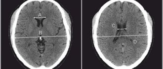
Carrying out the procedure for analyzing and studying SM fluid requires taking a sample as a punctate from the lumbar spinal cord. The collection is made through a small puncture in the desired area of the spine.
A complete analysis of CSF includes macroscopic and microscopic examination, as well as cytology, biochemistry, bacterioscopy and bacterial culture on a nutrient medium.
Indications for use
A study of cerebrospinal fluid is carried out when:
- the appearance of a risk of hemorrhage in the brain cavity;
- infections that affect the central nervous system and lead to inflammation of the membranes (meningitis, encephalitis, meningoencephalitis);
- malignant formations in the meninges;
- there is a suspicion of liquorrhea (loss of cerebrospinal fluid), the main cause of which is a violation of the integrity of the hard membranes of the brain.
The above indications are absolute, i.e. requiring mandatory analysis of the cerebrospinal fluid. The study can also be chosen as a diagnostic tool in other situations. Such relative indications may include:
- diseases caused by demyelination, i.e. destruction of the myelin sheath of neurons, in which the conductivity of nerve impulses deteriorates and various abnormalities occur (for example, multiple sclerosis);
- autoimmune diseases;
- blockage of blood vessels by pathogenic organisms (septic embolism);
- the child has a fever of unknown origin;
- systemic lesions of peripheral nerves (polyneuropathy) caused by inflammatory reactions.
In some cases, cerebrospinal fluid analysis is contraindicated, for example, in case of a large tumor, greatly increased cranial pressure, or edema.
The procedure is prescribed with caution in cases of bleeding disorders (due to the risk of bleeding) or thrombocytopenia (a decrease in the number of platelets, accompanied by bleeding).
Liquor is normal
Liquor is produced by the cells of the ventricles of the brain and performs a number of important functions
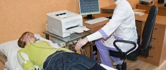
Liquor, like human blood, has a number of indicators that can be assessed using laboratory research methods. Cerebrospinal fluid for research is obtained using a lumbar puncture, during which up to 10 ml of fluid can be collected once without complications for the patient.
The following indicators are assessed:
- Color and transparency.
- Cerebrospinal fluid is normally colorless, transparent and odorless. 99% of liquor consists of water, the remaining 1% is dry residue.
- The normal relative density is 1.006-1.007.
- The amount of protein is 0.2-0.33 g/l.
- The amount of glucose is 2.8-3.9 mmol/l.
- The amount of Cl- (chlorides) is 120-130 mmol/l.
- The acidity of the cerebrospinal fluid (pH) is normally 7.28-7.32. If the permeability of the blood-brain barrier is not changed, then the pH of the cerebrospinal fluid remains within normal limits even when the pH of the blood changes.
- The number of cells in 1 μl of cerebrospinal fluid (cytosis) is up to 4 cells.
Cytology allows you to determine the total number of cells per 1 µl or 1 liter of liquid, as well as differentiate cellular elements (lymphocytes, neutrophils, in some cases, erythrocytes and other cells). In an adult, 1 liter of cerebrospinal fluid contains from 3*106 to 5*106 cells, and in children in the first three months of life their number reaches 20-25*106/l.
The content of lymphocytes is 80-85%, neutrophils 3-5%.
Analysis process
Typically, cerebrospinal fluid is obtained using a lumbar puncture to obtain a sample. Sometimes it is necessary to do a ventricular (ventricular) or suboccipital (occipital) puncture, but this is not common. The price of the procedure varies depending on the type of fluid taken; the cost also varies depending on the location and complexity of the study.
A sequential description of the procedure, which lasts on average 40-50 minutes, is as follows:
- The patient takes a horizontal position and pulls his legs to his stomach and his head to his chest (during lumbar puncture). In other types - lying or sitting.
- The desired area is treated with iodine and alcohol.
- For pain relief, novocaine is injected into the puncture area.
- The needle passes between the 3rd and 4th or 5th and 6th vertebrae (for lumbar puncture), between the temporal, parietal and frontal bones (for ventricular puncture), between the second cervical vertebra and the occipital bone (for occipital puncture).
- CSF is collected. By the way the liquid flows out, deviations can be judged. Normally, it is released in drops, but with an increase in intracranial pressure it runs rapidly. A pressure gauge is used to determine pressure.
- The needle is removed and the puncture area is covered with a sterile napkin.
- The patient is prescribed bed rest for a day.
The resulting liquor must be immediately delivered to the laboratory.
Pathologies of cerebrospinal fluid and their consequences
First of all, of course, experts pay attention to changes in the color of the liquor. So, with a yellow-brown or greenish-gray tint, a tumor neoplasm in the brain, or, less commonly, hepatitis, should be excluded. Whereas a reddish color indicates possible hemorrhage in the ventricles and subarachnoid space. Sometimes this result is a consequence of a traumatic brain injury.
Turbidity and the presence of sediment in the cerebrospinal fluid are an indication for emergency medical intervention. Most often, pathogenic microorganisms are implicated as the cause of infectious brain damage. An increase in cerebrospinal fluid pressure is an indication of its excessive accumulation in the brain cavities, for example, during concussions and bruises, fractures of the cranial bones or pressure on tumor tissue.
Detection of glucose in the cerebrospinal fluid is a harbinger or consequence of diabetes, encephalitis or even tetanus. The doctor will recommend additional examinations - magnetic resonance imaging, bacteriological culture of fluid, blood for tumor markers, PCR diagnosis of various infections. After all, establishing an accurate diagnosis contributes to the optimal selection of a treatment regimen. If you contact a doctor late, this is due to the functions of the cerebrospinal fluid, which aggravates the situation - metabolic disorders, paresis and paralysis, epilepsy and dementia, as well as death, develop.
To prevent various complications, doctors urge people to take care of their own health, give up bad habits, eat right and undergo preventive medical examinations in a timely manner.