Rheoencephalography of cerebral vessels is a simple but effective diagnostic method. As a result of this procedure, pathological processes such as circulatory disorders, as well as other deviations from the normal functioning of this important organ are identified. The method is popular with patients and doctors. This is explained not so much by the affordability of the examination, but by its high information content and the ability to quickly obtain accurate results. A big advantage over other methods of examining cerebral vessels is its minimal invasiveness, which becomes a factor favoring the use of this diagnostic even for pediatric patients.
General information about the method
Rheoencephalography (REG) makes it possible to identify circulatory disorders in the brain even in the early stages of pathology and thereby prevent the development of complications that pose a danger to the health and life of patients. Its invaluable advantage over MRI and CT is the ability to be examined without waiting in line, which in other places is about six months. Without detracting from the effectiveness of magnetic resonance and computed tomography, it should be noted that timely treatment is the key to victory over the disease, and in some cases, the ability to save the patient’s life.
What kind of procedure is this, who needs it, how to prepare for the examination - these are the questions that will be discussed in the article.
For what purpose is it carried out?
In addition to procedures related to the need to study pathological changes in the arteries and vessels of the brain, it is advisable to conduct REG for preventive purposes.
REG and USDG: what is the difference?
REG allows you to assess the tone, elasticity and blood supply of blood vessels, and reveals many pathologies of the brain. In addition, REG allows you to determine whether pathologies are functional or organic in nature. Doppler ultrasound makes it possible to judge pathologies by indirect signs: vascular patency, trajectory and speed of movement of red blood cells. REG is based on the electrical resistance of blood vessels and tissues, which is why it differs from Doppler ultrasound and other methods of ultrasound examination of the brain
.
Operating principle of the device
The essence of rheoencephalography is that with the help of a special device - a rheograph - an electric current of low frequency is passed through the brain, as a result of which the resistance of brain tissue is visualized on the monitor. In this way, abnormalities in the arteries, veins and small vessels are detected. The presence of six channels in the device makes it possible to examine several areas of the brain simultaneously. Metal electrodes are installed in the projection of the studied areas using an elastic rubber band, which transmit the image to the monitor.
Methodology of the procedure
To perform rheoencephalography, you need a special device ─ rheograph. The patient is placed on his back and asked to close his eyes. The doctor fixes the electrodes on the head of the patient using rubber bands. Next, a weak high-frequency current is passed through them, and this way the condition of the vessels is recorded. During the examination, the doctor may ask you to hold your breath or, conversely, to breathe frequently.
The main condition when undergoing REG is calmness
The main difficulty of the procedure is that if the patient worries too much during the examination, this affects vascular tone and the results may be unreliable.
The result is visualized in the form of a rheoencephalogram, which is deciphered by a neurologist. REG analysis can be carried out both from the monitor screen and in paper form.
When is an REG prescribed?
- patient complaints of dizziness;
- deterioration of condition with changes in atmospheric pressure;
- osteochondrosis;
- noise in ears;
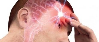
- debilitating headaches;
- suspicion of ischemic disease;
- memory losses;
- weakened vision;
- hearing loss;
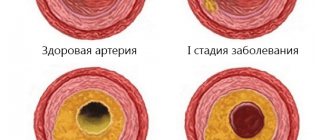
- atherosclerosis;
- hypertensive crisis;
- dystonia;
- hypertension of the cerebral arteries.
For all pathologies associated with a violation of the condition of blood vessels - their blood supply, changes in blood flow speed and viscosity, it is necessary to conduct an REG.
REG examination of cerebral vessels. Part 1. Lecture for doctors
Lecture for doctors “REG examination of cerebral vessels.” Part 1
Content
Ultrasound or REG. Aspects of the use of rheoencephalography to assess cerebral circulation. Part 2
Analysis of rheographic (REG) curve. Part 3
Methodology for rheoencephalography (REG). Study of cerebral blood flow. Part 4
Changes in REG in arterial hypertension. Part 5
Vascular dystonia. REG examination of cerebral vessels. Part 6
Changes in REG in atherosclerosis. REG examination of cerebral vessels. Part 7
Changes in the venous blood supply to the brain and intracranial hypertension. REG examination of cerebral vessels. Part 8
Intracranial hypertension. REG examination of cerebral vessels. Part 9
Cerebral circulation disorders. REG examination of cerebral vessels. Part 10
Changes in REG in case of impaired patency of the cerebral arteries. REG examination of cerebral vessels. Part 11
Closed craniocerebral injury. REG examination of cerebral vessels. Part 12
Functional tests in REG. REG examination of cerebral vessels. Part 13
Artifacts during registration of rheograms. REG examination of cerebral vessels. Part 14
Pathologies. REG examination of cerebral vessels. Part 15
Rheographic methods (REG)
Rheographic methods have virtually no contraindications and are suitable for long-term studies, including monitoring. The method allows long-term observation of patients while studying the effects of various pharmacological agents and assessing compensatory capabilities. The use of multichannel rheographs (polyreography) allows one to study the redistribution of blood and simultaneously assess the state of blood circulation in various organs under the influence of treatment and under functional loads.
This is a bloodless method for assessing the dynamic characteristics of blood circulation, based on graphical recording of changes in the electrical resistance of living tissues during the passage of high-frequency alternating current through them and reflecting changes in the pulse blood supply of the studied area of the body during the cardiac cycle, the functional state of blood vessels, and their tone.
Features of blood circulation in the brain
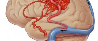
The blood circulation of the brain is characterized by specific features due to its complex structural and functional organization. The volume of blood flowing through the human brain usually accounts for a significant portion (in adults, approximately 15%) of the total blood volume. Of the total amount of oxygen entering the body with inhaled air, the brain consumes 20 - 25%.
In addition to the mass of circulating blood, an important factor determining the intensity of blood supply to the brain is the speed of blood flow. It is known that the speed of arterial blood flow in the brain is much higher than in other organs. This intensive blood supply is provided by a large and complex network of cerebral vessels with diverse angioarchitecture.
The blood supply to the brain is carried out by two pairs of main arteries - the internal carotid and vertebral, forming a circle of Willis at the base of the brain. The circle of Willis is a powerful collector that distributes blood in the brain. Due to the equality of pressure in the right and left, as well as in the anterior and posterior halves of the circle of Willis, “dead points” are formed in certain places of the anterior and posterior communicating arteries, in which there is no blood movement.
Consequently, blood from different vessels within the circle of Willis does not mix under physiological conditions, but enters the vascularization zone of each individual artery.
The posterior cerebral circulation is supported by blood flow from the vertebral arteries, and after their fusion into the basilar artery, blood from the right vertebral artery flows strictly along the right half, and from the left vertebral artery - along its left half. Perhaps the uniform distribution of blood along the homolateral sides is also facilitated by the vascular bundles extending from the dorsal sides of the vertebral arteries at their confluence.
However, even with a slight decrease in pressure in any of the main vessels (pressure of the arteries in the neck during sudden movements of the head or compression of the neck), blood immediately flows in the direction of the decreased pressure. From the above it is clear that the dynamics of blood supply to the brain, even under physiological conditions, depends on the state of collateral circulation. The circle of Willis is the most powerful and constantly operating system of anastomoses, providing collateral circulation in both hemispheres. In addition, there are two more systems of anastomotic connections that do not function under normal conditions, but become important in conditions of vascular pathology. These are the connections of the internal carotid and vertebral arteries with the external carotid artery and the anastomoses of the three cerebral arteries to each other on the surface of the brain.
The total mass of intracranial contents (medullary matter, arterial blood, venous blood and cerebrospinal fluid) is relatively constant. Arterial blood flow is an important factor in maintaining intracranial pressure. A change in the blood supply to the brain affects the pressure of the cerebrospinal fluid. Hemodynamics in the brain is maintained by the pulse movements of the blood. Rhythmic fluctuations in the volume of cerebral vessels (brain pulsation) are associated with active constriction and dilation of blood vessels and the movement of cerebrospinal fluid, and also depend on a number of influences, in particular on contractions of the heart and breathing (the suction action of the chest, promoting venous outflow from the brain).
The outflow of blood from the cranial cavity is carried out through a developed venous system (veins, sinuses, venous outlets), openly communicating with the extracranial veins. The anatomical and functional unity of the cerebral veins with the extracranial veins and the absence of valves in them provide the possibility of blood flow in different directions - depending on local conditions and the needs of the tissues for the inflow and outflow of blood. Using these features of the venous circulation of the head, A.A. Kedrov and A.I. Naumenko (1954), while studying the cerebral hemodynamics of dogs, obtained experimental data confirming the pulse nature of blood movement in the vessels of the brain in a closed skull. Constant pulse and respiratory fluctuations of intracranial pressure in a closed skull, according to their data, are possible due to the presence of peculiar adaptive mechanisms: on the one hand, the existence of a pulse venous outflow from the cranial cavity and, on the other, due to the movement of cerebrospinal fluid from the cranial cavity into the spinal cavity in connection with different phases of breathing. This was later confirmed in the studies of Yu.E. Moskalenko and A.I. Naumenko (1957). They determined not only the nature of these oscillations (pulse waves, respiratory waves and third-order waves), but also their absolute values. In the closed cavity of the skull, the volume of the brain fluctuates slightly due to the fact that it is surrounded on all sides by incompressible cerebrospinal fluid and during pulse fluctuations the blood pressure encounters counterpressure on all sides.
Cerebral hemodynamics, therefore, differs from the blood supply to other organs not only in its greater intensity and constancy, but also in the characteristics of collateral circulation, as well as its close relationship with cerebrospinal fluid circulation. The latter is manifested in the great interdependence between venous and liquor pressure. With venous stagnation of the brain, liquor hypertension develops.
Along with the existence of a relationship between the circulation of blood and cerebrospinal fluid, there is a close interdependence between the state of regional blood flow and the functional activity of various brain formations. Increased blood circulation in some structural formations of the brain during their increased activity is accompanied by a decrease in blood supply to others, which are at this time in a state of relative rest.
Due to the rich intracranial collateral blood flow - both arterial and venous - there is no area in both hemispheres that is supplied exclusively by one main artery or one main vein. This, along with the redistribution of blood in the brain depending on the functional activity of its various formations, determines the advisability of studying the regional hemodynamics of the brain simultaneously in several of its areas.
Mechanisms of rheoencephalogram (REG) formation
Changes in impedance between electrodes applied to the scalp are determined by a complex set of factors, which are presented in Fig. 1.1.
The leading factors, or disturbing influences, are fluctuations in systemic venous and arterial pressure, and the rest play a modulating role. The latter should be divided into three groups. The first is the factors of intracranial hemodynamics that determine the information content of the rheoencephalogram (REG). The second group includes factors not related to intracranial hemodynamics, i.e. factors that are a source of interference and reduce the information value of the REG. Therefore, it is necessary to find out the conditions under which the influence of intracranial factors will be most pronounced, and the influence of interfering factors will be minimal.
Based on the diagram in Fig. 1.1 it is obvious that intracranial hemodynamic and liquorodynamic factors can have a pronounced modulating effect on REG. Indeed, pulse changes in the passive electrical properties of intracranial contents are determined by an increase in blood supply to the cranial cavity due to pulse fluctuations in the arterial and venous systems of the brain. Due to the peculiarity of the biophysical structure of the intracranial hemodynamic system, the ability of the brain vessels to accommodate an additional volume of blood compared to other organs is very limited. In the mechanisms of compensation of systolic blood volume, factors such as fluctuations in intracranial pressure, acceleration of blood flow, transmission of arterial pulsation to the veins directly through the cerebrospinal fluid, redistribution of intracranial volume between arterial, venous blood and cerebrospinal fluid are of particular importance. The electrical conductivity of the cerebrospinal fluid differs from the electrical conductivity of the blood, and the latter is not the same in different parts of the vascular system of the brain. Thus, the REG pulse wave is a complex biophysical signal of a complex nature, the main information value of which lies in the ability to judge pulse changes in the blood supply to the brain tissue, which in turn depends on the extensibility of the walls of cerebral vessels. Consequently, REG can reflect both structural changes in the walls of cerebral vessels, for example in atherosclerosis, and dynamic changes in their tone in response to functional loads. The latter may be of interest as a non-invasive methodological approach for assessing the adaptive abilities of the vascular system of the brain under certain external influences on the body or pathological conditions.
Rice. 1.1. Scheme of REG wave formation
Influence of extracranial hemodynamic factors. The question of the relationship between extra- and intracranial factors is the most controversial in the physiological and biophysical justification of the REG method. As follows from Fig. 1, extracranial vessels are influenced by the same hemodynamic factors as intracranial ones. Moreover, their reactions to such influences as changes in the partial pressure of carbon dioxide in arterial blood, fluctuations in blood pressure, sympathetic stimulation and some other influences may be unequal and even multidirectional. The study of the relative role of extra- and intracranial vessels in the genesis of REH is carried out through biophysical analysis and through experimental physiological research.
Biophysical analysis of current distribution through extra- and intracranial tissues when applying electrodes to the skin of the head showed that it is not possible to completely avoid current shunting through extracranial tissues. Due to the high resistance of the skull bones, the best conditions for the passage of current to the brain are created when electrodes are applied near the large natural openings of the skull (orbits and foramen magnum).
The exact magnitude of the extracranial component of the REG signal is currently unknown, but is still significant. Therefore, for the REG method, as for all other methods of studying cerebral circulation, the problem of reducing this component remains very relevant. Standardization of the REG registration technique will make it possible to record the errors in question and make the research results comparable. Special methods for reducing the influence of extracranial factors when registering REG include the simultaneous removal of REG and a rheogram of the soft tissues of the head, followed by electronic comparison of them and obtaining the resulting curve, as well as the use of protective ring or shielding electrodes.
Thus, despite the significant modulating influence of fluctuations in blood supply to extracranial tissues, REG can retain its information value if this factor is properly taken into account.
The influence of changes in the electrical properties of tissues on REG readings. According to Fig. 1, REG pulse waves, especially their amplitudes, should depend on changes in the ratio between the passive electrical characteristics of the media and tissues filling the cranial cavity. It is known that the electrical resistance of blood depends on a variety of factors. The blood, cerebrospinal fluid, and intercellular fluid filling the cranial cavity are the main paths for conducting electric current, therefore both the basic resistance between the electrodes and its relative changes will be primarily determined by the ratio of the liquid and cellular phases in the area under study. This is evidenced by a significant increase in the amplitude of pulse fluctuations in resistance between the electrodes.
Changes in the electrical conductivity of blood during its movement have a certain significance for REG. A biophysical analysis of this phenomenon in a system of rigid tubes showed that changes in blood electrical conductivity are determined by the charge on the surface of red blood cells and the degree of their aggregation. Since the magnitude of the change in blood electrical conductivity during movement depends on the frequency of the measuring current, the frequency range recommended for REG registration was selected taking into account this phenomenon and the error due to speed changes in blood flow is no more than 8...10%. Studies have shown that the volumetric component of the rheographic signal is many times greater than the velocity component. Therefore, we can say that the REG pulse wave reflects volumetric changes in blood supply to the studied area of the brain.
All of the above indicates that the dynamics of REG indicators is determined not only by processes in the intracranial hemocirculation system, but also by changes in the electrical characteristics of blood and brain tissue, therefore this method should not be used for such effects on the body that have a significant impact on the electrical characteristics of blood and brain tissue. Taking into account the above facts will increase the information value of this technique.
What the study shows
- Based on rheoencephalography of the vessels of the head, specialists receive significant information about the condition of the object of examination. Among them is the possibility of studying vascular tone, their elasticity, blood circulation speed and blood inflow/outflow.
- The use of rheoencephalography makes it possible not only to identify abnormalities in the vessels of the brain, but also to monitor blood flow after complex operations or severe injuries.
- With the help of REG, various pathologies are detected, and the severity of the pathological process is established.
In this case, the high speed of obtaining results is of no small importance.
What problems are being identified?
- presence of traumatic brain injuries;
- localization of hematomas formed as a result of head trauma;
- pre-stroke condition;
- damage to blood vessels by atherosclerotic plaques (atherosclerosis);
- thrombus formation in the vessels of the brain;
- predisposition to increased blood pressure;
- diseases associated with circulatory disorders.
The procedure facilitates the task of making an accurate diagnosis, on the basis of which the doctor prescribes an adequate course of treatment. With the help of it, he subsequently monitors the effectiveness of therapy. Thanks to the complete safety of such an examination for the patient’s health, it can be carried out repeatedly. One of the most significant advantages of encephalography is the ability to distinguish between pre-stroke indicators, which have certain differences for men and women.
Other features of the method
Specialists receive even more information by conducting functional tests. The simplest and most accessible of them is with nitroglycerin. This substance helps reduce vascular tone. This test is used to differentiate organic and functional disorders.
What is the technique?
REG of the brain is one of the methods for diagnosing the condition of blood vessels, which can be used to assess the functioning of the veins and arteries of the head and cervical region.
This study is carried out by recording changes in the electrical resistance of tissues after a weak electric current is passed through them. Since blood is an electrolyte (a substance that conducts electric current), when the vessels are filled with blood, the electrical resistance in them decreases, and this is detected using REG. Taking into account the speed and time of change in resistance, the doctor draws conclusions regarding the patient’s health.
Rheoencephalography is a painless method for studying the condition of cerebral vessels
Using this method, you can evaluate the pulsation of blood flow in the head arteries, determine the degree of venous outflow from the skull, and study the tone and elasticity of the walls of blood vessels. REG is a non-invasive study.
Magnetic resonance imaging (MRI), unlike rheoencephalography, is more informative and can indicate the exact location of a damaged vessel, blood clot or any other abnormality in the vascular system.
How to decipher the results
When assessing the examination results, the patient’s age must be taken into account. This is explained by the fact that the walls of blood vessels lose their elasticity over the years, become more fragile, and react differently to various stimuli. An REG shows graphical wave fluctuations. The following indicators are taken into account:
The specialist reads the diagnostic results, taking into account the regularity of the waves, the appearance and rounding of the apex, as well as the location of the tooth and incisura. The normal fluctuations of the wave shown on the screen in adults differ from the manifestations of acceptable indicators in a child. Rheoencephalographic study makes it possible to classify the condition of the vessels into three types their behavior:
- Dystonic. Characterized by frequent manifestations of changes in vascular tone. Hypotonia with difficulty in venous outflow of blood and low pulse filling is more often observed.
- Angiodystonic. Its symptoms are similar to those of the previous type. The difference is that the cause of the tone disorder is a defect in the vessel wall.
- Hypertensive type according to REG. Significantly different from the species described above. Vascular tone is significantly increased. Venous outflow is impaired.
These types of behavior are not independent pathologies. They are only signs of other diseases and make it possible to identify them in the early stages of development.
You should not attempt to decipher the examination results yourself. It is better to leave this to qualified doctors who will do it professionally and establish an accurate diagnosis.
Decoding the results
To correctly assess the data obtained, the doctor takes into account the patient’s age, since the indicators will be very different for young and elderly people.
- MRI of cerebral vessels - what it shows, indications and contraindications, how it is performed
The resulting rheoencephalogram is studied, which has a wave-like appearance and consists of an anacrota (growing part), catacrota (falling part), incisura (bend between them) and a dicrotic tooth that appears immediately behind it.
Based on the waves of the resulting graph, the doctor evaluates the work of blood vessels
The doctor evaluates the regularity of the waves, the nature of the construction of their peaks, the appearance of anacrota and catacrota, the location of the incisura and the depth of the dicrotic tooth. The presence of additional waves is also being studied.
After evaluating the data obtained, we can talk about the following results based on the appearance of the rheogram:
- dystonic type of REG indicates possible hypotonic deviations, decreased pulse filling and problems with blood outflow through the veins;
- angiodystonic type indicates a decrease in the tone of the vascular walls and a decrease in blood flow speed;
- The hypertensive type indicates increased pressure and tone of the vessels through which blood flows to the head and their obstructed outflow.
The amplitude indicator of the rheogram (APR) indicates volumetric pulse filling:
- APR less than normal by no more than 40% indicates a moderate decrease in pulse blood supply;
- at 40–60% - a significant decrease;
- at 60–90% - pronounced;
- at 90–100% - critical.
The coefficient of asymmetry (CA), which indicates differences in blood supply to different parts of the brain, is very important for studying. Depending on the severity of CA, several degrees of asymmetry are distinguished:
- less than 7% - no pronounced asymmetry;
- 8–14% - weak asymmetry;
- 15–25% - moderate asymmetry;
- more than 26% - severe asymmetry.
What deviations do the external characteristics of waves show - table
| Possible diagnosis | Type of rheoencephalography |
| Cerebral atherosclerosis | |
| The waves take on a strongly pronounced dome shape. | |
| Arterial hypotonicity | Increased amplitude, sharp rise with a sharp apex, shortened anacrosis. |
| Arterial hypertonicity | Reduced amplitude, extended anacrotic with additional waves, displaced apex. |
| Vascular dystonia | Floating prongs, extra waves on catacrota. |
| Obstruction of blood outflow | Catacrota increased, many small waves before the next cycle. |
| Spasm of vascular walls | Rounding the top of the wave. |
How is the procedure performed?
The described diagnostic method is completely painless and safe. During the procedure, there is no impact on the patient’s skin, and no various instruments are used.
During the procedure, the patient is placed on a couch or offered to sit on a chair. To obtain more accurate information, the patient is asked to tilt his head forward, turn it to the right or left. The procedure lasts 10-15 minutes. The results of the study are displayed immediately on the monitor screen and are assessed by a neurologist.
Recommendations
To avoid distorting the results, you should consider some simple tips:
- Before installing the electrodes, some areas of the head are treated with alcohol. It is advisable not to stress and take it calmly.
- Eyes should be kept closed during the procedure.
- You need to completely relax. Anxiety can cause a sharp constriction of blood vessels. This will affect the wave oscillation performance.
- It is advisable to rest for 15-20 minutes before the procedure.
- The day before the scheduled examination, you should not take medications that can affect the speed of blood flow.
- The session should not be interfered with by any objects, so you need to remove chains, earrings, hairpins and let your hair down.
If you are examining a small child, you should tell him in advance everything about the upcoming procedure. You can pick him up and sit on a chair with him. Then he will not be afraid and nervous.
Electroencephalography (EEG)
Electroencephalography (EEG) is an accessible and safe method of studying the brain by recording brain biocurrents.
Neurons - the main elements of the central nervous system (and the brain, including) - are capable of generating and conducting electrical impulses that are recorded by an electroencephalograph. This method is of great importance for the early detection of injuries, tumors, vascular and inflammatory diseases of the brain, and epilepsy. In addition, this is the only neurological outpatient study that is performed during attacks of loss of consciousness.
In our clinic, electroencephalography is performed by highly qualified neurologists-neurophysiologists with extensive practical experience in working with patients of various age groups.
1 Electroencephalography (EEG)
2 Electroencephalography (EEG)
3 Electroencephalography (EEG)
EEG is absolutely harmless, has no contraindications, and therefore is used for patients of any age, both children and the elderly.
Electroencephalography is a highly informative study that reflects the functional state of the cortex, subcortical structures of the brain, as well as complex cortical-subcortical interactions, including the hidden pathology of diseases that have not yet manifested against the background of the complete clinical health of the subject.
EEG allows you to monitor the course of the disease over time, adjust treatment if necessary, and evaluate the effect of drug therapy (overdose or withdrawal of antipsychotics, tranquilizers, barbiturates, antidepressants) on brain activity. Unlike CT and MRI studies, EEG reveals structural and functional (reversible) changes that persist for a long time in the brain, for example, after a mild traumatic brain injury.
The role of EEG in the diagnosis of epilepsy
EEG is the most important diagnostic method for epilepsy. Every year, from 20 to 120,000 new cases of epilepsy are registered per year (on average - 70 - 100,000). In the CIS countries alone, about 2.5 million people suffer from this disease. Epilepsy is often combined with other diseases, such as cerebral palsy, chromosomal syndromes, and hereditary metabolic diseases. The incidence of epilepsy, for example, in patients with cerebral palsy is up to 33%.
An experienced neurologist/neurophysiologist can confirm the diagnosis of epilepsy based on the results of an EEG. In addition, diseases that occur without clinical manifestations have recently become widespread, but are characterized by pathological activity in the brain that significantly impairs its functioning (for example, epileptic encephalopathies). In such cases, EEG is the leading research method.
Electroencephalography technique
EEG is completely harmless and painless. During the examination, the patient sits comfortably in a chair. Using a special helmet, small electrodes are attached to his head, connected by wires to an electroencephalograph. The device amplifies the biopotentials received from the sensors hundreds of thousands of times and records them in the computer memory.
1 Electroencephalography (EEG) in MedicCity
2 Electroencephalography (EEG) in MedicCity
3 Electroencephalography (EEG) in MedicCity
No special preparation is required to conduct an EEG, but there are several recommendations. It is important that the patient is not hungry during the examination, as this may cause changes in the EEG. And you should wash your hair the day before the test - this will allow for better contact of the electrodes with the scalp and, accordingly, the results will be more reliable. You should not give up your usual medication intake, as this can provoke seizures and even epistatus. The interpretation of EEG results may depend on the patient’s age, medications he is taking, the presence of tremor of the head and limbs, visual impairment, skull defects, etc.
1 Electroencephalography (EEG) in MedicCity
2 Electroencephalography (EEG) in MedicCity
3 Electroencephalography (EEG) in MedicCity
Diagnostic capabilities of EEG
With EEG you can:
- distinguish epileptic seizures from non-epileptic ones and classify them;
- identify areas of the brain responsible for triggering seizures;
- track the dynamics of the action of drugs;
- assess the functional state of the brain (even in the absence of changes on a CT scan of the brain);
- resolve the issue of professional suitability (the detection of epileptiform phenomena serves as the basis for the selection of professions related to driving, requiring constant attention and quick response to sudden situations and stimuli in conditions of increased risk).
Indications for EEG:
- epilepsy and other types of paroxysms;
- brain tumors;
- traumatic brain injuries;
- vascular diseases;
- inflammatory diseases;
- degenerative brain lesions;
- headache;
- dysontogenetic diseases;
- hereditary diseases of the central nervous system;
- functional disorders of nervous activity (neuroses, neurasthenia, obsessive movement neurosis, somnambulism, etc.)
- psychiatric pathology;
- encephalopathy of various origins (vascular, post-traumatic, toxic);
- post-resuscitation conditions due to somatic pathology.
Our clinic uses the latest biofeedback-EEG training technique, which includes taking an electroencephalogram, which records the main rhythms of the brain (alpha, beta, delta, tetarhythms). An EEG (electroencephalogram) assessment is carried out by an experienced neurologist-neurophysiologist, and a conclusion is given about the characteristics of brain rhythms and the distribution of biopotentials in various areas of the cerebral cortex. Depending on the indications, the necessary course of biofeedback-EEG training is selected (relaxing, activating, etc.).
About contraindications
Due to the absolute absence of harm to the body, rheoencephalography has virtually no contraindications or side effects. This examination is contraindicated for newborn children. This is explained by the small amplitude of the reflected waves, the large size of the anacrote and the complete absence of incisura. Such indications do not provide an accurate picture of the condition of the vessels of the head. Rheoencephalography is an effective and affordable method for examining the vessels of the brain. Its widespread use is due to the presence of the device in every hospital and, of course, the absence of side effects and contraindications for use.