Cortical atrophy of the brain is a devastating, irreversible change that most often affects people between 50 and 55 years of age, although there are cases of cortical atrophy in newborn infants. Pathological disorders, as a rule, manifest themselves in the frontal lobes of the brain, which are responsible for the thinking process, human behavior, and control. The disease progresses slowly with a gradual increase in the main symptoms, which ultimately leads to the emergence and development of senile dementia.
Causes of the disease
The main factor that leads to brain atrophy in 95% of cases is genetic predisposition. With age, the volume and mass of the brain decrease. This pathology is most pronounced in older women. Provoking external factors can aggravate the situation. Mutations in chromosomes and infectious processes during pregnancy can lead to congenital brain atrophy in a newborn. The main reasons for the development of the disease include:
- Hereditary predisposition.
- Diseases of the mother that are transmitted to the fetus through the placenta, trauma during childbirth, hypoxia.
- Poor blood supply to the brain due to changes in blood vessels and a decrease in their capacity.
- Insufficient mental stress, as a result of which the brain atrophies.
- The influence of alcohol, drugs and medications, which has a detrimental effect on the cortex and subcortical formations of the brain. Radiation exposure.
- Chronic anemia, which leads to insufficient saturation of brain cells with oxygen, which ultimately leads to ischemia and atrophy.
- Injuries, including those resulting from neurosurgical intervention, lead to compression of blood vessels, which affect brain tissue, which can cause the development of atrophy. Slow development of neoplasms, which also compress the vessels.
- Acute and chronic infectious diseases of the brain.
Factors that contribute to the development of pathology include: excessive smoking, chronic arterial hypotension, use of vasoconstrictor drugs, chronic alcoholism.
It is important to note that cortical atrophy of the brain is changes in the nervous tissue of the brain after its normal formation. Primary underdevelopment of the central nervous system during intrauterine development cannot be considered atrophy.
The development of pathology in the cerebral cortex in elderly people very often depends on the level of their mental load in youth. People who think a lot and are engaged in intellectual work are less at risk of developing senile dementia.
Signs of brain atrophy
Pathology can be primary or secondary. The classification is determined by the causes of the disease. In primary atrophy, brain abnormalities and genetic disorders are detected. Deviations can manifest at any age, often progress quickly, and are difficult to correct.
Left-sided involution of brain tissue
Secondary atrophy is associated with exposure to external and internal unfavorable factors. Involution of nervous tissue can be a consequence of traumatic brain injury, vascular pathologies, metabolic disorders, bad habits, exposure to radiation, etc. In this case, eliminating the cause can help slow down the development of the disease. Proper drug therapy can ensure a stable, satisfactory condition and quality of life for the patient for many years.
Clinical manifestations of atrophy include disorders of higher nervous activity, disturbances in the synchronization of functions of various parts of the body.
Symptoms of the disease may include:
- problems with skeletal muscles (from decreased tone to complete loss of the ability to move);
- dizziness;
- convulsions;
- decreased hearing and vision up to complete loss;
- speech disorders (from poor vocabulary to the inability to express oneself);
- hallucinations;
- decreased or loss of sense of smell;
- changes in the sensitivity of different parts of the body;
- impaired coordination of movements;
- loss of spatial orientation;
- change in behavior (from irritability to paranoia);
- emotional instability (from excessive excitement to complete withdrawal).
The problem with diagnosing brain atrophy is the absence or mild manifestation of symptoms in the early stages of the disease. The following stages of pathology are distinguished:
- First. Organic changes in the brain are minimal, but mild neurological symptoms are already present. A decrease in the ability to concentrate, memory impairment, and emotional lability are noted.
- Second. Hearing, vision, and speech impairments appear. A person’s behavior changes significantly; illogical actions are possible, which the patient himself cannot explain or immediately forgets.
- Third. Personal degradation reaches its climax. The patient loses the ability to care for himself and becomes completely dependent on others. A person can live in this state for several years. Death occurs as a result of the destruction of the centers for controlling vital functions.
Clinical manifestations and diagnosis of the disease
Symptoms of cortical atrophy of the brain directly depend on the degree, site of damage and extent of the pathology. Cortical cerebral atrophy has five stages of development of the destructive process with its own characteristic features. It is also important to consider where exactly the harmful changes occur - in the cortex or in the subcortical zones. The first manifestations of the main signs of the disease depend on this. Depending on age and external factors, pathological changes may develop faster. The disease has the following stages of development:
- Grade 1 cortical cerebral atrophy usually occurs without any symptoms. From time to time there is a headache, weakness, and dizziness. This stage of the disease develops very quickly and moves into the next one.
- The second stage of pathology is characterized by the fact that a person becomes irritable, cannot calmly accept criticism, conflicts with the environment, and easily loses the thread of communication. Memory deteriorates, handwriting may change, old habits are forgotten and new ones are acquired. At the second stage of atrophy development, you can notice a disturbance in the coordination of movements and changes in gait. The patient may begin to copy other people, as his independence in thinking and movements is lost. The second stage should alert loved ones and encourage them to seek help from a specialist.
- Loss of control over behavior, sudden outbursts of anger, despondency are characteristic features of the third stage of the destructive process. Basic self-care skills are lost.
- In the last stages of the disease, a person reacts inadequately to the world around him, loses awareness of what is happening, and does not show any emotions.
A person in the last stage of pathology is often dangerous to society, and therefore such people may be isolated in a psychiatric or neurological clinic. The most famous pathology of the cerebral cortex is bihemispheric cortical atrophy (destructive changes occur in both hemispheres at once). This disease is better known as Alzheimer's disease. Another type of atrophic disease of brain tissue is Pick's disease, in which thinning of the cerebral cortex is observed. Its main symptoms include: inability to concentrate, increased distractibility, gluttony, hypersexuality. The pathology develops slowly, but progresses steadily. Unlike Alzheimer's disease, Pick's disease is treatable when detected early in its development.
Pathological changes in the cerebral cortex and the degree of damage can be diagnosed using additional studies, as well as computer or magnetic resonance therapy, the indications for which are the main symptoms of a possible disease. Dopplerography of the vessels of the neck and brain, and cognitive tests (to determine the stage of the pathology) are also performed. To identify neoplasms and lesions with structural changes, the patient undergoes X-ray examinations. The most accurate diagnostic method today is magnetic resonance therapy, which allows not only to identify pathological changes in brain tissue in the early stages of the disease, but also to monitor them over time.
Until recently, multiple sclerosis (MS) was traditionally regarded as a primary demyelinating disease. However, it is now generally accepted that both immuno-inflammatory and neurodegenerative components are involved in its pathogenesis, and it is the balance between inflammatory activity, progressive degeneration and reparative mechanisms that determines the clinical manifestations of MS at each specific stage [1].
The gold standard for diagnosing MS is magnetic resonance imaging (MRI). However, if standard MRI techniques are indispensable in terms of confirming the nature, determining the activity of the pathological process and monitoring the course of the disease, then with regard to correlations between clinical symptoms and neuroimaging parameters, their capabilities are extremely limited (the so-called clinical-radiological paradox). Various reasons for this phenomenon are discussed, including clinical assessment, lack of histopathological specificity (especially for axonal loss), insufficient attention to spinal cord involvement, underestimation of damage to apparently intact brain matter - both white (WM) and gray brain (GW), and also masking the effects of cortical adaptation [2]. At the present stage, MRI studies are carried out using new techniques (MR spectroscopy, functional MRI, MRI morphometry, SWI, DIR, MTC), which are aimed at clarifying the pathogenesis, pathophysiological mechanisms of the formation of neurological deficits, developing effective prognostic criteria and new monitoring markers the course of not only the immunoinflammatory, but also the neurodegenerative, atrophic component [3].
Initially, the presence of brain atrophy in patients with MS was identified qualitatively: dilation of the cerebral ventricles and subarachnoid spaces and a decrease in the volume of brain matter were described [4]. The next stage was a semi-automatic quantitative assessment of brain atrophy without differentiation into SV and WM. Currently, studies are using various MRI measurements to assess both global (brain as a whole) and regional atrophy. One of the most commonly used methods is the assessment of brain parenchyma fraction (BPF), which is determined by the ratio of brain matter to the sum of brain matter and cerebrospinal fluid (CSF). The advantage of this method is the fully automatic calculation of volumes and the possibility of individual assessment of parameters in each specific case, but it is difficult to conduct continued studies. Measuring the thickness of the cerebral cortex is also widely used - an automated technique is used, which includes the stages of SV and BV and recording not the volume as in morphometry, but the thickness of the cortex, but its disadvantage is the difficulty in obtaining clear boundaries of the SV due to the non-uniform magnetic field.
Detailed quantitative assessment became possible after the introduction of voxel-based MRI morphometry, a method of statistical analysis of structural MRI images using computerized segmentation of brain matter into SV and WM. Data on this technique were first published in 2000 [5]. Voxel-based MRI morphometry requires processing data obtained from an MRI study in T1-weighted images (WI) with a slice thickness of 1 mm. This preprocessing includes normalization, segmentation, and smoothing [6]. The normalization process involves comparing brain volume between subjects and normalizing it to the same stereotactic space. By segmentation, the main cerebral and extracerebral structures (SV, WM and CSF) are separated. With the help of smoothing, the elimination of the characteristics of the SV of each individual is achieved, which is necessary for further group analysis. MRI morphometry is used not only to calculate the volumes of the SV and WM as a whole, but also individual areas of interest when using so-called masks for each specific region of the brain. In morphometry, great importance is given to the choice of special applications for a personal computer, with the help of which the obtained MRI data is processed. The use of such programs allows you to accurately assess brain structures and avoid operator errors. There are several varieties of such programs, for example, statistical parametric mapping (SPM) [7].
When discussing atrophy of the central nervous system (CNS) and MRI morphometry data in MS, it is necessary to mention that in addition to the gradually increasing loss of brain matter in MS, short-term volume fluctuations may also be observed. Thus, inflammation and swelling as a result of the formation of new lesions lead to a temporary increase in brain volume, and conversely, taking corticosteroids leads to a short-term decrease in it - pseudoatrophy [8]. The mechanism of this process is not entirely clear, but it is believed that it occurs as a result of a decrease in inflammation in the central nervous system and associated edema.
Brain atrophy begins in the earliest stages of the disease and develops in both relapsing-remitting and progressive types of MS. Moreover, in all patients with MS (without division into types of course), brain volume decreases by an average of 0.5-1.3% per year, which is three times higher than normal brain aging (0.1-0.4%) [9]. The relationship between the degree of atrophy and the type of course, stage of the disease, genetic and toxic factors is shown. It was found that in patients with various types of MS and clinically isolated syndrome (CIS), the decrease in brain volume in general is 5.7% greater compared to controls, and in patients with the secondary progressive type of MS (SPMS) it is significant higher than in relapsing-remitting MS (RRMS).
In patients with apolipoprotein E-e4 in the genotype, the annual decrease in brain volume is 4 times higher than in patients with its absence. However, according to other studies, the genotype with the presence of apolipoprotein E-e4 does not affect the degree of atrophy [10]. Exposure to toxic exposures such as tobacco smoke also increases the rate of atrophy.
It has now been proven that atrophic processes in the central nervous system in MS are caused not only by the loss of myelin, but also by a neurodegenerative process with the loss of axons and neurons, which is reflected in a decrease in the volume of the cortex and subcortical structures. In this regard, when assessing the atrophic process in patients with MS using the voxel morphometry method, the following components are identified: atrophy of the WM, cerebral cortex and subcortical structures.
WM atrophy
Initially, in MS, it was the inflammatory demyelination of the BV that was given key importance and therefore it was believed that it was this that underwent atrophic changes. However, axonal damage within the WM lesion leads to atrophy, which most likely develops in two ways: loss of substance in the demyelination lesions themselves and further Wallerian degeneration of the pathways associated with the lesion. WM atrophy affects specific areas of the brain, including both hemispheres of the cerebrum, the brainstem, and the cerebellum. In patients with RMS, compared with the control group, atrophy of almost all parts of the WM is most pronounced: the corpus callosum, cingulate gyri on both sides, some parts of the frontal lobes (including the upper parts of the corona radiata and the upper longitudinal fasciculi), the temporal and occipital lobes (vault, lower and superior longitudinal fasciculi, inferior fronto-occipital tracts). In patients with RMS, WM atrophy (as opposed to SV atrophy) is also expressed in the brainstem and cerebellum and mainly affects the corticospinal tracts, superior cerebellar peduncles (mainly the right one), both cerebellar hemispheres, dorsal pons and rostral medulla oblongata. [11, 12]. Some studies have shown that WM atrophy is less noticeable compared to SV atrophy due to more pronounced inflammatory processes that may mask atrophy [13, 14].
According to the results of some studies, there is a relationship between the degree of focal damage to the WM and the total volume of the brain, as well as with atrophy of the cortex and subcortical structures [12]. Moreover, a connection was revealed with lesions visualized in T2-WI mode, a lower degree of correlation was noted with lesions in T1-WI and its complete absence was observed with lesions accumulating contrast agent [15]. When conducting correlations between the number and location of lesions in T2-WI and atrophy of the SV, the following results were obtained: in patients with RMS, the total volume of lesions in T2-WI correlated with atrophy of the thalamus; in patients with a benign type of MS (BTMS) - with atrophy of the thalamus, bodies of the caudate nuclei and the right putamen; in patients with SPMS, lesion volume correlated with atrophy of the thalamus, caudate nuclei, left inferior parietal lobule, parahippocampal gyri, and superior and inferior colliculus. It was also found that in patients with SPMS and the primary progressive type of MS (PPMS), cortical atrophy corresponded to the number of foci in the corresponding lobes or caudally located parts of the WM, and the degree of atrophy of subcortical structures correlated with the number of foci in all lobes of both cerebral hemispheres.
However, according to other studies, the relationship between the degree of focal damage to the PV and atrophy of the PV is quite insignificant, since other processes also lead to a decrease in its volume.
Cortical atrophy
Studies have been carried out both measuring the volume of CO and assessing the thinning of the cerebral cortex. According to in vivo studies, it is the loss of DM that accelerates as the disease progresses. For example, in a study by E. Fisher et al. [16] in patients with the transition of CIS to RRMS, a 3.4-fold increase in the degree of SV atrophy was found, and in patients with the transformation of RRMS into SPMS, a 14-fold increase. However, there is still no clear opinion whether SV atrophy is more significant in the development of MS progression compared to WM atrophy.
In patients with MS (without division by type of course), diffuse and local atrophy of the cerebral cortex is noted. The average cortical thickness in MS patients is 2.3 mm, compared to 2.48 mm in healthy people. Predominant atrophy of the cortex of the frontal (2.37 mm compared to 2.73 mm in the control group) and parietal lobes (2.65 compared to 2.95 mm) is revealed even in the early stages of the disease. In patients with a long duration of the disease, the motor cortex predominantly atrophies (2.35 mm compared to 2.74 mm normally) [12].
In the course of a meta-analysis, in patients with various types of MS, without dividing into groups, predominant atrophy of the cortex of the pre- and postcentral gyri (more pronounced in the left hemisphere), the left middle frontal gyrus, the inferior parietal lobule of the right hemisphere of the brain, the cingulate gyrus, predominantly the right posterior cingulate and left parahippocampal gyri [17].
Atrophy of the subcortical SV
Recent evidence suggests that thalamic atrophy develops earlier than cortical atrophy, as demonstrated in a follow-up study of patients with RMS and PPMS [18, 19]. Atrophy of the thalamus is observed in all types of MS, to the greatest extent in SPMS, which is likely due to the duration of the disease.
In the work of A. Cifelli et al. [20] showed that, compared with the control group, in patients with SPMS, the volume of the thalamus decreased by 17%, and the transverse size of the third ventricle doubled, and a clear inverse relationship between their volumes was revealed. The above-described MRI data are confirmed by histological studies: a reduction in the number of neurons in the medial posterior thalamic nucleus and a decrease in the total volume of the thalamus by an average of 22% were revealed. Overall, the loss of brain neurons was estimated at 30-35%. Similar data were obtained in patients with RRMS when assessing the concentration of N-acetylaspartate (NAA) in the thalamus, and it was its decrease that correlated with thalamic atrophy and disease duration.
In the aforementioned meta-analysis, patients with RMS and CIS showed the area of greatest atrophy in the right thalamus, and there was also bilateral involvement of subcortical structures, including the left thalamus.
According to some data [21, 22], in patients with CIS there were no areas of significant reduction in the volume of SV, in particular the thalamus, compared to controls. However, other studies using voxel-based MRI morphometry [23, 24] and T1-weighted magnetic resonance imaging have reported thalamic atrophy [25]. The discrepancy in data may be due to the fact that T1-weighted imaging using a magnetization transfer pulse is more sensitive to changes in CO that precede atrophy. The conflicting results from the studies described above could also be due to the different techniques used to measure atrophy. According to a meta-analysis, it was revealed that in patients with RMS and CIS, the right thalamus is the most affected area. When studies that included patients with CIS were excluded from the list of all studies, there was little change in the distribution patterns of SV atrophy. The peak of atrophy shifted to the left thalamus.
In patients with SPMS, compared with RMS, there was a significant decrease in the volume of some subcortical structures (thalamus, caudate nuclei), superior and inferior colliculi of the quadrigeminal roof. Compared with patients with PPMS, those with SPMS had more severe thalamic atrophy on both sides of the superior and inferior colliculi. At the same time, in patients with PPMS, no areas of greater atrophy were identified in relation to SPMS.
Severe atrophy of the thalamus in MS can have several mechanisms: firstly, due to inflammatory changes in the thalamus itself and a direct neurodegenerative process, and secondly, as a result of indirect damage to the brainstem (the thalamus is the so-called barometer of the diffuse neurodegenerative process in MS, so how, thanks to well-developed reciprocal connections with the cortex and subcortical SV, the thalamus is sensitive to their damage) [26]. Also, the presence of more pronounced atrophy of subcortical structures compared to the cerebral cortex, especially in patients with CIS, indicates the probable atrophy of the deep parts of the SV as a result of axonal damage rather than direct impact [27]. This is also confirmed by the fact that when the optic radiation is affected, atrophy of the lateral geniculate body is noted [28].
Patterns of brain atrophy in different types of MS
When assessing global brain atrophy in progressive types of MS, simultaneous expansion of the ventricles of the brain and the subarachnoid space was revealed, while in RMS the expansion of the ventricular system of the brain, mainly the bodies, anterior and posterior horns of the lateral ventricles and the third ventricle, is more determined, which indicates atrophy of subcortical structures [29]. As for the atrophy of the cerebral cortex in patients with RMS compared with patients with CIS and the control group, significant atrophy of the right pre- and postcentral gyri was noted in the absence of pronounced changes in WM. In SPMS, compared with RMS, the greatest atrophy of both WM and SV was revealed: the cortex of all lobes of the cerebral hemispheres (mainly the superior frontal, postcentral gyri, right superior and inferior parietal lobes, parahippocampal gyri, cuneus in both hemispheres, left middle occipital gyrus) , anterior lobes of the cerebellum, superior and inferior colliculus and subcortical structures [30]. Compared with PPMS, SPMS showed atrophy of the cortex of the postcentral gyri (mainly the left one), middle occipital gyri, left cuneus, thalamus, anterior lobes of the cerebellum and colliculi of the quadrigeminal roof.
DTRS deserves special attention. This diagnosis is made retrospectively when the duration of the disease is 10 years or more from the onset of MS with a slight degree of disability (no more than 3 points on the EDSS scale). According to a study by S. Mesaros et al. [31], in patients with TDRS, compared with the control group, no changes in brain volume as a whole were detected, but there was a local decrease in the volume of the brain: predominantly in subcortical structures and, to a lesser extent, in the cortex of the frontal and parietal lobes of the left cerebral hemisphere. Some patients had severe cognitive impairment. When comparing patients with SPMS and patients with DTRS with cognitive deficits, no differences were found in changes in the volume of brain structures, while when comparing patients with SPMS and patients with DTMS without cognitive changes, the former showed pronounced atrophy of the cerebellar cortex. These results confirm that irreversible damage to the cerebellum may play a significant role in the development of motor disorders [32, 33].
Spinal cord atrophy
The clinical picture of MS is extremely heterogeneous, and the symptoms that arise as a result of spinal cord injury (SpI) often dominate and are observed at some stage in 90% of patients [34]. In the SpM, both local and diffuse foci of demyelination are detected. In patients with CIS and DTMS, the number of lesions is smaller than in RRMS, SPMS and PPMS, which probably leads to a milder course of the disease. At the same time, studies have shown [35, 36] that there is no correlation between the number of lesions detected on T2-WI and atrophy, which indicates their small role in the development of SpM atrophy.
Atrophic changes are most often detected in the cervical spine, and to a greater extent in patients with a progressive type of MS. There is a relationship between the degree of SpM atrophy and the severity of clinical symptoms [37, 38]. Given the small size of the SpM and the structure of the surrounding tissues, measuring its volume is quite difficult. However, with the use of MRI morphometry this became possible. Severe SpM atrophy is observed in patients with SPMS, PPMS, and even DTMS [34]. This confirms that atrophy develops with a progressive and long-term disease. With these types of MS, a similar decrease in volume is observed throughout the entire length of the cervical spine, while at each individual level it is more pronounced in the posterior and lateral sections of its diameter. This is also confirmed using other methods and the T1-VI mode using a magnetization transfer pulse. Patients with DTMS have a lower degree of atrophy overall compared to patients with SPMS, which suggests that the less pronounced the degenerative processes, the more benign the MS is. This is also confirmed by the presence of correlations between the severity of SpM atrophy in PPMS and SPMS and the degree of disability on the EDSS functional scale.
In PPMS, there is a pronounced atrophy of the SpM, especially in its posterior parts, compared to patients with CIS, RRMS and the control group, despite the short duration of the disease. However, SpM atrophy is less severe in PPMS than in SPMS; Although both of these types of MS are progressive, the identified differences confirm different mechanisms of their development.
Correlations of atrophic changes and clinical symptoms
Currently, it is atrophy of the central nervous system (brain and spinal cord) that is considered responsible for the steady progression of both focal neurological symptoms and cognitive impairment [8]. Using histological and neuroimaging techniques, the greatest correlation of a number of clinical symptoms with changes in the SV compared with the WM or the brain as a whole has been proven [39-41].
Correlations of brain atrophy with the development of disability
According to some data [42], there are correlations between the EDSS score and the total thickness of the cerebral cortex, the cortex of the left pre- and postcentral, right parahippocampal and left lateral occipital gyri, as well as with the volume of the right caudate nucleus and right nucleus accumbens (a group of neurons in the ventral part of the striatum). Some studies have shown that patients with persistent progression of disability according to the EDSS scale have a significantly higher rate of atrophic processes compared to patients with stable neurological symptoms. It is also believed that SV volume, compared with WM volume, is a more sensitive predictor of disability assessed by the EDSS scale [43]. Opposite data were obtained in a study by A. Ceccarelli [44], in which no relationship was found between local SV lesions and EDSS scores in patients with MS. In patients with DTRS, there was also no association between SV atrophy, disease duration and EDSS score [45]. There were also no correlations between changes in WM volume and clinical symptoms.
A study that provided new insight into the low degree of correlation between the clinical picture of the disease and MRI data was performed in 2009 [46]. Over the course of 6 years, patients with different types of MS were assessed for clinical manifestations using the EDSS scale and the MS functional scale - MSFC, which included 3 tests. As a result, it was revealed that progression on the MSFC scale correlated with the volume of the brain as a whole, the volumes of the SV and WM, and the lack of connection with the EDSS scale was confirmed. These results, according to the authors, are associated with the relatively low level of agreement between the methods of measuring the degree of disability on these two scales (62%). The lack of correlation between brain atrophy and the EDSS may be due to the following factors: the insensitivity of the EDSS in detecting disease progression in patients with high disability, and errors in assessing disease progression in patients with stable MSFC scores and low MSFC scores. EDSS. Also, discrepancies in the results may be due to the fact that other factors lead to the development of disability, such as changes in SpM. In a study of patients with SPMS, similar results were obtained, namely the presence of correlations between whole brain atrophy, CO and SpM with the MSFC scale.
Correlations of brain atrophy with cognitive impairment
Cognitive impairment, including decreased memory, attention, and speed of information reproduction, is observed in 70% of patients with MS, and they occur already in the early stages of the disease (within the first 3 years). In patients with RRMS with cognitive impairment, compared with patients without them, a decrease in brain volume in general, as well as CO in the cerebral cortex, is detected. Indeed, cortical atrophy is a predictor of cognitive impairment because even mild cognitive changes are associated with significant thinning of the cerebral cortex [47]. A significant correlation was also found with thalamic atrophy.
In different types of MS with the presence of cognitive impairment, different patterns of damage to the brainstem are observed: in patients with MS and cognitive changes, atrophy of the cortex of the left superior temporal gyrus, left insula, thalamus and right middle occipital gyrus is detected; in patients with SPMS, atrophy of the anterior cingulate cortex, hippocampal insula and right superior frontal gyrus is noted; and in PPMS - atrophy of the anterior cingulate cortex and right superior temporal gyrus.
There is evidence that brain atrophy has less of an impact on cognitive impairment in patients with high “cognitive reserve,” which is a result of high levels of education and intelligence.
Correlates of brain atrophy with fatigue
In patients with MS, a common symptom (up to 80% of cases) is fatigue. It is not associated with disease progression, degree of disability, or location of lesions, but is likely to be associated with the frontothalamic-basal network [48]. Research on this topic is scarce. When comparing patients with and without fatigue, the former showed atrophy of the substance of the supratentorial parts of the brain, including the cortex, the adjacent WM and the heads of the caudate nuclei, i.e. areas that are functionally associated with attention control [49]. The authors suggest that impairment of central motor activation may be associated with damage to cortical-subcortical pathways including the motor cortex. Thus, 222 patients with RRMS with low EDSS scores (no more than 2) were divided into two groups: patients with a high degree of fatigue (FSS no less than 5) and patients with a low degree of fatigue (FSS no more than 4). In group 1, there was a smaller volume of SV and BV and the largest number of lesions in T2-WI and T1-WI modes. The authors suggested that atrophy of the SV and WM may be a risk factor for fatigue, which is not associated with the degree of disability.
Conclusion
Thus, modern MRI techniques, including voxel-based MRI morphometry, significantly expand our understanding of the pathogenesis of MS. Numerous studies show that, in addition to WM atrophy, SW atrophy in MS is observed already in the early stages of disease development and progresses faster than in healthy people, being a significant MRI predictor of the development of disability. Studies showing atrophy of different areas of the brain in different types of MS make new contributions to the understanding of the pathophysiological mechanisms of the degenerative process. However, the sequence of development of atrophic processes has not been fully established. Additional detailing of the patterns of atrophy in MS using MRI morphometry will play an important role in research in MS, both in a fundamental sense - clarifying the pathophysiological mechanisms, and in practical terms - developing prognostic criteria for the course of the disease, monitoring progression and assessing the effectiveness of therapy in terms of prevention /slowing down the neurodegenerative process.
Methods for treating atrophic brain pathology
Cortical atrophy of the brain cannot be completely cured. The most important thing is to stop the progression of the pathology, which can be done with greater success at a young age than in older people. Therapy for atrophy, depending on the situation, can be carried out using the following medications:
- nootropics are used to improve nutrition of brain cells. In addition, these drugs help normalize the thinking process;
- the action of antioxidants is aimed at improving metabolism, slowing down the atrophy process, and counteracting oxygen free radicals;
- drugs to improve blood microcirculation.
In addition, if a person suffers from headaches, he is prescribed to take analgesics or anti-inflammatory non-steroidal drugs. For increased excitability, irritation, and insomnia, sedative medications are indicated. The support of the patient’s relatives, their help and understanding is very important. It is undesirable for the patient to change the usual way of life; it is necessary to create the most calm and familiar environment for the person. Walking in the fresh air and feasible physical activity are very important. Daytime sleep is undesirable for a person with atrophic pathology of the brain. If the doctor allows, the patient may be prescribed a course of therapeutic massage to improve the patient's blood flow. If necessary, the treating specialist can prescribe sedatives, antidepressants, and tranquilizers. In cases where relatives are not able to cope with the patient on their own, due to his aggressive behavior, specialized nursing homes and boarding schools are provided for patients with impaired brain function.
The prognosis of people with cortical atrophy of the brain is far from rosy. The disease, one way or another, progresses at a slow or rapid pace, leading to irreversible changes in the cerebral cortex, personality degradation, which ultimately leads to death. The main factors that can stop the development of pathology in a patient are:
- positive psycho-emotional mood, complete absence of stressful situations;
- daily mental stress, working on your memory;
- eating healthy foods;
- complete cessation of habits harmful to the body, such as smoking, alcohol, and taking drugs;
- blood pressure control;
- daily walks, moderate physical activity.
The role of nutrition in the treatment of disease
Proper nutrition is very important to support the normal functioning of the brain and saturate its cells with all the necessary vitamins and minerals. A person diagnosed with cortical atrophy of the brain must necessarily include omega acids, unsaturated fats, and fat-soluble vitamins in their diet. It is necessary to remove everything fatty, fried, smoked and floury. Eating walnuts, fruits and vegetables has a good effect on brain nutrition. You should introduce fatty fish into your diet. Proper nutrition and a healthy lifestyle can stop the death of nerve cells and enable the patient to carry on with his usual life activities.
Treatment of pathology using traditional medicine
Brain atrophy is characterized by irreversible processes in the body that cannot be cured. However, with the help of traditional methods, it is possible to slow down the rate of development of pathological processes and improve the patient’s well-being. Herbal teas and tinctures are used with great success in the treatment of brain atrophy. Here are the best options for such fees:
- Take oregano, horsetail, motherwort and nettle in equal proportions. Pour boiling water into a thermos and leave overnight. Take 3 times a day;
- A tincture of young rye and chickweed, steamed with boiling water, can be drunk after meals in any quantity. This tea helps a lot after an injury.
- A tincture of viburnum, rosehip and barberry, steamed with boiling water and infused for 8 hours, is taken instead of tea in unlimited quantities, or with honey.
Preventive examinations, careful attitude towards one’s health, and strict adherence to the recommendations of specialists and the attending physician are very important for every person. This will help you maintain good health and live many more years of a happy and fulfilling life.
Cortical atrophy is a destructive process observed in the cerebral cortex. Most often, the disorder occurs in fairly old age, and it may also be associated with pathological changes occurring in the body.
Deviations inherent in this disease in most cases are located in the frontal lobes of the brain, which are responsible for thinking processes, and also thanks to them, control of behavior and planning is exercised.
In some cases, the process can affect other areas of the brain, but this is much less common. Typically, an extremely extensive process can lead to outright dementia. Manifestations of a focal nature do not have a pathological effect on the abilities of the brain.
The atrophy process is most dangerous for older people (over 50 years), since it is at this time that its slow progression occurs. The percentage of women susceptible to this problem is much higher than men. Ultimately, cortical atrophy leads to senile dementia.
Cases of cortical atrophy also occur in newly born babies, but this deviation is associated with severe heredity.
Reasons provoking the violation
In the development of brain atrophy, and as a result of senile dementia, there is a complex of reasons that cause atrophic processes in the brain. The formation of the disease is influenced by the following factors:
- blood oxygen saturation deteriorates;
- the body's ability to regenerate deteriorates;
- as a result of atherosclerosis, there is a disruption in the blood supply to brain tissue;
- processes of genetic predisposition to atrophic pathologies are activated;
- reduction of mental load and thought processes.
What is typical, even if a person has a predisposition to brain atrophy (genetic), but in the process of life he studied foreign languages, was interested in science, art, read a variety of literature, developed intellectually in every possible way and used the work of the intellect in practice, that is, made the most of his potential brain, he is less susceptible to developing dementia.
The development of congenital cortical cerebral atrophy is essentially brain hypoplasia, since its normal formation does not occur. Despite this, the process is also called atrophic.
Also in adulthood, the destructive process can develop for a number of reasons:
- atrophy can occur as a result of toxic poisoning (alcoholism); with frequent consumption of alcoholic beverages, neurons die off, and due to ongoing intoxication, their restoration does not occur, which leads to this problem;
- constant low blood pressure;
- use of vasoconstrictor drugs;
- lack of mental stress;
- injuries resulting in compression of blood vessels and development of cerebral edema;
- cysts and tumors lead to compression of blood vessels; neoplasms that have stopped growing are especially dangerous; they influence the development of atrophy; growing neoplasms have a less negative impact;
- in rare cases, neurosurgery may be the cause.
Magnetic resonance imaging in the diagnosis of brain atrophy
Atrophy is an intravital decrease in the volume of a tissue or organ due to a decrease in the size of each cell, and subsequently in the number of cells making up the tissue, accompanied by a decrease or cessation of their function. [5]
Cerebral atrophy is understood as a decrease in the volume of brain matter with a reduced density, due to a generalized (external, internal, mixed) or focal defect of brain tissue. [6]
Physiological cerebral atrophy develops, according to researchers, between the ages of 40 and 60 years, reaching its maximum severity by 70 years [7]. The aging brain shrinks by an average of 1.9 percent every 10 years.
According to Shah SA, Doriswmy PM, Husain ML (1991), aging is associated with a significant increase in the volumes of the lateral and third ventricles (by 3% annually).
In patients aged 65 to 95 years, Coffey CE, Wilkinson WE, Parashos IA (1992) determined the volume of the lateral and third ventricles; an increase in their volume of approximately 0.95 ml/year was detected.
Quantitative studies of the subarachnoid space show an increase in size with age in most people.
According to Zatz LM, Jernigan TL (1982), the volume of cerebrospinal fluid increases from 40 years by 1 ml annually, to 40 ml in the ninth decade. In the period from 40 to 90 years, the ratio of cerebrospinal fluid volume to intracranial volume increases from 3 to 10%.
A study of 76 healthy volunteers showed that increasing age is accompanied by a decrease in the volume of the cerebral hemispheres by 0.23% per year.
In contrast to the hemispheres as a whole, the rate of decrease in the volume of the frontal lobe is 0.55% per year. The volumetric loss rate for the temporal lobe is 0.28% per year, the amygdala-hippocampal complex is 0.30% per year.
Pathological atrophy is considered in relation to degenerative diseases of the brain that are genetically determined, i.e., primary degenerative and secondary CA.
Clinical manifestations of CA are considered mainly from the standpoint of disorders of higher cortical functions, and exclusively in elderly and senile patients. [7] In this regard, it is noted that cognitive deficits are detected in almost all patients, starting from the early stages of CA, regardless of the genesis of atrophy. [2]
Clinical manifestations of secondary cerebral atrophy syndrome are polymorphic. At the same time, there are typical clinical variants, identified taking into account the prevailing symptom complexes. The most common is the variant of neurological disorders (57%), the neurotic (22%) and mixed (15%) variants are less frequently recorded, and the rarest is the variant of cognitive disorders (6%). [6]
Since the second half of the 20th century. All over the world there is a change in the age structure of the population with a constant increase in the proportion of elderly and old people in the population. Therefore, in modern medicine, the prevention and treatment of a large number of age-dependent diseases are becoming increasingly important. Due to the increasing number of elderly people (it is expected that in 2030, 20% of the population will be over 65 years of age), brain atrophy, leading to dementia, is becoming one of the main public health problems. [10]
The problem of brain atrophy is closely related to the development of dementia. According to European epidemiological studies, dementia affects 6–7% of people over 65 years of age, and Alzheimer's disease alone ranks 2–3rd in terms of health and social care costs among neurological and mental disorders. [2,8] The incidence of dementia increases with age; it is 5 times more common in people over 80 than in 70-year-olds.
Despite this spread, currently there are no specific methods for diagnosing cerebral atrophy [1,2,3,4]. In the diagnosis of CA, the main place belongs, of course, to neuroimaging methods: computed tomography and magnetic resonance imaging [6], as well as the method of functional neuroimaging - single-photon emission computed tomography, positron emission tomography, MRI spectroscopy. Additional diagnostic methods include ultrasound dopplerography of the brachiocephalic arteries, as well as transcranial Dopplerography, duplex scanning of the brachiocephalic arteries, thoracic or selective angiography, EEG, evoked potential method, pathomorphological methods, a number of laboratory techniques, including studies of cerebrospinal fluid (CSF) and plasma blood.
Both physiological and pathological atrophy do not have standard diagnostic criteria that would allow one to verify the presence of CA, determine its neurovisualization type and the severity of the atrophic process. [6]
Magnetic resonance imaging is one of the priority and highly informative modern methods of non-invasive diagnostics. Of all the radiation methods, MRI provides a picture that is closest to the anatomical one.
MRI is preferable for neurodegenerative dementias, allowing to determine the topography of atrophy. [9]
The purpose of this study was to improve the diagnosis of diseases leading to brain atrophy based on MRI studies.
An MRI study was performed on 60 patients aged 45 to 80 years. Of these, 20 were healthy volunteers, 13 patients with presumed Alzheimer's disease (based on clinical manifestations), 27 patients with cerebrovascular insufficiency of atherosclerotic origin.
In the group with suspected Alzheimer's disease, according to data obtained from an MRI study of the brain, a decrease in the size of the temporal lobes of the brain by 10–56% and the hippocampus by 10–40% was revealed (the decrease correlates with the severity of dementia (mild, moderate, severe), expansion Sylvian fissures in 13 (100%) patients, which is associated with atrophic changes mostly in the temporal and frontal lobes of the brain, involutional changes in the cerebral cortex with the expansion of the fissures up to 1.5–5.0 mm, and the subarachnoid space of the convextal surfaces in the frontal -parietal and temporal areas in 13 (100%) patients.
In patients with cerebrovascular insufficiency, a decrease in the size of the temporal lobes did not exceed 2% and was observed in 5 (18.5%) patients, which was regarded as natural age-related changes. It was also revealed that the boundaries of the white and gray matter of the brain were erased in 18 (67%) patients, the presence of post-ischemic cysts - microcysts up to 3-5 mm in diameter in 23 (85%), macrocysts over 5 mm in diameter in 16 (59%), widening of the Sylvian fissures in 20 (74%) patients, which is associated with general atrophic changes in the cortex, involutional changes in the cerebral cortex, in particular widening of the fissures to 1.5–2.0 mm with a uniform expansion of the subarachnoid space of the convexital surfaces in 18 (67% ) patients.
In the group of healthy volunteers, 8 (40%) showed generalized atrophy with a relatively uniform expansion of the subarachnoid spaces.
Studies have shown that the use of magnetic resonance imaging plays an important role in the diagnosis and differentiation of various types of cerebral atrophy. Thus, it was revealed that in patients at risk of developing Alzheimer's disease, as well as with an established diagnosis, atrophic changes develop in the frontoparietal and temporal regions of the brain. These changes are characterized by a decrease in the temporal lobes and hippocampus, expansion of the Sylvian fissures and subarachnoid space in the corresponding sections. The severity of these changes is directly dependent on the severity of the patient's condition. The combination of such changes was not identified among other groups, which indicates that in other pathological conditions of the brain the complex of these structural changes does not occur.
Literature:
1. Vavilov S. B. Computed tomography of the brain in psychiatry: morphometric methods / Computed tomography and other modern diagnostic methods (opportunities and prospects). Mat. international Symposium.-1989.-P.60–67.
2. Damulin I. V. Alzheimer's disease: Clinical MRI study / I. V. Damulin, O. S. Levin, N. N. Yakhno / Nevrol. magazine -2005, pp.-34–38.
3. Kolykhalov I. V. Clinical and computed tomographic comparisons in dementia of the Alzheimer’s type. Abstract of thesis - 1993, S-25.
4. Maksimovich I.V. Features of brain microcirculation in individuals with an increased risk of occurrence and early stages of Alzheimer’s disease/I. V. Maksimovich, S. A. Ovsyannikov, L. N. Gotman//Angiology and Vascular Surgery - 2006, T.4, No. 3, P-20–24.
5. Strukov A.I. Pathological anatomy.
6. Cherkasova V. G. Secondary cerebral atrophies. Clinical characteristics, diagnosis and treatment options, 2005
7. Yakhno N. N. Current issues in neurogeriatrics\\ In the book. "Advances in neurogeriatrics." Edited by N. N. Yakhno, I. V. Damulin-M.1995.-Part 1.-P.9–29.
8. Jochemsen HM, Muller M, Visseren FL. Blood pressure and progression of brain atrophy: the SMART - MR Study. JAMA Neurol. 2013 Aug: 70(8):1046–53.
9. Lehericy, Delmaire. Neuroimaging in Dementia, Presse Med 2007 oct 36 (10 Pt 2):1453–63
10. Prestia, Baglieri, Pievani. The in vivo topography of cortical changes in healthy aging and prodromal AD. SupplClinNeurophysiol. 2013: 62:67–80.
Symptoms of each stage of the destructive process
Cortical cerebral atrophy goes through five stages of development, each of which has its own symptoms and manifestations:
- The initial stage is characterized by a complete absence of manifestations, but its development occurs rapidly and quickly moves into the next stage.
- At the second stage, communication with others deteriorates very quickly. A person cannot adequately perceive criticism, becomes irritable, conflicts involving him are a normal phenomenon that recurs regularly. Very often the thread of the conversation gets lost.
- The third stage - the patient loses control over his behavior in several stages, which becomes outrageous. Frequent manifestations of despondency or the occurrence of causeless outbursts of anger.
- four : awareness of the essence of events is completely lost. The patient cannot adequately respond to the demands of others.
- Fifth stage - the patient does not take pictures of the events taking place at all, and, accordingly, this does not cause any emotions in him.
Depending on who and what lobe of the brain was affected by atrophy, some speech disorders arise in the early stages, unreasonable euphoria or indifference, lethargy, sexual hyperactivity, and some types of mania appear.
Diagnostic criteria and research methods
Cortical cerebral atrophy has a number of symptoms that should prompt you to visit a doctor and diagnose the degenerative process.
- decreased ability to analyze and think;
- impaired hand motor skills;
- memory impairment, up to the inability to remember a minimum of information;
- change in speech features: tone, tempo, etc.
When different parts of the brain are affected, different symptoms appear:
- if the cerebellum , muscle tone and coordination of movements are impaired;
- pathology of the diencephalon leads to problems with metabolism, problems with thermoregulation, homeostasis;
- problems with the forebrain lead to the loss of all reflexes;
- atrophy of the medulla oblongata causes disturbances in breathing, digestion, functioning of the cardiovascular system, and protective reflexes; the reaction to stimuli from outside disappears due to the death of the midbrain cortex.
When visiting a doctor, it is necessary to outline the symptoms on the basis of which further tests are prescribed:
- X-ray examination (makes it possible to detect neoplasms, lesions with structural changes);
- cognitive tests (the severity of the disease is determined);
- Dopplerography of the vessels of the neck and brain (to visualize the lumens of blood vessels);
- MRI (gives the most accurate diagnosis).
Brain atrophy: causes, symptoms, diagnosis
Atrophic changes in the cerebral cortex lead to the destruction of neural connections and a decrease in the activity of functional centers.
The condition leads to disruption of intracerebral metabolism, dementia, and the formation of a number of mental diseases (Alzheimer's, amyotrophic lateral sclerosis, dementia). Clinical symptoms depend on the type, stage, and degree of the disease. The multisystem form is accompanied by diffuse death of neurons and gradual loss of body functions.
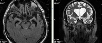
Brain atrophy on MRI
How to help a patient
Cortical cerebral atrophy cannot be completely cured. The main task is to prescribe comprehensive treatment aimed at slowing the development of symptoms. Atrophies that appear at a young age can be effectively corrected if the etiological factor is excluded.
For the treatment of cortical atrophy, patients are prescribed drugs from the following groups:
- To improve blood microcirculation . The most popular drug is Trental. A drug with a vasodilating effect, increasing the lumen of capillaries, improving gas exchange through the walls of blood vessels and blood microcirculation.
- Nootropics . Drugs in this group improve blood circulation and brain metabolism. Highly effective drugs: Piracetam, Ceraxon, Cerepro, Cerebrolysin. Medicines in this group have a beneficial effect on the patient’s thinking abilities.
- Antioxidants . Drugs in this group stimulate regeneration processes, increase metabolic rate, slow down atrophy, and reduce the impact of oxygen free radicals.
- In order to relieve associated symptoms such as headache, the use of non-steroidal anti-inflammatory drugs .
An important factor in the treatment of cortical atrophy is monitoring the patient’s neuropsychic state. This diagnosis should be accepted adequately by family members, as should their attitude towards the patient:
- the patient needs walks in the fresh air daily;
- moderate physical activity;
- the patient must be entrusted with all possible self-care procedures;
- in case of a neurasthenic state, the use of mild sedatives is permissible.
The disease progresses very quickly and the result of this process is personality degradation. The manner of communication, speech, and behavior takes on an ornate connotation. The vocabulary becomes significantly scarcer, which leads to the use of monosyllabic phrases in speech.
The prognosis for patients with cortical atrophy of the brain is always unfavorable: whether the process progresses slowly or quickly, it always leads to degradation and, ultimately, death.
There are currently no effective methods for preventing the destructive process. The only thing that can slow down the process is:
- timely treatment of all existing diseases;
- active lifestyle;
- working on your memory;
- development of intelligence from youth;
- positive attitude.
Brain atrophy is destructive changes that provoke depletion of organ tissue, deterioration of vitality, and loss of function. Accompanied by necrosis of nerve cells and severance of neural connections within chemically or functionally related groups. The volume of brain tissue decreases. Destructive processes spread to different parts - the cortex and subcortical (subcortical) areas. Often occurs in patients over 50 years of age. Diagnosed in newborn infants and children under one year of age.
The death of the cells that make up the brain provokes serious consequences. There is a violation of cognitive abilities, which include speech, spatial orientation, understanding, logical thinking, reasoning, calculation and learning. The disease causes neurological disorders and motor dysfunction.
Doctors give a negative answer to the question of whether cerebral atrophy, which occurs in the brain, affects life expectancy. Neurons die gradually. From the initial signs of pathology to a state where a large area of the brain atrophies with the subsequent development of dementia, more than 20 years can pass. The death of a patient is usually caused by other diseases that cause disruptions in the functioning of the body that are incompatible with life.
Discussions on how long patients with atrophic lesions live do not correctly reflect the characteristics and influence of the pathology. Cerebral atrophy does not reduce life expectancy, but significantly impairs its quality. Leads to dementia and disability. The person is not capable of self-care and needs constant medical supervision and care. Often forced to spend the rest of his life in a specialized dispensary.
Magnetic resonance imaging (MRI) in St. Petersburg
MRI of the brain. Demonstration of atrophy and normality in color treatment.
The term “neurodegenerative diseases” (NDDs) defines a large group of diseases, predominantly of late age, which are characterized by slowly progressive death of certain groups of nerve cells and, at the same time, gradually increasing atrophy of the corresponding parts of the brain and/or spinal cord. The development of these diseases is based on metabolic disorders and changes in the conformation of cellular proteins with their subsequent accumulation and aggregation in certain groups of neurons. In NDD, predominantly neurons and glial cells of the basal ganglia and stem structures that produce acetylcholine, dopamine, and serotonin are affected.
The classification divides NDD into 2 large groups – sporadic and irritative.
- Sporadic NDD:
- Progressive supranuclear palsy (Steele-Richardson-Olszewski disease).
- Multiple system atrophy.
- Dementia with Lewy bodies.
- Parkinsonian dementia (Guam syndrome).
- Corticobasal degeneration.
- Alzheimer's disease.
- Irritative NDZ:
- Huntington's disease.
- Hallerwarden-Spatz disease.
- Wilson-Konovalov disease.
- Farah's disease.
- Bessen-Kornzweig disease.
Alzheimer's disease is a progressive neurodegenerative disease characterized by the gradual development of dementia. The origin of the disease is precisely unknown. Biochemical changes consist of a decrease in the activity of choline acetyl transferase in the cerebral cortex and hippocampus. Pathological manifestations include the formation of specific amyloid plaques, neurofibrillary strands and reactive gliosis. Atrophy develops, predominantly affecting the cortex around the Sylvian fissures and the hippocampus, with secondary expansion of the ventricles, especially the temporal horns
The disease was first described by Alois Alzheimer in 1907. The process resembles natural aging, but sharply accelerated. It begins with memory impairment, then loss, inability to perform daily self-care, and repeated questions. Later, profound mental and speech disorders, weight loss, and convulsions appear.
The incidence is 0.51% for individuals aged 70–74 years, with a progressive increase in incidence with age. Clinical manifestations include memory impairment, depression, behavioral disturbances and hallucinations. In the later stages, extrapyramidal symptoms are added to mental disorders. The disease ranks 4th in mortality. Diagnosis is made on the basis of clinical and neurophysiological examination, as well as neuroimaging. Typical CT findings include diffuse atrophy (especially of the temporal lobes), secondary widening of the sulci and ventricles. Sensitivity (without volume measurements) in comparison with the normal age group is about 80%, specificity is about 70%. Measuring hippocampal volumes using thin-slice MRI increases accuracy to 85%.
MRI of the brain. T2-weighted sagittal MRI. Pick's disease. Color processing of the image.
MRI of the brain is the method of choice for assessing structural changes. Atrophic changes are expressed throughout the mediobasal region of the temporal lobe. The sensitivity and specificity of MRI for early dementia is about 80%. Measuring hippocampal and amygdala volumes improves accuracy to 85%.
MRI of the brain. T1-weighted coronal MRI. Diffuse atrophy in Alzheimer's disease.
Differential diagnosis with MRI of the brain should be carried out with Parkinson's disease, multi-infarct dementia and frontoparietal dementia (Pick's disease).
These same areas show hypoperfusion and decreased activation on fMRI. In addition to MRI, [18F]fluoro-2-deoxyglucase (FDG) PET is important in the study of Alzheimer's disease. Hypometabolism correlates well with the severity of the disease and predicts its development.
Parkinson's syndromes include a group of diseases clinically close to Parkinson's disease. Parkinson's syndromes include rapidly progressive dementia with Lewy bodies. In MRI of the brain, a low signal is observed not only from the compact part of the substantia nigra, but also from the putamen, which becomes even darker than the globus pallidus. With olivopontocerebellar atrophy, sagittal MRI of the brain shows a decrease in the volume of the pons and cerebellum. With progressive supranuclear palsy, atrophy of the quadrigeminal plate is detected. The characteristic symptoms of MRI are described - “penguin”, “Mickey Mouse” and others, the meaning of which is to describe the signs of atrophy.
In dementia associated with Parkinson's disease, MRI of the brain shows a decrease in the thickness of the cortex in the projection of the parahippocampal part of the left middle occipitotemporal gyrus and a decrease in the volume of the left inferior longitudinal fasciculus. Decreased left parahippocampal cortical thickness is associated with a high risk of depression. It was noted that daytime sleepiness correlates with a decrease in the thickness of the fusiform cortex, determined by MRI.
To track dynamics and forecast, various measurements are also used with MRI of the brain:
- The average brain-pontine coefficient is normally 0.24, and with progressive supranuclear palsy it becomes less than 0.12.
- Parkinsonism index - the ratio of the width of the superior cerebellar peduncle in the coronal plane to the area of the midbrain in the midsagittal plane multiplied by the ratio of the width of the middle cerebellar peduncle to the width of the superior cerebellar peduncle - more than 13.55 indicates in favor of parkinsonian syndromes. MRI reveals atrophy of the caudate nuclei with secondary expansion of the anterior horns; atrophy of the putamen and cortex of the frontal lobes. The ratio of the width of the anterior horns to the distance between the caudate nuclei (along their edges), measured in the transverse plane, decreases from 2.2-2.6 to values close to 1.0. Another coefficient - the distance between the caudate nuclei (between their heads to the width of the skull along the internal plates) - increases (normally 0.09-0.12). MRI of the brain reveals diffuse brain atrophy, expansion of the Virchow-Robin perivascular spaces and leukoaraiosis. The latter is a consequence of stenosis and occlusion of the deep veins of the brain. On T2-dependent MRI images, leukoaraiasis appears as small foci of hyperintensity. In general, these signs are nonspecific and reflect aging of the brain. In MRI of the brain, tomograms of both types of weighting reveal an increased signal from the pons and tegmentum of the cerebellum. A typical signal change is from the periphery of the bridge. Unlike tumors, there is no swelling or mass effect on MRI. The earliest manifestations are detected on diffusion-weighted MRI of the brain, approximately 24 hours after the onset of tetraparesis.
Progressive supranuclear palsy manifests as disturbances in upward gaze, extrapyramidal symptoms, and mental impairment. The disease develops in people around 60 years of age. The etiology is unknown, almost all cases are sporadic. The frequency is 1-1.5 cases per 100 thousand population. The disease is characterized by an abnormal accumulation of tau protein in the brain. MRI of the brain shows diffuse atrophy, with a characteristic “penguin” sign noted on sagittal T1-weighted MRI. Atrophic changes in the pons and midbrain lead to expansion of the aqueduct and the third ventricle, the contour of which resembles the outline of a penguin.
MRI of the brain. T12-weighted sagittal MRI. Progressive supranuclear palsy. Penguin symptom.
Central pontine myelinosis (osmotic dementia) is an acquired metabolic disorder. More common in alcoholics. Hyponatremia results in demyelination visible on MRI. Central pontine myelinosis is often accompanied by extracentral myelinosis, when there is a lesion above the trunk. Clinical manifestations are limited to lethargy (even lethargy), spastic tetraparesis and damage to the lower cranial nerves.
MRI of the brain. T2-weighted axial MRI. Central pontine myelinosis,
Binswanger's disease (subcortical atherosclerotic encephalopathy, small vessel dementia). This is a condition associated with multiple infarctions of small branches, which are visible on MRI of the brain as lacunar strokes. The disease gradually progresses. A variant of Binswanger's disease can be considered an inherited familial arteriopathic leukoencephalopathy.
MRI of the brain. T2-weighted FLAIR MRI. Binswanger's disease.
Huntington's disease is an inherited disease that appears in middle age and progresses rapidly. The clinical picture is dominated by choreoathetosis and dementia.
MRI of the brain. T1-weighted coronal MRI. Huntington's disease.
Fahr's disease is a very rare hereditary disease manifested by calcification of the basal ganglia and dentate nucleus. On T2-weighted MRI, the nuclei are sharply hypointense, consistent with calcifications clearly visible on CT. Often, small hyperintense foci are found in the area of the visual tuberosities.
MRI and CT scan of the brain in the axial plane. Farah's disease.
You can also read about MRI in St. Petersburg for neurodegenerative diseases on the page of our other website. We perform the study in a closed machine, but it is also possible using an open MRI. MRI St. Petersburg allows you to choose the location of the MRI, but in this case we recommend that you be examined by us.
Leave feedback.
MRI in St. Petersburg USA
What is atrophy that occurs in the brain
Atrophic changes occurring in the brain look like a compensatory increase in the volume of cerebrospinal fluid against the background of a decrease in the proportion of neurons (brain parenchyma). The condition resembles hydrocephalus with the difference that it does not reflect a focal loss of tissue volume, but progressive pathological changes in them. It is expressed in the partial loss of physical and mental functions, provoked by local damage to a certain area of \u200b\u200bbrain tissue. There are 4 stages of the disease.
Grade 1 atrophy occurring in the brain is characterized by the absence of pronounced symptoms. A person may experience headaches, be depressed, emotionally unstable, become irritable and tearful. Copes with the usual tasks of professional activity and lives a full life. If treatment is not started, the mild initial form gradually develops into stage 2, when a person loses communication abilities and emotional connections with others.
Neurological symptoms are more pronounced - motor dysfunction, movement coordination disorder. Pathological processes lead to inevitable and irreversible dementia. The third degree is accompanied by death - necrosis of areas of gray and white matter from which the brain is built. The patient does not control behavior and often requires hospitalization and constant medical supervision. The picture of cerebral atrophy occurring in the brain in adults and elderly patients is illustrated by the following symptoms:
- incoherent, uninformed speech;
- loss of professional skills;
- loss of orientation within space and time period;
- loss of self-service skills.
The number of complaints about poor health decreases as the destructive processes of cortical atrophy increase. This is an alarming signal indicating a deterioration in the adequate perception of one’s own physical and mental state.
Hippocampal atrophy in Parkinson's disease
The atrophy of brain regions seen in Alzheimer's disease is also found in the cognitive decline associated with Parkinson's disease, a new study has found.
Atrophy of the hippocampus, temporal and parietal lobes, and decreased volume of the prefrontal cortex are found in Parkinson's patients with cognitive decline, including subjects with mild cognitive decline, the scientists note.
«The same areas that undergo neurodegeneration in Alzheimer's disease are associated with all stages of cognitive decline in Parkinson's disease
“said Dr. Daniel Weintraub (Perelman School of Medicine, University of Pennsylvania, Philadelphia).
«Patients with Parkinson's disease without dementia with specific patterns of atrophy diagnosed by brain MRI may be at increased risk of long-term cognitive decline, possibly with subsequent development of dementia.
", he added.
The study appears in the December issue of Archives of Neurology.
Dr. Weintraub and colleagues assessed areas and patterns of brain atrophy in 84 patients with Parkinson's disease and 23 healthy controls. In the Parkinson's disease group, 61 subjects had normal cognitive status, 12 subjects had mild cognitive decline, and 11 subjects had Parkinson's disease-related cognitive decline.
All study participants underwent brain MRI, with image quantification through morphometric analysis of specific brain regions to identify patterns of atrophy in different stages of cognitive dysfunction.
In patients with Parkinson's disease and normal cognitive status, no significant brain atrophy was found, the scientists report, and the volumes of certain brain regions were similar to those in healthy controls.
«This suggests that in Parkinson's disease, in the absence of concomitant cognitive decline, there is no significant atrophy of brain regions
"say the authors.
Compared with patients with Parkinson's disease and normal cognitive status, subjects with Parkinson's disease and mild cognitive decline showed hippocampal atrophy, and subjects with Parkinson's disease-related dementia showed hippocampal and medial temporal lobe atrophy.
In patients with Parkinson's disease and mild cognitive decline, the pattern of brain atrophy found was different from the MRI findings in patients with Parkinson's disease and normal cognitive status, and was similar to that found in patients with Parkinson's disease dementia. The pattern was characterized by reductions in gray and white matter in the hippocampal cortex, prefrontal cortex, and white matter in the parietal lobe.
«The findings suggest that hippocampal neurodegeneration is associated with the initial stages of cognitive decline in Parkinson's disease, with more severe cognitive decline associated with additional atrophy in the medial temporal lobe structures
", write the authors.
The scientists also found a positive correlation between memory performance and hippocampal volume in Parkinson's patients without dementia.
These observations support emerging evidence of early hippocampal involvement in memory decline in Parkinson's disease.
Dr. Weintraub and colleagues say that using morphometric analysis can identify diffuse areas of gray and white matter atrophy early in cognitive decline in patients with Parkinson's disease. The different patterns also differentiate between patients with Parkinson's disease with associated dementia and those with normal cognitive status.
Despite some limitations of the study, the findings support "recent evidence that memory decline may be an initial manifestation of cognitive decline and is common in patients with Parkinson's disease," Dr. Weintraub said.
Therefore, " it is important to develop consistent diagnostic criteria, validate assessment tools for use in clinical practice and research, and evaluate approaches to treat symptomatic and disease-modifying effects" in patients with Parkinson's disease.
- the authors conclude.
«Validation of biomarkers of neurodegeneration associated with mild cognitive decline and distinguishing mild cognitive decline in Parkinson's disease from early stages of dementia with Lewy bodies will allow for a better understanding of the neuropathophysiological features of the early stages of cognitive decline in Parkinson's disease
", write the scientists.
Types of pathology
The generalized form of cerebral atrophy involves multiple areas of nerve cells in the brain tissue. Diffuse brain atrophy is the uniform death of neurons in all areas of the brain structures. It develops as a result of arterial hypertension, which is characterized by damage to small vessels located in each part of the brain.
The initial symptoms of diffuse atrophy resemble dysfunction of the cerebellum. The progressive course leads to a rapid increase in symptoms, which makes it possible to differentiate the pathology at later stages. Unlike the cortical type, with diffuse atrophy the symptoms of damage to the controlling, dominant hemisphere are clearly expressed. With cortical subatrophy occurring in the brain, destruction and tissue destruction are only just beginning.
Subatrophy, which occurs in the brain, is a condition that precedes the stage of neuronal death. The mechanism of the disease has already been launched, destructive processes have begun, but the body independently compensates for the violations that have arisen. Subatrophic changes are accompanied by mild symptoms. Bihemispheric cortical atrophy occurs in the tissues of both hemispheres. Manifested by Alzheimer's syndrome.
Alcohol atrophy developing in the brain
Organic damage to the structures of the brain matter, which develops against the background of constant exposure to ethanol, is called toxic encephalopathy. Affects all parts of the brain. The cortical layers and cerebellum are especially sensitive to the negative effects of alcohol. Often leads to cranial nerve palsy. The frontal lobes are responsible for behavior, intelligence, emotions and moral qualities - properties that characterize a conscious personality.
Developing pathology causes atrophic changes in tissues and is one of the main causes of dementia. Dementia, as a consequence of alcoholism, is diagnosed in 10-30% of patients who abuse alcoholic beverages. A person becomes infantile and loses the ability for abstract logical thinking. As the disease progresses, the patient loses basic skills - the ability to brush teeth, tie shoelaces, and hold cutlery.
Carrying out MRI diagnostics
MRI of the brain can reveal:
- Disorders of the frontotemporal cortex (a sign of Alzheimer's disease). As the disease progresses, the cells of these zones die, accompanied by inappropriate behavior, Pique's disease.
- Moderate cortical and cerebral atrophy, which affects people over the age of 55. Tissue changes can appear in all areas of the brain, and subatrophy of the hemispheres is a sign of dementia. Small lesions do not affect a person’s behavior and mental abilities.
- Diffuse atrophy, provoked mainly by brain injuries. In the first stages, the disease manifests itself as a nervous disorder concentrated in the cerebellum, later spreading to other areas of the brain.
- Focal subatropy, which manifests itself in those suffering from epilepsy and aggravates this disease. The cause may be injury that interferes with normal blood flow. Once the underlying cause has been ruled out, the attacks stop.
- Cortical atrophy with reduction of cortical gyri. The disease is genetic in nature. Often accompanied by vascular insufficiency.
- Generalized atrophy, which affects newborns. The cause is a lack of oxygen or an infection of the fetus. Such babies are placed in perinatal hospitals and undergo a number of rehabilitation measures.
- Subatrophy of the parietal frontal lobes, manifested against the background of trauma. Treated with therapeutic methods.
In general, all methods of treating brain atrophy are aimed at eliminating the factor in the development of pathological processes and are based on drug therapy.
Symptoms
Initial signs of atrophy affecting the tissues and structures of the brain usually appear in people over 45 years of age. Pathology is more often diagnosed in female patients. Characteristic symptoms:
- Changing personality type. Apathy, indifference, narrowing of interests.
- Psycho-emotional disorder. Mood swings, depression, increased irritability.
- Impaired memory function.
- Reducing vocabulary.
- Motor dysfunction, impaired coordination of movements and fine motor skills.
- Deterioration of mental activity.
- Decreased performance.
- Epileptic seizures.
The body's regenerative reactions weaken. Reflexes are depressed. Symptoms become brighter and more expressive. Atrophic changes are manifested by Parkinson's and Alzheimer's syndrome. The following signs indicate a specific affected area:
- Medulla. Deviations in the functioning of the respiratory, digestive, and cardiovascular systems. Defense reflexes are suppressed.
- Cerebellum. Weakness of skeletal muscles, malfunctions of the musculoskeletal system.
- Midbrain. Inhibited or absent reactions to external stimuli.
- Diencephalon. Pathological deviations in the functioning of the thermoregulation system, disruption of the hemostatic and metabolic systems.
- Frontal lobes. Secrecy, aggression, demonstrative behavior.
Signs such as impulsiveness, previously unusual rudeness, increased sexuality, decreased self-control, and apathy indicate malfunctions in the functioning of the main organ of the central nervous system.
Causes of brain atrophy
After age 50, the risk of neurodegenerative conditions increases. Provoking factors increase the likelihood of the occurrence of a nosological form:
- Decreased kidney function (failure);
- Long-term increase in intracranial pressure (hydrocephalus);
- Frequent use of alcohol, drugs;
- Infectious damage to the cerebral cortex (retroviruses, poliomyelitis, encephalitis);
- Traumatic brain injury;
- Vascular diseases (thrombosis, atherosclerosis, aneurysm);
- Metabolic conditions;
- Mental illnesses - Alzheimer's, Itsenko-Cushing's syndrome, Parkinson's, Whipple, Gellervorden-Spatz.
Increases the likelihood of nosology - metabolic disorders, birth injuries, sexually transmitted infections, lack of B vitamins, folic acid.
The main causes of atrophy of the cerebral cortex
Scientific studies show a high probability of damage to cortical and subcortical structures in people 50-55 years old due to genetic predisposition. Cortical atrophy develops in patients suffering from hereditary Huntington's chorea.
Other reasons:
- Traumatic brain injuries accompanied by hematoma, neuronal death, and cyst formation;
- Chronic alcoholism, drug addiction, and the use of certain medications reduce the thickness of the cerebral hemispheres and subcortical sphere. Long-term alcohol intoxication disrupts intracellular metabolism and ensures the gradual death of neurons;
- Chronic cerebral (brain) ischemia is caused by vascular diseases (atherosclerosis, hypertension). Lack of oxygen contributes to irreversible tissue death;
- Congenital hydrocephalus in newborns leads to increased intracranial pressure and atrophy of the brain matter;
- More than seventy percent of cases of the disease in people over 55 years of age are due to neurodegenerative diseases - Pick, Lewy, Alzheimer's, Parkinson's. Nosologies form senile dementia.
Less common etiological factors of nosology are hypoxia of newborns, hydrocephalus, multiple congenital cysts in a child.
Causes of cerebral atrophy in newborns
The main etiological factor in reducing the thickness of the hemispheres of newborns is intrauterine hypoxia and problems during childbirth. Damage to the baby's head when passing through the birth canal provokes a traumatic brain injury and contributes to the appearance of hydrocephalus (dropsy).
Causes of atrophic cerebral changes in newborns:
- Damage to the bones of the skull;
- Increased amount of cerebrospinal fluid (hydrocephalus);
- Intrauterine infections (cytomegaly, herpes, meningitis).
There are no effective treatments for neonatal atrophy. Timely detection using MRI allows you to prescribe maintenance therapy and reduce the progression of the disease. Moderate changes are correlated with drug therapy. The child will be able to attend kindergarten and study in a special school.
Causes of the disease
Understanding the topic of what atrophy that occurs in the brain is, it should be noted that this is always a secondary diagnosis that develops against the background of long-term damaging effects on the central nervous system. Doctors name several reasons why brain cells die:
- Genetic predisposition. The most important factor.
- Intoxication of the body, repeated with high frequency, associated with the use of alcoholic beverages and drugs.
- Injuries to the skull and soft tissues inside the skull.
- Insufficient blood supply to tissues, cerebral ischemia.
- Chronic anemia – insufficient oxygen supply. The condition occurs as a result of low concentrations of hemoglobin protein and red blood cells in the blood, which deliver oxygen to the tissues.
- Infections that affect the nervous system - polio, meningitis, Kuru disease, leptospirosis, brain tissue abscess.
- Diseases of the cardiovascular system - ischemia of the heart muscle, heart failure, atherosclerotic vascular pathologies.
- Decortication due to coma.
- Intracranial pressure. It is often the cause of cerebellar atrophy in newborns.
- Large tumors that compress surrounding tissues and interfere with normal blood supply to parts of the brain.
- Cerebrovascular disease is destructive changes in the vessels located in the brain.
If a person avoids mental activity, the risk of developing atrophic diseases occurring in the brain increases. Among the factors that increase the likelihood of the death of neurons located in the brain are smoking, low mental stress, chronic arterial hypertension, hydrocephalus, and long-term use of drugs that constrict blood vessels.
Why is it developing?
Naturally, many are interested in what are the reasons for the development of such degenerative processes. The risks of brain shrinkage and deterioration increase with older age. Triggers for atrophy are:
- renal failure;
- long-term increased intracranial pressure;
- alcohol abuse and taking illegal drugs;
- infections of the cerebral cortex, such as polio, encephalitis and others;
- traumatic brain injury;
- vascular diseases, such as atherosclerosis, aneurysms;
- metabolic failures;
- the presence of mental illness, in old age this may be Alzheimer's disease, as well as a neurodegenerative condition - Parkinson's disease.
- birth injuries, lack of B vitamins and folic acid;
- heredity.
Let go and don't forget. How the body saves memory when there is poor blood supply Read more
Diagnostics
To determine after which cases of atrophied areas of brain tissue appear, diagnostic studies are prescribed. The difficulty of diagnosing in the early stages prevents correct, timely treatment and full restoration of functions. During the examination, the doctor determines the level of reflexes and reactivity - the ability to respond to external stimuli. Instrumental and hardware methods:
- MRI, CT. Allows you to identify cystic and tumor formations, hematomas, and local lesions.
- Ultrasound, neurosonography - in newborns.
- Dopplerography. Reveals the condition and patency of elements of the vascular system.
- Angiography is an X-ray examination of blood vessels.
Neurophysiological studies, including electroencephalography (determining the degree of brain activity), rheoencephalography (determining the state of cerebral circulation), diagnostic punctures, are carried out to identify the causes that led to damage to the cells that make up the brain tissue.
Treatment prescribed for brain atrophy
It is impossible to completely cure. Complex therapy is carried out with the aim of restoring the normal functioning of the nervous system, regulating metabolic processes occurring in the cells of the medulla, normalizing blood flow and blood supply to tissues. The disease is treated with conservative methods. Correct drug therapy inhibits the development of the disease. Taking into account the symptoms, the neurologist prescribes drugs from the following groups:
- Sedatives (calming agents).
- Tranquilizers.
- Antidepressants.
- Nootropic drugs that stimulate thinking abilities.
- Neuroprotectors that protect neurons from damage.
- Antihypertensive drugs and antiplatelet agents that lower blood pressure and improve blood counts.
The regimen is maintained simultaneously with drug therapy. The patient is advised to take walks in the fresh air, dosed physical activity, a healthy, balanced diet, activities related to mental activity to improve mental abilities, and memory training.
Prevention
Pathology is often a consequence of arterial hypertension and atherosclerosis. In order to prevent negative consequences, it is recommended to promptly treat diseases that provoke atrophic processes in the tissues of the brain matter. Doctors advise giving up bad habits, leading a healthy lifestyle, loading the brain with logical tasks, and stimulating intellectual activity.
Brain atrophy is a long-term pathological process that, in the absence of correct therapy, leads to dementia, disability, and complete dependence on staff. Often the patient requires hospitalization. In order to promptly identify and stop the development of the disease, at the first alarming symptoms it is better to consult a neurologist.