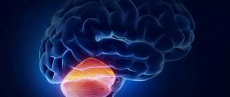
The human brain is the main organ of the central nervous system. Its surface layer consists of many nerve cells that are interconnected by synaptic connections.
Only 7% of the total number of neurons are in working condition; the rest are waiting for their “turn”. Even from a school biology course, it is known that some brain cells replace others in case of damage or complete death.
However, there are also anatomical deviations that negatively affect working and non-working neurons, thereby killing them and destroying the connection between them. This pathology leads to loss of brain mass and its functional abilities.
The death of nerve cells in the brain is a completely normal process that occurs every day. But the catch is that with neurological abnormalities, the atrophy process covers a significantly larger number of neurons than normal. This almost always leads to the emergence and progression of serious diseases that end in death.
How cerebral neurons die...
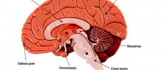
Cerebral atrophy affects the frontal areas of the brain (cortex and subcortex). It is this zone that is responsible for intellectual and mnestic functions and emotions. But this disease is classified into several types, which have different locations:
- Cortical atrophy . Here destruction occurs in the tissues of the cerebral cortex. And most often this appears during the aging process of nerve cells, but other pathological effects on the brain (BM) cannot be ruled out.
- Multisystem extinction . Characterized by damage to the cerebellum, brainstem trunk, and basal ganglia. Has a build-up effect.
- Diffuse mortality affects a variety of processes in opposite places. The disease begins its action in the cerebellum area, and then symptoms appear that are characteristic of other areas of the brain.
- Cerebellar death . Characterized by cerebellar disorders with additional pathological processes in other parts of the brain.
- Posterior cortical . Causes atrophy of neurons in the occipital and crown areas. Clusters of plaques and tangles of neurofibrillary tangles form, which contribute to death.
What is meant by neuronal death?
In general, atrophic changes in the brain are not considered a disease. Atrophy can appear not only as a result of the progression of the disease, but can itself become the cause of the development of the disease.
More precisely, brain atrophy is a pathological activity that smoothes the tissue of the cerebral cortex, reduces size, weight, and kills brain neural networks. Consequently, it affects the intellectual activity and other functions of a person.
This pathology is most common in people of old age. Everyone knows that people aged 70-80 years begin to suffer from dementia, partial memory loss and similar problems. But it cannot be said that this cannot happen to a young man or even a newborn child.
Such changes lead to changes in a person’s character and life. From here we will draw a conclusion. The death of brain neurons means:
- deterioration or complete loss of memory;
- speech disorders;
- motor impairment;
- decreased intelligence.
Lack of nutritional factors for nerves
Many theories regarding the death of motor neurons mainly describe disturbances in intracellular processes. There is evidence that motor neurons become more susceptible to ALS due to lack of nutrients. One group of such substances is a group of chemical compounds called neurotrophic factors, which literally means “nutrient factors for the neuron.”
Clinical trials using such factors to treat ALS have so far failed. No new drugs were invented. However, a lesson was learned that will lead to better research results in the future.
What causes brain cell death
It is often believed that external factors such as traumatic brain injury, alcohol or drug abuse are the main provocateurs of the development of atrophic activity.
But there are also physiological factors that no less actively accompany this:
- CNS infections . GM cells can die as a result of the presence of bacteria, infections and other parasites. This can happen within a few hours or over many years, depending on the type of infection.
- Immunity . Sometimes diseases appear in which the immune system begins to fight against a person, or more precisely against his nerve cells. This is usually observed in multiple sclerosis, when the myelin sheath of nerve cells is destroyed.
- Plaques . As mentioned above, plaque accumulation plays a role in the death of cerebral cells. The process takes many years.
- Radiation exposures . Inside, they have a strong effect on the body, which negatively affects the neurons of the brain and leads them to death.
- Biochemical damage to the nervous system.
There are also several pathological groups of diseases, the progress of which results in brain atrophy:
- Genetic diseases . In most cases, the disease develops due to a hereditary predisposition. There are several genetic diseases with which atrophy goes hand in hand (neurofibromatosis, dementia).
- Chronic ischemia . Here, the death of neurons occurs due to oxygen starvation and lack of nutrients. This is caused by vascular damage during disease activity;
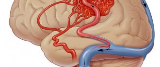
- Vascular diseases . They are divided into several subtypes: weakening of blood vessels - leads to their leakage, as a result of which blood enters the brain tissue; growth of blood vessels (Hippel-Lindau disease, appearance of hemangioblastomas); A stroke is a pathological phenomenon that is caused by blood clots. This leads to a shortage of blood supply to certain parts of the brain.
- Diseases of the nervous system . Diseases whose pathological action is aimed at the death of nerve cells.
“Editors” strike (RNA processing)
The human genetic code consists of more than three billion characters and contains “instructions” for how everything should be built in our body. "Editors" are needed to copy, assemble and transfer short pieces of code called RNA, which contain the "instructions" for assembling new proteins.
By studying the functions of the TDP-43 and FUS genes (these are the same “editors”, violations of which, as recently revealed, are the causes of familial ALS), the researchers came to the conclusion that their roles in the “editing” process are very similar. Therefore, it was suggested that a malfunction of these “editors” plays a certain role in the development of the disease.
What does it look like?
At first, it is very difficult to notice anything suspicious, since externally only changes occur in the person’s character. The person becomes distracted, lethargic, sometimes aggressive and indifferent. After a short time, a person develops memory problems, decreased logic, loss of meaning in actions, and depleted vocabulary.
In addition, over time, the death of brain cells is accompanied by the following symptoms:
- constant aggression;
- selfishness;
- lack of self-control;
- frequent irritability;
- asociality;
- abstract thinking is lost;
- mental disorders;
- depression;
- lethargy.
Symptoms will vary depending on the location of the atrophy in the brain.
Diagnostics and differential diagnostics
Cerebral arthrosis is a disease for which diagnosis may require a detailed history of the patient.
The medical history is studied and the patient is interviewed about his living conditions and well-being.
But to make an accurate diagnosis, professionals send the patient to undergo the following diagnostic tests:
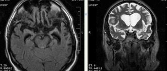
- CT head;
- diffuse optical tomography;
- MEG (measurement and visualization of magnetic fields);
- two-photon or single-photon emission tomography;
- MRI of the head.
And also in rare cases, differential diagnosis is possible. Having a patient's medical history, using a specially created computer program, a diagnosis can be made by exclusion. Based on the facts and symptoms that appear in the patient, the computer reduces the range of all possible diseases to one.
If it is impossible to carry out a complete diagnosis, a partial differential diagnosis can be made.
How to stop or slow down cell death
In order to stop the disease, it is necessary to eliminate its causes. In most cases, this is very difficult to do, especially considering the fact that nerve cells are not restored - this is impossible.
If cell death was diagnosed at the initial stage, then it is possible to stop it or at least minimize the consequences for the brain with the help of vitamin complexes that strengthen cells and antioxidants that block the oxidation process. This treatment is aimed only at eliminating symptoms. Atrophy itself cannot be treated with modern drugs.
If we talk about the patient’s lifestyle, then all responsibility now falls on the shoulders of loved ones. They must provide the person with constant care. The patient needs to be surrounded with care, ensure comfort and absence of stressful situations.
The patient should not be spared from homework; on the contrary, it would be better if he went about his usual activities. As for inpatient treatment, this will only worsen the situation. When focusing on the problem, the patient worries more, which leads to the progression of cell death.
A calm and stable environment without changes can slow down the development of the disease, and in the best case, stop it. In addition, you can use antidepressants or tranquilizers, thereby avoiding outbursts of aggression.
Clinical picture
At the initial stages, symptoms are invisible. However, as it progresses, extremely unpleasant signals may appear:
- convulsions or even epileptic seizures;
- loss of criticality towards one's actions;
- speech impairment;
- problems expressing thoughts;
- memory problems;
- deterioration of the psycho-emotional state - when depression develops, increased irritability is noted, and mood swings are noticeable;
- loss of empathy for other people and much more.
If such symptoms appear that should alert, if not the person himself, then his loved ones, you should contact a specialist to determine the cause of the problem. For example, they conduct a hardware examination - they often talk about MRI and CT diagnostics in order to accurately understand the areas of brain damage, volumes and other important factors. In addition, they may offer examination of cerebral vessels and other options. Next, a decision on therapy is made.
How to keep neurons safe and sound
All preventive measures consist of treating and preventing diseases that contribute to the death of cerebral cells. According to statistics, this phenomenon is quite often observed in people with diseases of the nervous system. From this we can conclude that it is necessary to think positively and lead a healthy and active lifestyle. It has been scientifically proven that positive people live longer and do not have such health problems.
The main “friend” of dementia, especially in old age, is vascular atherosclerosis. Its appearance adds to the death of cells and atrophy of the cortex, which is fraught with functional disorders of other vital organs.
In order to prevent “friends from reuniting”, it is necessary:
- to live an active lifestyle;
- balance your diet so as not to gain excess weight;
- give up nicotine and alcohol products;
- strengthen the immune system;
- avoid stressful situations and worry less;
- control blood sugar levels.
You should also reduce your cholesterol intake and increase the amount of fruits and vegetables in your diet. Such nutritional trends bring the body’s condition back to normal.
Chronic cerebral ischemia
Cerebrovascular diseases are one of the main problems of modern medicine. It is known that in recent years the structure of vascular diseases of the brain has changed due to the increase in ischemic forms. This is due to an increase in the proportion of arterial hypertension and atherosclerosis as the main cause of cerebrovascular pathology. When studying individual forms of cerebral circulatory disorders, chronic ischemia ranks first in prevalence.
Chronic cerebral ischemia (CHI) is a special type of vascular cerebral pathology caused by a slowly progressive diffuse disorder of the blood supply to the brain with gradually increasing various defects in its functioning. The term “chronic cerebral ischemia” is used in accordance with the International Classification of Diseases, 10th revision, instead of the previously used term “dyscirculatory encephalopathy”.
The development of chronic cerebral ischemia is promoted by a number of reasons, which are commonly called risk factors. Risk factors are divided into correctable and non-correctable. Uncorrectable factors include old age, gender, and hereditary predisposition. It is known, for example, that a stroke or encephalopathy in parents increases the likelihood of vascular diseases in children. These factors cannot be influenced, but they help to identify in advance those at increased risk of developing vascular pathology and help prevent the development of the disease. The main correctable factors in the development of chronic ischemia are atherosclerosis and hypertension. Diabetes mellitus, smoking, alcohol, obesity, insufficient physical activity, poor nutrition are the reasons that lead to the progression of atherosclerosis and the deterioration of the patient’s condition. In these cases, the blood coagulation and anticoagulation system is disrupted, and the development of atherosclerotic plaques is accelerated. Due to this, the lumen of the artery decreases or is completely blocked (Fig.). At the same time, the crisis course of hypertension is especially dangerous: it leads to an increase in the load on the blood vessels of the brain. Arteries modified by atherosclerosis are unable to maintain normal cerebral blood flow. The walls of the vessel gradually become thinner, which can ultimately lead to the development of a stroke.
| Drawing. MR angiogram: occlusion of the right middle cerebral artery |
The etiology of CCI is associated with occlusive atherosclerotic stenosis, thrombosis, and embolism. A certain role is played by post-traumatic dissection of the vertebral arteries, extravasal compression due to pathology of the spine or neck muscles, deformation of the arteries with permanent or periodic disturbances in their patency, hemorheological changes in the blood (increased hematocrit, viscosity, fibrinogen, platelet aggregation and adhesion). It must be borne in mind that symptoms similar to those that occur with chronic ischemia can be caused not only by vascular, but also by other factors - chronic infection, neuroses, allergic conditions, malignant tumors and other reasons with which a differential diagnosis should be made . If the described disorders are supposed to have a vascular origin, instrumental and laboratory confirmation of damage to the cardiovascular system is necessary (ECG, Doppler ultrasound of the main arteries of the head, MRA, MRI, CT, biochemical blood tests, etc.).
To make a diagnosis, one must adhere to strict diagnostic criteria: the presence of cause-and-effect relationships (clinical, anamnestic, instrumental) of brain lesions with hemodynamic disturbances with the development of clinical, neuropsychological, psychiatric symptoms; signs of progression of cerebrovascular insufficiency. The possibility of subclinical acute cerebral dyscirculatory disorders, including small-focal, lacunar infarctions, which form symptoms characteristic of encephalopathy, should be taken into account. For the main etiological reasons, atherosclerotic, hypertensive, mixed, and venous encephalopathies are distinguished, although other causes leading to chronic cerebral vascular insufficiency (rheumatism, vasculitis of other etiologies, blood diseases, etc.) are also possible.
The pathomorphological picture of CCI is characterized by areas of ischemically altered neurons or their loss with the development of gliosis. Small cavities (lacunae) and larger lesions develop. With multiple lacunae, the so-called “lacunary state” is formed. These changes are predominantly observed in the area of the basal ganglia and have a typical clinical expression in the form of amyostatic and pseudobulbar syndromes, dementia, described at the beginning of the twentieth century. French neurologist P. Marie. The development of status lacunaris is most characteristic of arterial hypertension. In this case, changes in blood vessels are observed in the form of fibrinoid necrosis of the walls, their plasmatic impregnation, the formation of miliary aneurysms, and stenoses.
The so-called criblures, which are dilated perivascular spaces, are distinguished as changes characteristic of hypertensive encephalopathy. Thus, the chronic nature of the process is pathomorphologically confirmed by multiple zones of brain ischemia, especially its subcortical regions and cortex, accompanied by atrophic changes developing against the background of corresponding changes in the cerebral vessels. Using CT and MRI, in typical cases, multiple microfocal changes are detected, mainly in the subcortical zones, periventricularly, often accompanied by cortical atrophy, dilation of the cerebral ventricles, and the phenomenon of leukoaraiosis (“periventricular glow”), which is a reflection of the demyelination process. However, similar changes can be observed during normal aging and primary degenerative-atrophic processes of the brain.
Clinical manifestations of CCI are not always detected by CT and MRI studies. Therefore, the diagnostic significance of neuroimaging methods cannot be overestimated. Making a correct diagnosis for a patient requires an objective analysis of the clinical picture and instrumental examination data from the doctor.
The pathogenesis of cerebral ischemia is caused by insufficiency of cerebral circulation in its relatively stable form or in the form of repeated short-term episodes of discirculation.
As a result of pathological changes in the vascular wall, developing as a result of arterial hypertension, atherosclerosis, vasculitis, etc., autoregulation of cerebral circulation is disrupted, and there is an increasing dependence on the state of systemic hemodynamics, which also turns out to be unstable due to the same diseases of the cardiovascular system. Added to this are disturbances in the neurogenic regulation of systemic and cerebral hemodynamics. Brain hypoxia itself leads to further damage to the mechanisms of autoregulation of cerebral circulation. The pathogenetic mechanisms of acute and chronic cerebral ischemia have much in common. The main pathogenetic mechanisms of cerebral ischemia constitute the “ischemic cascade” (V.I. Skvortsova, 2000), which includes:
- decreased cerebral blood flow;
- increase in glutamate excitotoxicity;
- calcium accumulation and lactic acidosis;
- activation of intracellular enzymes;
- activation of local and systemic proteolysis;
- emergence and progression of antioxidant stress;
- expression of early response genes with the development of depression of plastic proteins and a decrease in energy processes;
- long-term consequences of ischemia (local inflammatory reaction, microcirculatory disorders, damage to the BBB).
A condition called “oxidative stress” plays a major role in damage to brain neurons. Oxidative stress is an excessive intracellular accumulation of free radicals, activation of lipid peroxidation (LPO) processes and excessive accumulation of lipid peroxidation products, aggravating overexcitation of glutamate receptors and enhancing glutamate excitoxic effects. Glutamate excitotoxicity is understood as hyperstimulation by mediators of excitation of NDMA receptors of N-methyl-D-aspartate, provoking dilatation of calcium channels and, as a consequence, massive influx of calcium into cells, with subsequent activation of proteases and phospholipases. This leads to a gradual decrease in neuronal activity, a change in the neuron-glia ratio, which causes a deterioration in brain metabolism. Understanding the pathogenesis of CCI is necessary for an adequate, optimally selected treatment strategy.
As the severity of the clinical picture increases, pathological changes in the vascular system of the brain intensify. If at the beginning of the process stenotic changes in one or two main vessels are detected, then most or even all of the main arteries of the head turn out to be significantly changed. Moreover, the clinical picture is not identical to damage to the great vessels, due to the presence in patients of compensatory mechanisms of autoregulation of cerebral blood flow. The condition of intracranial vessels plays an important role in the mechanisms of compensation for cerebral circulatory disorders. With well-developed and preserved collateral circulation pathways, satisfactory compensation is possible, even with significant damage to several great vessels. On the contrary, individual structural features of the cerebral vascular system may be the cause of decompensation (clinical or subclinical), aggravating the clinical picture. This may explain the more severe clinical course of cerebral ischemia in middle-aged patients.
Based on the main clinical syndrome, several forms of CCI are distinguished: with diffuse cerebrovascular insufficiency; predominant pathology of the vessels of the carotid or vertebrobasilar systems; vegetative-vascular paroxysms; predominant mental disorders. All forms have similar clinical manifestations. In the initial stages of the disease, all patients complain of headache, non-systemic dizziness, noise in the head, memory impairment, and decreased mental performance. As a rule, these symptoms occur during a period of significant emotional and mental stress, requiring a significant increase in cerebral circulation. If two or more of these symptoms are often repeated or exist for a long time (at least the last 3 months) and there are no signs of an organic nature, instability when walking, or damage to the nervous system, a presumptive diagnosis is made.
The clinical picture of CCI has a progressive development and, according to the severity of symptoms, is divided into three stages: initial manifestations, subcompensation and decompensation.
Stage 1 is dominated by subjective disorders in the form of headaches and a feeling of heaviness in the head, general weakness, increased fatigue, emotional lability, dizziness, decreased memory and attention, and sleep disturbances. These phenomena are accompanied, although mild, but quite persistent objective disorders in the form of anisoreflexia, discoordination phenomena, oculomotor insufficiency, symptoms of oral automatism, memory loss and asthenia. At this stage, as a rule, the formation of distinct neurological syndromes (except for asthenic) has not yet occurred, and with adequate therapy, it is possible to reduce the severity or eliminate both individual symptoms and the disease as a whole.
The complaints of patients with the 2nd stage of CCI more often include memory impairment, loss of ability to work, dizziness, instability when walking, and manifestations of an asthenic symptom complex are less often present. At the same time, focal symptoms become more distinct: revival of reflexes of oral automatism, central insufficiency of the facial and hypoglossal nerves, coordination and oculomotor disorders, pyramidal insufficiency, amyostatic syndrome, increased mnestic-intellectual disorders. At this stage, it is possible to identify certain dominant neurological syndromes - discoordination, pyramidal, amyostatic, dysmnestic, etc., which can help in prescribing symptomatic treatment.
At the 3rd stage of CCI, objective neurological disorders in the form of discoordination, pyramidal, pseudobulbar, amyostatic, and psychoorganic syndromes are more pronounced. Paroxysmal conditions—falls, fainting—are more common. In the stage of decompensation, cerebral circulation disorders are possible in the form of “small strokes” or prolonged reversible ischemic neurological deficit, the duration of focal disorders in which ranges from 24 hours to 2 weeks. At the same time, the clinical picture of diffuse insufficiency of blood supply to the brain corresponds to that of moderate encephalopathy. Another manifestation of decompensation may be a progressive “complete stroke” and residual effects after it. This stage of the process with diffuse damage corresponds to the clinical picture of severe encephalopathy. Focal symptoms are often combined with diffuse manifestations of brain failure.
In chronic cerebral ischemia, there is a clear correlation between the severity of neurological symptoms and the age of the patients. This must be kept in mind when assessing the significance of individual neurological signs that are considered normal for elderly and senile people. This dependence reflects age-related manifestations of dysfunction of the cardiovascular and other visceral systems, affecting the state and functions of the brain. To a lesser extent, this dependence is observed in hypertensive encephalopathy. In this case, the severity of the clinical picture is largely determined by the course of the underlying disease and its duration.
Along with the progression of neurological symptoms, as the pathological process develops in the neurons of the brain, an increase in cognitive disorders occurs. This applies not only to memory and intelligence, which are impaired in the 3rd stage to the level of dementia, but also to such neuropsychological syndromes as praxis and gnosis. Initial, essentially subclinical disorders of these functions are observed already in the 1st stage, then they intensify, change, and become distinct. The 2nd and especially the 3rd stages of the disease are characterized by pronounced impairments of higher brain functions, which sharply reduces the quality of life and social adaptation of patients.
In the picture of CIM, several main clinical syndromes are distinguished: cephalgic, vestibulo-ataxic, pyramidal, amyostatic, pseudobulbar, paroxysmal, vegetative-vascular, psychopathological. A feature of the cephalgic syndrome is its polymorphism, inconstancy, lack of connection in most cases with specific vascular and hemodynamic factors (excluding headaches during hypertensive crises with high blood pressure), and a decrease in the frequency of occurrence as the disease progresses.
The second most common syndrome is vestibulo-ataxic syndrome. The main complaints of patients are: dizziness, instability when walking, coordination disorders. Sometimes, especially in the initial stages, patients, complaining of dizziness, do not notice coordination problems. The results of otoneurological examination are also insufficiently indicative. In later stages of the disease, subjective and objective discoordination disorders are clearly interrelated. Dizziness and unsteadiness when walking may be partly due to age-related changes in the vestibular apparatus, motor system and ischemic neuropathy of the vestibulocochlear nerve. Therefore, to assess the significance of subjective vestibulo-ataxic disorders, their qualitative analysis during a patient interview, neurological and otoneurological examination is important. In most cases, these disorders are caused by chronic circulatory failure in the blood supply of the vertebrobasilar arterial system, so it is necessary to rely not on the subjective sensations of patients, but to look for signs of diffuse damage to the parts of the brain that are supplied with blood from this vascular system. In some cases, in patients with stages 2–3 CCI, ataxic disorders are caused not so much by cerebellar-stem dysfunction, but rather by damage to the frontal-stem pathways. There is a phenomenon of frontal ataxia, or apraxia of walking, reminiscent of hypokinesia in patients with parkinsonism. A CT examination reveals significant hydrocephalus (along with cortical atrophy), i.e., a condition similar to normal pressure hydrocephalus occurs. In general, the syndrome of circulatory failure in the vertebrobasilar system is diagnosed with CCI more often than insufficiency of the carotid system.
A feature of the pyramidal syndrome is its moderate clinical manifestation (anisoreflexia, facial asymmetry, minimally expressed paresis, revitalization of oral automatism reflexes, hand symptoms). A clear asymmetry of reflexes indicates either a previously existing cerebral stroke or another disease occurring under the guise of CCI (for example, large intracranial processes, consequences of traumatic brain injury). Diffuse and fairly symmetrical revival of deep reflexes, as well as pathological pyramidal reflexes, often combined with a significant revival of oral automatism reflexes and the development of pseudobulbar syndrome, especially in old age, indicates multifocal vascular damage to the brain (subject to the exclusion of other possible causes).
In patients with clinical manifestations of circulatory failure in the vertebrobasilar system, paroxysmal conditions are often observed. These conditions may be caused by a combined or isolated effect on the vertebral arteries of vertebrogenic factors (compression, reflex), which is associated with changes in the cervical spine (dorsopathies, osteoarthritis, deformities).
Mental disorders are quite characteristic and varied in form at different stages of CCI. If in the initial stages they are of the nature of asthenic, asthenodepressive and anxiety-depressive disorders, then in the 2nd and especially in the 3rd stage they are joined by pronounced dysmnestic and intellectual disorders, forming the syndrome of vascular dementia, which often comes first in the clinical picture .
Electroencephalographic changes are nonspecific for CCI. They consist of a progressive decrease in the β-rhythm, an increase in the proportion of slow θ- and δ-activity, accentuation of interhemispheric asymmetry, and a decrease in EEG reactivity to external stimulation.
CT characteristics undergo dynamics from normal indicators or minimal atrophic signs in the 1st stage to more pronounced small-focal changes in the brain substance and atrophic (external and internal) manifestations in the 2nd stage to sharply defined cortical atrophy and hydrocephalus with multiple hypodense foci in the hemispheres - in the 3rd stage.
A comparison of clinical and instrumental characteristics in patients with atherosclerotic, hypertensive and mixed forms of CCI does not reveal any clear differences. In severe cases of hypertension, a faster rate of increase in psychoneurological disorders, early manifestation of cerebral disorders, and a greater likelihood of developing lacunar stroke are possible.
Treatment of CCI should be based on certain criteria, including the concepts of pathogenetic and symptomatic therapy. To correctly determine the pathogenetic treatment strategy, one should take into account: the stage of the disease; identified mechanisms of pathogenesis; the presence of concomitant diseases and somatic complications; age and gender of patients; the need to restore quantitative and qualitative indicators of cerebral blood flow, normalize impaired brain functions; the possibility of preventing recurrent cerebral dysgemia.
The most important direction of CCI therapy is the impact on existing risk factors, such as arterial hypertension and atherosclerosis. Treatment of atherosclerosis is carried out according to generally accepted regimens using statins, in combination with correction of the diet and lifestyle of patients. The selection of antihypertensive drugs and the order of their prescription is carried out by a general practitioner, taking into account the individual characteristics of patients. Complex therapy for CCI includes the prescription of antioxidants, antiplatelet agents, drugs that optimize brain metabolism, and vasoactive drugs. Antidepressants are prescribed for severe asthenodepressive manifestations of the disease. Antiasthenic drugs are prescribed in the same way.
An important component of the treatment of CCI is the administration of drugs with antioxidant activity. Currently, the following drugs of this series are used in clinical practice: Actovegin, Mexidol, Mildronate.
Actovegin is a modern antioxidant, which is a deproteinized extract of the blood of young calves. Its main effect is to improve the utilization of oxygen and glucose. Under the influence of the drug, the diffusion of oxygen in neuronal structures significantly improves, which makes it possible to reduce the severity of secondary trophic disorders. There is also a significant improvement in cerebral and peripheral microcirculation against the background of improved aerobic energy exchange of vascular walls and the release of prostacyclin and nitric oxide. The resulting vasodilation and decrease in peripheral resistance are secondary to the activation of oxygen metabolism of the vascular walls (A. I. Fedin, S. A. Rumyantseva, 2002).
In case of CCI, it is advisable to use Actovegin, especially in the absence of effect from other treatment methods (E. G. Dubenko, 2002). The method of application consists of drip administration of 600–800 mg of the drug for 10 days, followed by switching to oral administration.
A constant in the treatment regimen for CCI is the use of drugs that optimize cerebral circulation. The most commonly used drugs are: Cavinton, Halidor, Trental, Instenon.
Halidor (bencyclane) is a drug that has a multidirectional mechanism of action due to phosphodiesterase blockade, antiserotonin action, and calcium antagonism. It inhibits the aggregation and adhesion of platelets, prevents the aggregation and adhesion of erythrocytes, increasing the elasticity and osmotic resistance of the latter. Halidor reduces blood viscosity, normalizes intracellular metabolism of glucose and ATP, affects phosphokinase and lactate dehydrogenase, and enhances tissue oxygenation. It has been proven that the use of this drug for 8 weeks eliminates the clinical manifestations of chronic cerebral vascular insufficiency in 86% of patients. The drug has a positive effect on a person’s emotional environment, reduces forgetfulness and absent-mindedness. Halidor is prescribed in a daily dose of 400 mg for 6–8 weeks.
Instenon is a combined drug with neuroprotective action, including a vasoactive agent from the group of purine derivatives, a substance that affects the state of the ascending reticular formation and cortical-subcortical relationships, and, finally, an activator of tissue respiration processes under hypoxic conditions (S. A. Rumyantseva, 2002; B V. Kovalchuk, 2002).
The three components of instenon (etophylline, etamivan, hexobendine) jointly act on various parts of the pathogenesis of ischemic brain damage.
Etophylline, a vasoactive component of the purine series, activates myocardial metabolism with an increase in stroke volume. The transition from a hypokinetic type of blood circulation to a normokinetic one is accompanied by an increase in cerebral blood flow. An important effect of the component is an increase in renal blood flow and, as a consequence, dehydration and diuretic effects.
Etamivan has a nootropic effect in the form of a direct effect on the processes of memory, attention, mental and physical performance as a result of increased activity of the reticular formation of the brain.
Hexobendine selectively stimulates metabolism based on increased utilization of oxygen and glucose, due to increased anaerobic glycolysis and pentose cycles. At the same time, the physiological mechanisms of autoregulation of cerebral and systemic blood flow are stabilized.
Instenon is used intramuscularly 2.0 ml, course - 5-10 procedures. Then oral administration of instenon-forte continues, 1 tablet 3 times a day for a month (S. V. Kotov, I. G. Rudakova, E. V. Isakova, 2003). A clear regression of neurological symptoms is observed by the 15th–20th day of treatment. A particularly good effect is observed with the combined use of Actovegin (drops) and instenon (intramuscular injections or oral administration). Instenon therapy has a positive effect on cognitive functions, especially on the regulation of mnestic activity and psychomotor functions.
In the complex therapy of CCI, much attention is paid to nootropic drugs that increase the resistance of brain tissue to various adverse metabolic influences (ischemia, hypoxia). The actual “nootropic” drugs include derivatives of piracetam (nootropil, lutetam), encephabol.
Piracetam increases the synthesis of high-energy phosphates (ATP), enhances aerobic metabolism under hypoxic conditions, facilitates impulse conduction, normalizes the ratio of phospholipids of cell membranes and their permeability, increases the density and sensitivity of receptors, improves interaction between the cerebral hemispheres, improves metabolic processes in the central nervous system, facilitates neuronal transfer.
Piracetam improves microcirculation due to its disaggregant properties, facilitates the conduction of nerve impulses, and improves interaction between the hemispheres of the brain. The drug normalizes the ratio of phospholipids of cell membranes and increases their permeability, prevents the adhesion of red blood cells, reduces platelet aggregation, reduces the levels of fibrinogen and factor VIII, and relieves spasm of arterioles. The drug is prescribed in a daily dose of 2.4–4.8 g for 8–12 weeks.
Encephabol is a derivative of pyritinol. The drug increases the density and sensitivity of receptors, normalizes neuroplasticity. It has a neuroprotective effect, stimulates learning processes, improves memory, memorization and concentration. Encephabol stabilizes the cell membranes of neurons by inhibiting lysosomal enzymes and preventing the formation of free radicals, improves the rheological properties of blood, increases the conformational ability of red blood cells, increasing the ATP content in their membrane. For adults, the average daily dose is 600 mg for 6–8 weeks.
Antiplatelet drugs include acetylsalicylic acid and its derivatives (cardiomagnyl, thrombo ACC). Given the presence of contraindications when prescribing acetylsalicylic acid, other drugs with antiplatelet activity (Curantil, Tiklid, Plavix) are often used.
Symptomatic therapy for CCI includes the prescription of drugs that reduce the manifestations of various symptoms of the disease. For all patients with stages 2–3 of the disease, it is advisable to prescribe anti-anxiety or antidepressant drugs. Benzodiazepine drugs are the safest for long-term use.
Grandaxin is an atypical benzodiazepine derivative, a selective anxiolytic. The drug effectively eliminates anxiety, fear, and emotional stress without sedation or muscle relaxation. The drug has a vegetative-corrective effect, which makes it possible to use it in patients with severe vegetative-vascular syndrome.
In neurological practice, a daily dose of 50–100 mg is used, the duration of use is determined individually for each patient.
The prevalence of chronic vascular pathology of the brain, the progression of its course, and the high degree of disability of patients determine the social and medical significance of the problem of CCI therapy. Currently, in clinical practice there is a trend towards increasing the use of non-drug treatment methods. This is due to the absence in patients of the phenomenon of addiction to medicinal substances with a long period of therapeutic aftereffect.
Considering the complexity of the pathogenetic mechanisms of CCI, during therapy it is necessary to achieve normalization of systemic and cerebral circulation, adjust the metabolism in the brain tissue, and the state of hemorheology. Currently, the possibilities for pharmacological correction of the manifestations of CCI are quite extensive; they allow the use of various drugs that affect all parts of the pathogenesis of post-ischemic and post-hypoxic damage to nervous tissue.
Thus, recognizing the causes, identifying risk factors and, therefore, the real possibility of effective targeted treatment and preventing the development of chronic cerebral vascular pathology requires accurate knowledge of the structural, physiological and clinical features of the manifestation of the disease. This becomes possible thanks to a systematic approach to the study of etiology, pathogenesis, clinical picture and modern methods of therapy.
M. V. Putilina , Doctor of Medical Sciences, Professor
RGMU, Moscow
The larger the lesion, the worse the manifestation
As for the consequences that threaten the death of brain cells, the rule is relevant here: the larger the damage, the worse the manifestation.
And also in the absence of treatment, a person’s condition worsens faster. As a result, convulsions, loss of muscle function, or respiratory depression may occur. The simultaneous manifestation of such consequences can lead the patient into coma or stupor.
Nothing good can be expected here, since such a process can no longer be stopped, and when a significant part of the cells for life dies, death occurs.
Treatment tactics
Treatment of organic brain damage can be very diverse and depends on the pathogenetic mechanism of development of the damage and the immediate cause.
Treatment of organic brain damage can be surgical and conservative. For example, the development of high intracranial pressure, which poses a threat to life, can be treated both surgically and conservatively. Surgical treatment - the application of a burr hole for brain decompression is applicable in the formation of a severe hematoma due to trauma or hemorrhagic stroke, and conservative therapy is possible with a moderate increase in intracranial pressure without brain dislocation. For conservative therapy, diuretics are used that cause forced diuresis, allowing for the rapid elimination of edema.
Treatment of cerebral artery atherosclerosis can also be either surgical or conservative. Surgical – angiography with the installation of stents to widen the lumen of the arteries. Conservative – antithrombotic therapy and correction of dyslepidemia.