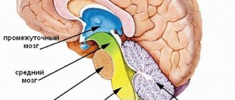
Stroke is a serious condition that is characterized by an acute disruption of the blood supply to parts of the brain and leads to morphological and functional damage to the central nervous system. Depending on the type of damage, hemorrhagic and ischemic types are distinguished. The brain stem includes the medulla oblongata, midbrain and pons. It is a connecting structure between the spinal cord and the brain and is of great importance, since the structures that ensure the vital functions of the body are concentrated in it. In case of damage to the brain stem, the statistics are disappointing: no more than 25% of such patients survive, and only 2% of them can count on a full recovery without severe complications.
Brainstem stroke is the least favorable type of cerebral stroke. The structures of the trunk are responsible for providing the following life support functions: breathing, blood circulation and cardiac activity, vital reflexes. Patients who have had this stroke have a poor prognosis, which is further worsened by hemorrhage. Mortality reaches 75%. Diagnosis is based on clinical symptoms and MRI data.
Among the clinical signs, disturbances of consciousness and bulbar syndrome prevail. The standard for diagnosis is MRI, which gives an idea of the topography and volume of the lesion. At the same time, rehabilitation and partial restoration of lost functions are possible with timely diagnosis and immediate initiation of therapy. The Neurology Clinic of the Yusupov Hospital is equipped with a ventilator, necessary during the acute period of a stroke, and simulators for restoring motor skills.
The effectiveness of treatment most of all depends on the timing and quality of the treatment measures.
Causes of brainstem stroke
The following factors contribute to the development of the disease:
- Hypertonic disease. It is the most common cause of vascular damage to the brain stem. With high blood pressure, the walls of the arteries become brittle, which in turn leads to their rupture and hemorrhage.
- Atherosclerosis, thrombosis. When the lumen of a vessel is blocked by an atherosclerotic plaque or thrombus, the blood supply to the surrounding tissue is disrupted, which leads to the development of ischemia.
- Aneurysms, collagenosis, vascular malformations. Pathological changes in the structure of the vascular wall increase the risk of its rupture. This anomaly can cause a stroke at a young age.
- Diabetes mellitus, rheumatological diseases, heart pathologies, blood clotting disorders, etc. Features of the course of some diseases lead to the development of atherosclerosis, blood clots, macro- and microangiopathy.
- Lifestyle. Smoking, alcohol abuse, physical inactivity, poor diet, stress, and chronic fatigue increase the risk of vascular accidents.
- Age. Each subsequent 10 years increases the likelihood of developing ischemia or hemorrhage by 5-8 times.
- Genetic predisposition. Cohort studies have revealed that the risk of stroke increases by 30% if a person has a history of this disease in their family.
Make an appointment
How does a stem lesion differ from other types of stroke?
Depending on the location and extent of the pathological process, disruption of the blood supply to the trunk can have a variety of clinical manifestations.
The first signs of the disease may be severe pain in the back of the head, dizziness, and in 70-80% of cases loss of consciousness occurs. The clinical feature of vascular damage in this particular department is expressed in the appearance of alternating syndromes - damage to the cranial nerves on one side in combination with motor and sensory disorders on the opposite. The functioning of the cardiovascular and respiratory systems and thermoregulation are disrupted, and paralysis of the muscles of the face, pharynx, and muscles of the limbs develops. The development of “locked-in person” syndrome is possible. All symptoms are severe and, if timely medical care is not provided, lead to death in the first two days of the disease. An imbalance of blood circulation in this part of the brain brings serious consequences associated with a person’s life. Basic health parameters are affected. In particular, the following symptoms result from a stroke:
- dysfunction of the heart muscle. There is a change in the heartbeat rhythm, which causes bradycardia, arrhythmia or fibrillation. A distinctive feature of damage to the stem structure is a poor prognosis for rehabilitation;
- failure in the respiratory system. Shortness of breath and inability to take in air appear. In such a situation, the presence of an artificial respiration apparatus is of great importance, otherwise death is possible;
- speech and swallowing problems. The first is more harmless in nature due to the inability to pass saliva. The latter leads to profuse salivation, and some body positions allow fluid to enter the oxygen-receiving organs, thereby provoking the onset of pneumonia.
In addition to all of the above, there is a possibility of problems with vision and coordination, and a decrease in muscle activity.
If timely assistance is not provided, the outcome can be fatal. In ICD-10, brainstem stroke is designated by code G 46.3. According to WHO statistics, stroke occupies a leading position in the structure of morbidity and mortality. Every year, cerebrovascular accidents are detected in 3 people per 100 thousand population. Death at the onset of the disease occurs in 15–30% of cases. With a second episode of stroke, the mortality rate is 70%. According to statistics, the five-year survival rate after a stroke is 40–60%. The prognosis is influenced by the form of cerebrovascular accident. Brainstem stroke affects the brainstem. Doctors consider this type of disease to be the most dangerous.
To diagnose stroke, the Yusupov Hospital uses modern equipment. CT and MRI are considered the most informative ways to determine the localization of a pathological focus in the brain. Therapy includes methods of drug treatment and surgical intervention if indicated. The drugs used meet quality and safety standards. The duration of rehabilitation is determined by the severity of the condition.
Thanks to an individual approach to each patient, positive results are achieved in the shortest possible time.
Expert opinion
Author: Andrey Igorevich Volkov
Neurologist, Candidate of Medical Sciences
Brainstem stroke is the least favorable type of cerebral stroke. The structures of the trunk are responsible for providing the following life support functions: breathing, blood circulation and cardiac activity, vital reflexes. Patients who have had this stroke have a poor prognosis, which is further worsened by hemorrhage. Mortality reaches 75%.
Diagnosis is based on clinical symptoms and MRI data. Among the clinical signs, disturbances of consciousness and bulbar syndrome prevail. The standard for diagnosis is MRI, which gives an idea of the topography and volume of the lesion.
At the same time, rehabilitation and partial restoration of lost functions are possible with timely diagnosis and immediate initiation of therapy. Patients with brainstem stroke are less likely to lose interaction and communication skills, allowing them to participate in their own recovery.
The Neurology Clinic of the Yusupov Hospital is equipped with a ventilator, necessary during the acute period of a stroke, and simulators for restoring motor skills. The effectiveness of treatment most of all depends on the timing and quality of the treatment measures.
Types of brain lesions
Brain damage can be either focal or disseminated. Let's figure out what focal brain damage is. This is a condition in which there is a clearly demarcated single focus with necrotic tissue, i.e. This is a local brain lesion. This type of damage often occurs during acute cerebrovascular accident.
Disseminated or multifocal brain damage is a type of injury in which multiple scattered foci of damage to brain tissue are detected. The multifocal form occurs in infectious diseases of the brain, for example, when an infectious agent is introduced hematogenously into the medulla or with an oncological lesion.
Characteristic symptoms
The fact that the pathological process has affected the brainstem can be understood in 95% of cases from examination data.
Focal neurological manifestations can be observed in the first hours of the disease and during the recovery period. The clinical picture of brainstem damage appears suddenly and develops at lightning speed, compared to other parts. The prognosis for recovery is usually worse. Signs of brain stem pathology are directly related to its purpose. It belongs to the nervous system and is its central part. Responsible for perspiration, heart function, body temperature, swallowing and chewing. Therefore, brainstem strokes are extremely dangerous, even fatal. The defeat of the department manifests itself sharply. The state of health quickly deteriorates, temperature fluctuations begin, nausea, vomiting, tachycardia, loss of consciousness, and coma often occurs.
In medicine, indicators for a disorder of this type are divided into two groups, depending on the form of the ailment, but there is a more general list suitable for all types:
- speech dysfunction;
- failure to regulate body temperature;
- poor coordination;
- muscle paralysis;
- change of voice after swallowing;
- change in pulmonary ventilation;
- inflammation of the respiratory tract;
- bradycardia or tachycardia;
- deterioration of vision of space;
- clouding of consciousness;
- poor sensitivity of the limbs;
- loss of consciousness;
- coma.
Hemorrhages occur suddenly, and the speed of assistance plays a big role. As soon as the severity of symptoms becomes obvious to others, you need to immediately call an ambulance. Most often, loss of consciousness or complete paralysis occurs. Particular attention should be paid to the quality of breathing.
Dizziness and loss of coordination
The harbinger is a feeling of pain in the skull or dizziness, accompanied by deterioration in coordination of movements and loss of balance.
This is often the earliest symptom of a pathological process in the trunk. Due to a persistent feeling of dizziness and pain in the back of the head, it is difficult for a person to stand on his feet and maintain balance. In such cases, the patient may suddenly fall or be forced to take a horizontal position. Coordination of movements suffers: the patient may not understand the position of body parts, an unsteady gait develops, hand movements are uneven and slow, and handwriting changes. All manifestations have a similar picture, but there are signs that are more noticeable than others or appear earlier. Unfortunately, a symptom such as dizziness will not allow you to accurately determine the cause of its occurrence, but will help you prepare for all possible outcomes. If the patient exhibits the described signal, place him in a lying or sitting position and wait for improvement. If none are noted, call a team of medical workers.
Make an appointment
Movement disorders
A brainstem stroke causes motor problems that become noticeable in the period following the stroke.
It occurs due to damage to the area of the brain responsible for cognitive activity. At the initial stage, programmatic decay manifests itself, which is expressed in changes in gait, frequent motor errors and focusing increased attention on arbitrary objects. Morphological damage in the nervous tissue of the trunk leads to impaired motor activity. This defect can progress and provoke structural disintegration of the statolocomotor system, which leads to disorganization of dynamic control of movement. At the final stage of development, the basic characteristics of the central generator of the step rhythm are affected. In this case, the patient experiences asymmetry in movements of both legs and arms, as well as freezing in place while walking. In other cases, while consciousness is preserved, a person is struck by paralysis of one half of the body (hemiparesis) or spreads to all limbs (tetraparesis). The prognosis for the restoration of motor functions is more favorable in the first 2-3 months of the disease.
Swallowing disorders - dysphagia
This is a severe manifestation of an acute disorder of cerebral blood supply.
It is observed in 65% of patients with brain stem stroke. When the nerve center is damaged, vital parameters, for example, the swallowing of saliva, are significantly affected. These tasks involve a significant number of muscles that can fail due to illness. Imperfect swallowing is called dysphagia. The danger of this complication is associated with a high risk of developing respiratory disorders and aspiration pneumonia. Most of these patients require tube feeding. The prognosis is usually unfavorable. In approximately 85% of cases, the ability to swallow is restored on its own within 2-3 weeks after the incident. Rarely does this defect remain for the rest of one's life. In this case, complications will follow:
- poor nutrition will lead to a deterioration in overall health;
- constant inflammation of the lungs due to unwanted microorganisms entering the respiratory tract;
- suffocation.
Timely treatment will allow you to get rid of all possible consequences and recover. To do this, it is recommended to contact a speech therapist, he will determine the severity and prescribe appropriate complex therapy.
Speech disorders - dysarthria
The cause of this disorder is a violation of the motor functions of the muscles of the tongue, facial muscles, and pharynx.
It is observed in approximately 30% of cases and manifests itself in changes in sound pronunciation, timbre, and intonation. Speech is distorted, losing intelligibility and articulation. In severe cases, speaking skills are completely lost. Dysfunction is accompanied by paralysis of the facial muscles located on the face and responsible for the ability to speak. This phenomenon is medically called dysarthria. The ability to communicate is lost due to disturbances in the brain stem. Associated symptoms are as follows:
- poor facial mobility;
- distorted, unintelligible speech;
- uneven speed of pronunciation;
- stops in the middle of a phrase;
- lack of emotional coloring.
To restore speech functions, the supervision of a speech therapist is required. Based on clinical information about the patient, he will select a personal plan of classes and exercises, after which he will begin rehabilitation. It is worth noting that the speed of recovery directly depends on the moment you contact a specialist.
Eye symptoms
If the part where the oculomotor center is located is damaged, the patient loses the ability to control the movements of one or both eyeballs.
This is reflected in the inability to fixate on an object, double vision, strabismus, outward deviation of the eyes and drooping eyelids. Sometimes fields of vision may disappear. Vision plays one of the main roles in the perception of surrounding information. It allows you to distinguish colors, objects and significantly affects the quality of life. Damage to critical areas leads to deterioration of visual perception. The described disadvantage can be determined by the following characteristics:
- black spots before the eyes;
- difficulty concentrating on one object;
- impossibility of recognition.
A problem with seeing the environment can haunt the patient throughout his life and cause disability, but it is not uncommon for him to be restored through medical practice.
Alternating syndrome - paralysis and distorted face
This symptom complex occurs as a result of dysfunction of the cranial nerves on the side of the lesion and conduction disorders on the opposite side of the body. Due to paralysis of the facial muscles, the patient on the affected side experiences sagging skin, drooping of the corner of the mouth and upper eyelid. In this case, the upper and lower limbs from the opposite area of the body remain immobilized. Paralysis or distortion of the face is one of the main symptoms of the disease. This has a significant impact on the ability to speak, causing dysarthria or dysphagia. To confirm, ask the victim to smile. If asymmetry is observed, then specialists should be called immediately.
Disorders of breathing, heartbeat and thermoregulation
Damage to the respiratory, vasomotor centers and thermoregulation center in a large percentage of cases ends in death.
Impaired respiratory function during a trunk stroke manifests itself in the form of severe shortness of breath or complete loss of the ability to breathe independently. Such patients are completely dependent on a ventilator. In other cases, when the center is not completely affected, the period of wakefulness may be accompanied by spontaneous breathing, but during sleep periods of apnea are observed. A change in the functioning of the cardiovascular system is expressed in an increase in heart rate, which is then replaced by bradycardia and a drop in blood pressure. Arrhythmias and fibrillations may develop. Taking traditional antiarrhythmic drugs in such cases is ineffective. Unstable thermoregulation is observed in rarer cases and is characteristic of the most severe degree of the disease. It manifests itself in the form of a sharp and persistent increase in body temperature to 39ᵒC and above, and cannot be corrected with medication.
In other cases, critically low temperatures may occur. Both options have a disappointing prognosis for recovery.
What is brain damage?
Organic brain damage is primarily a symptom of a disease, manifested by disruption or loss of a number of functions as a result of the pathogenic effect of some factor on brain tissue. The etiology of brain damage can be very diverse, and this will be discussed in more detail later in the article. Organic damage means that brain cells - neurons - are exposed to a variety of influences, which lead to the formation of dystrophic processes inside neurons and disrupt their functional activity. In the most serious cases, neurons simply undergo first necrobiosis and then necrosis, i.e. are dying. The death of a large number of neurons localized in a single anatomical space leads to the loss of one or another function in the body of the affected person, and the identification of a disturbed function allows specialists to understand in which part of the brain the catastrophe occurred - this is called topical diagnostics. Symptoms of organic brain damage in children manifest themselves differently than in adults, since the full activity of the higher nervous system has not yet been formed. Children may experience delays in mental, mental and physical development, unstable mood and behavioral abnormalities.
Pathogenetic mechanisms of neuronal damage
A number of mechanisms of different nature can lead to organic damage to the brain. This pathological condition can be provoked by both external and internal factors, and this must be taken into account, since therapeutic measures, depending on the pathogenetic type of development of damage to brain neurons, will differ radically.
Violation of energy supply
The most common pathogenetic variant of brain damage is associated with an imbalance between the need of neurons for energy and its entry into the cell. Energy deficiency can develop due to insufficiency:
- Nutrients in the victim's body, for example as a result of hypoglycemia, when there is not enough glucose in the blood;
- Oxygen, which causes a condition such as hypoxia. Brain hypoxia causes damage to nervous tissue and often occurs in acute ischemic or hemorrhagic cerebrovascular accidents. In children, brain hypoxia can develop in the antenotal period and during childbirth, which leads to anoxic brain damage in the child.
- When the concentration of potassium, calcium, sodium and chlorine ions increases or, conversely, excessively decreases, transmembrane proteins may malfunction, which also entails energy deficiency inside the cell.
It is worth noting that energy deficiency leads to rapidly progressive damage to brain tissue and within 5-7 minutes, in the absence of sufficient oxygenation, neurons begin to experience acute hypoxia and die. Damage to the blood vessels of the brain has the following symptoms:
- The patient notes memory impairment;
- There is a decrease in vision and hearing;
- The synthetic activity of the brain slows down;
- When performing angiography of cerebral vessels, multiple stenoses of cerebral vessels can be detected;
- MRI of the brain shows dystrophic disorders and a decrease in the volume of the cerebral cortex.
All of the above symptoms are signs of systemic atherosclerosis, which affects most older people. Atherosclerosis leads to the formation of discirculatory encephalopathy.
Traumatic injuries
Injuries are always associated with mechanical damage to the brain and the subsequent development of edema, which leads to an increase in intracranial pressure. Since the brain is located in the cranium and literally floats in the cerebrospinal fluid - intracerebral fluid, the consequences of blows and bruises become serious. Despite. The cerebrospinal fluid plays a protective and shock-absorbing role; with the development of a brain contusion, an increase in intracranial pressure occurs, since the fluid is not physically compressible. Brain cells are exposed to excessive pressure and begin to die. Brain tissues occupy up to 96% of the volume of the cranial cavity, which makes this organ very sensitive to changes in intracranial pressure.
It is very important to note that quite often injuries are accompanied by internal hemorrhage, which can lead to the formation of an extensive hematoma and displacement of the brain. Dislocation of the brain leads to wedging of its subcortical structures into the foramen magnum, which inevitably leads to the death of neurons located in the nuclei of the vasomotor and respiratory centers, without which the life of the victim is impossible.
Infectious
Brain damage can be caused not only by physical factors. But also biological. Conditions such as meningitis, encephalitis, and ventriculitis can significantly impair the functional activity of the brain.
- Meningitis is an inflammation of the lining of the brain. Etiological factors can be very diverse, so the brain can be affected by many bacterial and viral diseases. Inflammation of the membranes of the brain can occur primarily through direct infection through the wound gate. And secondarily – as a result of an immunodeficiency state.
- Encephalitis is inflammation of the brain tissue itself. Encephalitis is an even more severe infectious disease than meningitis. As a result of encephalitis, purulent melting and liquefaction of areas of the brain can occur, which leads to the formation of persistent disturbances in the functioning of various organs of the victim. With encephalitis, brain damage very often leads to disability or even death.
- Ventriculitis is an inflammation of the integumentary tissues lining the ventricles of the brain. This disease occurs in newborns and infants and leads to increased intracranial pressure and the development of hydrocephalus due to insufficient drainage function of the cerebrospinal fluid.
The brain can be affected by both specific and nonspecific infectious agents; this is important to consider when prescribing treatment, since antibacterial therapy regimens will vary.
Congenital pathology
Anomalies of brain development can form in the very early stages of a child’s development. The first trimester of pregnancy is the most dangerous for the woman and the fetus, since the pregnant woman’s body and the fetus are unprotected from external factors, and at the time of the initiation and formation of organs, the most dangerous anomalies and gross developmental pathologies can form, for example micro or acephaly.
Toxic damage
Not the most common type of brain damage, but nevertheless it does occur. Brain damage occurs if the chemical has neurotoxic properties and is able to cross the blood-brain barrier. Neurotoxic agents lead to organic damage in various parts of the nerve cell; most often, neurons suffer from disruption of the transmembrane transfer of nutrients and disturbances in the synthesis of neurotransmitters. Toxic injuries of varying severity can lead to both persistent encephalopathy and complete loss of some functions of the person affected by intoxication. Most often, gross organic damage to the brain is caused by substances such as arsenic and nitrogen metabolism products, with excessive accumulation of the latter in the blood plasma.
Oncological diseases
Brain damage in oncology can be primary. When a tumor develops directly from brain tissue or secondary - when atypical tumor cells are metastatically introduced into the brain.
Diagnosis of brainstem stroke
Diagnosis of the disease begins with examination data and collection of the patient’s medical history. Based on the characteristic symptoms, a brain stem stroke can be suspected in the first minutes. Such patients are hospitalized on an emergency basis. In a hospital setting, a number of procedures are performed to confirm the diagnosis. The following have the greatest diagnostic capabilities:
- Computed tomography is an x-ray method for studying the layer-by-layer structure of organs and tissues. With a high probability it helps to recognize foci of hemorrhage in the brain.
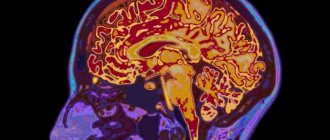
- Magnetic resonance imaging is an effective method for detecting pathological lesions using nuclear magnetic resonance. Allows you to assess the degree and volume of tissue damage.
- Doppler ultrasound. Using ultrasound of the vessels of the neck and brain, pathological changes (stenosis, occlusion) can be detected in the carotid and vertebral arteries, as well as in their branches.
- Electroencephalography. It is also a non-invasive diagnostic method. By recording bioelectrical activity, it reflects qualitative and quantitative changes in the function of the cortex and deep structures of the brain.
In addition, it is possible to conduct laboratory tests of the composition of blood and cerebrospinal fluid. According to indications, a neurologist is able to draw up an almost complete clinical picture, which reflects the depth and localization of the damage that has occurred. But to confirm the diagnosis, additional studies are required using CT or MRI, which will show:
- type of disorder;
- the extent of the pathology;
- presence of hematoma or ischemia.
The more fully and accurately the factors of change are identified, the easier it is to draw up a rehabilitation plan.
Alalia is an organic disorder (underdevelopment) of speech of a central nature. With alalia, there is a delay in the maturation of nerve cells in certain areas of the cerebral cortex. Nerve cells stop their development, remaining at a young immature stage - neuroblasts. This underdevelopment of the brain can be congenital or early acquired in the pre-speech period - organic brain damage with alalia occurred in the prenatal or early postnatal period. Conventionally, the pre-speech period is considered to be the first three years of a child’s life, when the cells of the cerebral cortex are intensively formed and when the child’s experience of using speech is still very small. The development of the brain systems most important for speech function does not end in the prenatal period, but continues after the birth of the child.
Underdevelopment of the brain or its early damage leads to a decrease in the excitability of nerve cells and to changes in the mobility of basic nervous processes, which entails a decrease in the performance of cells in the cerebral cortex.
The study of the pathophysiological mechanisms underlying alalia reveals a wide irradiation of the processes of excitation and inhibition, inertia of the main nervous processes, and increased functional exhaustion of cells in the cerebral cortex (I. K. Samoilova, 1952). Researchers note a lack of spatial concentration of excitatory and inhibitory processes in the cerebral cortex. A study of the electrical activity of the brain in children with alalia revealed clear local changes in biopotentials mainly in the temporo-parieto-occipital regions, in the frontotemporal and temporal branches of the dominant hemisphere (L. A. Belogrud, 1971; A. L. Lindenbaum, 1971; E. M. Mastyukova, 1972).Recent studies show that with alalia there are mild but multiple damage to the cerebral cortex of both hemispheres, i.e., bilateral lesions. Apparently, with unilateral brain damage, speech development occurs due to the compensatory capabilities of a healthy, normally developing and functioning hemisphere. With bilateral injuries, compensation becomes impossible or severely difficult. Thus, the previously existing point of view about the narrow local nature of damage to the speech areas of the brain (the cortical end of the speech-auditory and speech-motor analyzers) is not confirmed.” (Volkova L.S.)
Alalia forms.
Alalia motor (expressive). “Alalia is an underdevelopment or gross disturbance of the development of speech in a child, occurring in the pre-speech period, having a systemic nature and caused by pathology of the central nervous system of certain areas of the cerebral cortex….
The fact that alalia is determined by the pathology of the central nervous system in the pre-speech period indicates that alalia is a consequence of some early pathological influences on the child’s brain. The attribution of the pathology primarily to the level of the cortex indicates that the pathological process involves mainly not the elementary, musculomotor or sensory, but the higher parts of the central nervous system, which are closely related to thinking.”
Expressive speech is realized through different levels of the brain. At the gnostic-praxic level, articulatory praxis is carried out: afferent (kinesthetic) is associated with the functioning of the lower parietal (postcentral) zone, efferent (kinetic) articulatory praxis is provided by the premotor cortex of the brain. At the symbolic (linguistic) level, the brain mechanisms of speech are relevant for the phonological (phonemic) system of language, and also for the lexical and syntactic system. Within the framework of the lexical system of language, the main type of speech activity is naming - a function that is carried out primarily by the tertiary (temporo-occipital) zone on the left (according to E.P. Kok).
The brain organization of the syntactic system of language (phrasal speech) has the most complex multi-level structure. At the level of deep (“nuclear”) syntax, the frontal lobes of the brain play a major role. The core syntactic structure of a phrase is, “essentially”, its extremely compressed program.
It is distinguished by a high degree of logic and, therefore, is close to mental activity as a whole. The surface syntactic structure of a phrase is a “reversal” of its nuclear part. It is carried out mainly due to the posterior frontal parts of the left hemisphere, where typical models of phrases are “stored”, as well as due to the parietal lobes of the brain, responsible for morphological language operations.” (Wiesel T.G.)
Main directions of correctional work:
· Early start of correctional speech therapy work (from 2-2.5 years old) gives the best results. This allows you to avoid the appearance of secondary symptoms - arrest of intellectual development, the appearance of speech negativism, psychological layers. · Work with a non-speaking alalik child should begin with the formation in the child of a desire to use verbal speech.
· The main purpose of speech therapy work for motor (expressive) alalia is the formation of the lexical and grammatical side of speech, learning to use independent coherent speech. · Since the mechanism of motor alalia is the unformation or inferiority of various neural connections in the CGM (interhemispheric and interanalyzer), recruitment, expansion and clarification dictionary, mastering all possible grammatical models of the Russian language requires very long work. It is recommended to use an integrated approach, when specialists and parents work with the child not only on speech, but also on general motor skills, the development of non-verbal intelligence, the development of visual perception, accompanying all the child’s activities with speech.* Read more in the language concept
.Alalia sensory (impressive).
“Impressive speech (speech perception) is carried out primarily through the left temporal cortex. At the same time, the primary fields of this area, being the cortical end of the auditory analyzer, provide (together with the primary fields of the right temporal lobe) physical hearing. Due to secondary fields, the function of speech auditory gnosis is acquired and used in the future, i.e. the ability to recognize (distinguish) speech signals. Thanks to the activity of the cortex at the level of tertiary fields, the formation and further use of the phonemic system of the language is ensured. This is accomplished by the overlap zone of the temporal, parietal and occipital lobes (TPL). She is also responsible for understanding complex logical and grammatical figures of speech.” (Wiesel T.G.)
Main directions of correctional work:
· Corrective work should begin as early as possible. · The main task of speech therapy work for sensory alalia is the development of understanding of addressed speech. · Corrective work should begin by limiting auditory information around the child (radio, TV).
· The initial set of understandable vocabulary is carried out through familiarity with real objects and actions.
· New words are presented to the child in an unchanged form. Inflection begins when the word is well mastered, thus the child is introduced to each form of the word. · Corrective work with the sensory alalik can be based on the visual analyzer. Therefore, a child with sensory alalia begins to be taught global reading and the development of phrasal speech with the help of diagrams as early as possible.
· Attention should be paid to the development of phonemic hearing (perception, analysis, attention), the development of thinking, memory, perception, attention through intact analyzers.
Providing emergency first aid
A stroke occurs suddenly and requires immediate outside help.
The first step is to call a team of medical workers, and while it is on its way, take care of the victim, providing him with support. The process is often accompanied by vomiting, and the accumulated masses interfere with breathing. If problems with perspiration are detected, it is necessary to clear the pathways through which oxygen enters the lungs. To do this, the person should be laid on his side and with a finger wrapped in gauze or a piece of clothing, clean the oral cavity. If you have instruments with you that can measure pressure or blood sugar levels, it is recommended to record the values of these parameters and report them to the arriving doctors.
An important indicator is the moment of onset of symptoms. It would not be amiss to note the time of its occurrence. The use of drugs aimed at lowering blood pressure is contraindicated. Increased pressure after a circulatory disturbance in the brain stem is a normal reaction of the body necessary to adapt to an emergency situation. Eating food or water is also contraindicated; there is a possibility of a negative effect that will aggravate the matter. If a person loses consciousness, it is allowed to be moved, despite existing myths that state the opposite.
The place should be arranged in such a way that the upper body and head are raised, and clothes are unbuttoned to facilitate the flow of oxygen into the lungs. If the incident occurred in a confined space, it is advisable to open the windows and provide fresh air and peace.
If the victim experiences convulsive muscle contractions and foamy discharge from the mouth, it is necessary to place a pillow or rolled up clothing under the head, and also monitor the position of the arms and legs so that the patient does not injure himself and others. Holding the limbs while contracting is strictly prohibited; this will lead to worsening of the situation. The person providing assistance must be prepared for repeated attacks and remove foam from the mouth in a timely manner.
You cannot take measures to restore consciousness, much less use ammonia for this. Its effects can cause lung failure and ultimately death. During the period of pre-medical measures, it is necessary to monitor the heartbeat and pulse. If cardiac arrest is determined, immediate resuscitation is required through chest compressions and mouth-to-mouth or mouth-to-nose artificial respiration.
Neurokinesiology
Structure and functions of the medulla oblongata: symptoms of damage
Admin
09.01.2019
Uncategorized
The medulla oblongata is a section of the central nervous system, also called bulbus, bulbus or medulla oblongata in Latin.
Located between the spinal region, the pons and the cerebellum, it is part of the brain stem. Performs many important functions: regulation of breathing, blood circulation, digestion. It is the oldest formation of the central nervous system. Its defeat often leads to death, since vital functions are switched off.
Location and anatomy of the medulla oblongata The posterior part of the central nervous system is where the medulla oblongata is located. From below it passes into the dorsal, and from above it is adjacent to the bridge. The cavity of the fourth ventricle, filled with fluid (cerebrospinal fluid), separates the bulbus from the cerebellum. It ends approximately where the head meets the neck, that is, its lower border is located at the level of the occipital inlet (hole).
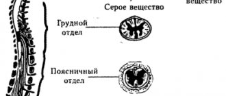
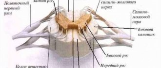
The anatomy of the medulla oblongata is similar to the spinal and cephalic portions of the central nervous system. The bulb consists of white and gray matter, i.e. pathways and nuclei, respectively. It has formations (pyramids) that control motor function and pass into the anterior dorsal pathways.
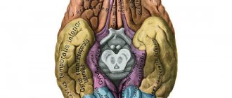
To the side of the pyramids are olive trees - oval formations separated by a groove. On the posterior surface of the medulla oblongata there are median, intermediate and lateral borders. Posteriorly, cranial fibers of the ninth, tenth and eleventh pairs emerge from the lateral border. It is useful to know what asymmetry of the lateral ventricles of the brain is and what the consequences are. All about the basal ganglia: anatomy and physiology.
The bulbus of the central nervous system consists of the following gray matter formations:
- Olive nucleus, which has connections with the dentate nucleus of the cerebellum.
- Provides balance.
- The reticular formation is a switch that integrates various parts of the central nervous system with each other, ensuring the coordinated functioning of the nuclei.
- Vasomotor and respiratory centers.
- Nuclei of the glossopharyngeal, vagus, accessory and hypoglossal nerve fibers.
White matter (nerve fibers of the medulla oblongata) provides conductive function and connects the head part of the central nervous system with the spinal part. There are long and short fibers. The pyramidal tracts and the wedge-shaped and thin fasciculus tracts are formed by long conducting fibers.
Functions of the medulla oblongata.
The bulbus, as part of the trunk of the central nervous system, is responsible for the regulation of blood pressure and the work of the respiratory muscles. These functions of the medulla oblongata are vital for humans. Therefore, its defeat during injuries, strokes, and other injuries often leads to death.
Main functions:
- Regulation of blood circulation and breathing.
- Presence of sneezing and coughing reflexes.
- The nucleus of the glossopharyngeal nerve provides swallowing.
- The vagus nerve has autonomic fibers that influence the functioning of the heart and digestive system.
- Balance is ensured by communication with the cerebellum.
Breathing is regulated through the coordinated work of the inspiratory (responsible for inhalation) and expiratory (responsible for exhalation) departments.
Sometimes the respiratory center is suppressed by shock, trauma, strokes, poisoning, and metabolic disorders. It is also suppressed during hyperventilation (increased oxygen levels in the blood). The nucleus of the 10th pair of cranial nerves is also involved in breathing.
Blood circulation is regulated by the work of the vagus nerve nucleus, which affects both cardiac activity and vascular tone. This center receives information from the heart, digestive system and other parts of the human body. The tenth pair of nerves emanating from it reduces the heart rate.
The vagus nerve enhances the functioning of the gastrointestinal tract. Stimulates the release of hydrochloric acid, pancreatic enzymes, accelerates peristalsis of the large intestine. Its sensitive fibers come from the pharynx and eardrum. Motor fibers ensure coordination of swallowing processes, in which the muscles of the pharynx and soft palate participate.
The glossopharyngeal nerves, the ninth pair, ensure the act of swallowing, pushing the bolus of food from the oral cavity into the pharynx, then the esophagus.
The hypoglossal nerve has motor fibers that regulate the functioning of the tongue muscles. Provides sucking, licking, swallowing, articulation (speech).
Symptoms of bulbus damage
Sometimes, as a result of injuries, intoxications, metabolic diseases, hemorrhages, ischemia, and shock conditions, the activity of medulla oblongata is disrupted, which leads to bulbar syndrome.
The main causes of the pathology:
- Strokes (hemorrhages).
- Syringomyelia (presence of cavities).
- Porphyria.
- Botulism.
- Dislocation syndrome in injuries, hematomas.
- Diabetes mellitus, ketoacidosis.
- Effect of antipsychotic drugs.
It is important to find out what the pons is: structure, functions, symptoms in pathological conditions. What do gliosis changes in the brain lead to: treatment, diagnosis, prevention. Note: what the pituitary gland is responsible for and what disruption of its functions leads to.
Symptoms of medulla oblongata damage include:
- Circulatory disorders: bradycardia, decreased blood pressure.
- Respiratory dysfunction: Kussmaul breathing in ketoacidosis, shortness of breath.
- Coma state.
- Swallowing and chewing disorders.
- Movement disorders.
- Loss of taste.
- Impaired reflexes.
- Speech disorder.
If this part of the brain is damaged, the function of the respiratory center may be turned off, leading to asphyxia (suffocation). Pressor dysfunction causes a drop in blood pressure.
Bulbar symptoms include difficulty swallowing and choking on food. A person's heart rate slows down and shortness of breath occurs. Since the activity of the hypoglossal nerve is disrupted, the patient loses the ability to pronounce words and chew. Saliva may leak from the mouth. As can be seen from the article, the medulla oblongata is important in ensuring human life. Blood circulation and breathing are its most important functions. Damage to this section can lead to death.
Source: https://golmozg.ru/stroenie/prodolgovatyy-mozg.html Blog Brain
You can sign up online or get the schedule of the nearest master classes at the link: https://qviz.matomba.ru/1c06710015fbab389df6
Drug therapy
Drug treatment is an integral part of patient resuscitation, prevention and recovery.
The therapy provided should be comprehensive and aimed at preserving the vital functions of the body. The main directions for this: Correction of disorders of the respiratory system. If necessary, connection to a ventilator. In the acute period, infusion therapy with plasma preparations and rheopolyglucin is carried out. Normalization of blood pressure indicators. At high blood pressure levels, antihypertensive drugs are prescribed, making sure that changes to normal values occur smoothly and gradually. The main drugs used are from the group of ganglion blockers and ACE inhibitors. In case of severe hypotension, glucocorticosteroids and dopaminomimetics are administered. Reducing body temperature is carried out using non-steroidal anti-inflammatory drugs.
Reducing intracranial pressure and cerebral edema - with the help of diuretics. In some cases, a hypertonic saline solution is administered.
Restoring blood supply to the affected areas and preventing thrombosis. Here they use drugs from the group of antiplatelet agents and anticoagulants.
Improving the nutrition of brain tissue and intracellular metabolism is carried out with the help of nootropics, antioxidants, and cytoprotectors.
The treatment of hemorrhagic and ischemic stroke has its own characteristics. In case of hemorrhage, the use of anticoagulants and antiplatelet agents is contraindicated. The main treatment is aimed at stopping the bleeding and removing the hematoma. Treatment with medications is carried out in the intensive care unit. The following substances are prescribed:
- diuretics aimed at preventing edema;
- sedatives;
- oxygen therapy - if the patient is able to breathe on his own, catheters are installed, otherwise a special support device is connected;
- muscle relaxants to prevent seizures;
- blood thinners that increase blood flow;
- antihypertensive drugs to prevent recurrent vascular ruptures.
At first, all drug therapy is aimed at eliminating repeated attacks and severe consequences.
Doctors keep an eye on the patient until the body’s functioning is completely normalized, and when the patient’s well-being improves, they are transferred to a ward for further recovery. Make an appointment
Surgical intervention
Any surgical intervention on the brain is a high risk and can often lead to serious complications, including the death of the patient. Therefore, surgical treatment is carried out in the presence of strict indications:
- Swelling or compression by hematoma of the medulla oblongata, which leads to the progression of neurological symptoms. In this case, to save the patient’s life, craniotomy is performed using a certain technique.
- Pathological changes in cerebral vessels (aneurysms, malformations), accompanied by bleeding. For treatment, special clips are used, which are surgically applied to the neck of the aneurysm.
- For ischemic stroke, when conservative therapy is ineffective, carotid endarterectomy followed by stenting can be performed. The operation is performed endovascularly (without incisions).
In the postoperative period, the patient's condition remains unstable for a long time. It can often be complicated by swelling of the brain tissue, which lasts up to two weeks. In the future, therapeutic measures are carried out aimed at restoring lost functions and preventing a recurrent stroke.
Prognosis after brainstem stroke
The type of stroke, the extent of the pathological process, and the timeliness of medical care are of great importance for the prognosis.
The ischemic type is more favorable - mortality is about 25%. In case of hemorrhage, according to statistics, every second patient dies within a month. The main reasons: cerebral edema with entrapment in the foramen magnum, death of stem structures. Even if life is preserved, the majority of such patients remain deeply disabled. According to research, a negative outcome is more often observed in cases of respiratory failure, persistent bradycardia, changes in thermoregulation, and loss of speech. An uncertain prognosis is given if the patient has dysphagia, movement disorders, or impaired eye movement functions. In any case, treatment of patients with brainstem stroke requires competently selected therapy and the use of all possible rehabilitation methods.
The prognosis of brainstem stroke and further condition is determined by the degree of manifestation of symptoms. Doctors see the most favorable recovery in patients with less pronounced signs of the disease, but, of course, there are always exceptions. The most skeptical view of complete recovery is captured by the following observations:
- problems with speech and breathing;
- dysregulation of blood pressure and body temperature.
A more positive prognosis can be heard when:
- weakness of swallowing;
- complete or incomplete paralysis of the limbs;
- problems with eye rotation;
- frequent dizziness.
Important factors when forming a prognosis are the speed of referral to specialists and the age of the patient. The older a person is, the more difficult the recovery process is.
Structure and functions of the medulla oblongata: symptoms of damage
The medulla oblongata is a section of the central nervous system, also called bulbus, bulbus or medulla oblongata in Latin.
Located between the spinal region, the pons and the cerebellum, it is part of the brain stem. Performs many important functions: regulation of breathing, blood circulation, digestion. It is the oldest formation of the central nervous system. Its defeat often leads to death, since vital functions are switched off.
Location and anatomy of the medulla oblongata The posterior part of the central nervous system is where the medulla oblongata is located. From below it passes into the dorsal, and from above it is adjacent to the bridge. The cavity of the fourth ventricle, filled with fluid (cerebrospinal fluid), separates the bulbus from the cerebellum. It ends approximately where the head meets the neck, that is, its lower border is located at the level of the occipital inlet (hole).
The anatomy of the medulla oblongata is similar to the spinal and cephalic portions of the central nervous system. The bulb consists of white and gray matter, i.e. pathways and nuclei, respectively. It has formations (pyramids) that control motor function and pass into the anterior dorsal pathways.
To the side of the pyramids are olive trees - oval formations separated by a groove. On the posterior surface of the medulla oblongata there are median, intermediate and lateral borders. Posteriorly, cranial fibers of the ninth, tenth and eleventh pairs emerge from the lateral border. It is useful to know what asymmetry of the lateral ventricles of the brain is and what the consequences are. All about the basal ganglia: anatomy and physiology.
The bulbus of the central nervous system consists of the following gray matter formations:
- Olive nucleus, which has connections with the dentate nucleus of the cerebellum.
- Provides balance.
- The reticular formation is a switch that integrates various parts of the central nervous system with each other, ensuring the coordinated functioning of the nuclei.
- Vasomotor and respiratory centers.
- Nuclei of the glossopharyngeal, vagus, accessory and hypoglossal nerve fibers.
White matter (nerve fibers of the medulla oblongata) provides conductive function and connects the head part of the central nervous system with the spinal part. There are long and short fibers. The pyramidal tracts and the wedge-shaped and thin fasciculus tracts are formed by long conducting fibers.
Functions of the medulla oblongata.
The bulbus, as part of the trunk of the central nervous system, is responsible for the regulation of blood pressure and the work of the respiratory muscles. These functions of the medulla oblongata are vital for humans. Therefore, its defeat during injuries, strokes, and other injuries often leads to death.
Main functions:
- Regulation of blood circulation and breathing.
- Presence of sneezing and coughing reflexes.
- The nucleus of the glossopharyngeal nerve provides swallowing.
- The vagus nerve has autonomic fibers that influence the functioning of the heart and digestive system.
- Balance is ensured by communication with the cerebellum.
Breathing is regulated through the coordinated work of the inspiratory (responsible for inhalation) and expiratory (responsible for exhalation) departments.
Sometimes the respiratory center is suppressed by shock, trauma, strokes, poisoning, and metabolic disorders. It is also suppressed during hyperventilation (increased oxygen levels in the blood). The nucleus of the 10th pair of cranial nerves is also involved in breathing.
Blood circulation is regulated by the work of the vagus nerve nucleus, which affects both cardiac activity and vascular tone. This center receives information from the heart, digestive system and other parts of the human body. The tenth pair of nerves emanating from it reduces the heart rate.
The vagus nerve enhances the functioning of the gastrointestinal tract. Stimulates the release of hydrochloric acid, pancreatic enzymes, accelerates peristalsis of the large intestine. Its sensitive fibers come from the pharynx and eardrum. Motor fibers ensure coordination of swallowing processes, in which the muscles of the pharynx and soft palate participate.
The glossopharyngeal nerves, the ninth pair, ensure the act of swallowing, pushing the bolus of food from the oral cavity into the pharynx, then the esophagus.
The hypoglossal nerve has motor fibers that regulate the functioning of the tongue muscles. Provides sucking, licking, swallowing, articulation (speech).
Symptoms of bulbus damage
Sometimes, as a result of injuries, intoxications, metabolic diseases, hemorrhages, ischemia, and shock conditions, the activity of medulla oblongata is disrupted, which leads to bulbar syndrome.
The main causes of the pathology:
- Strokes (hemorrhages).
- Syringomyelia (presence of cavities).
- Porphyria.
- Botulism.
- Dislocation syndrome in injuries, hematomas.
- Diabetes mellitus, ketoacidosis.
- Effect of antipsychotic drugs.
It is important to find out what the pons is: structure, functions, symptoms in pathological conditions. What do gliosis changes in the brain lead to: treatment, diagnosis, prevention. Note: what the pituitary gland is responsible for and what disruption of its functions leads to.
Symptoms of medulla oblongata damage include:
- Circulatory disorders: bradycardia, decreased blood pressure.
- Respiratory dysfunction: Kussmaul breathing in ketoacidosis, shortness of breath.
- Coma state.
- Swallowing and chewing disorders.
- Movement disorders.
- Loss of taste.
- Impaired reflexes.
- Speech disorder.
If this part of the brain is damaged, the function of the respiratory center may be turned off, leading to asphyxia (suffocation). Pressor dysfunction causes a drop in blood pressure.
Bulbar symptoms include difficulty swallowing and choking on food. A person's heart rate slows down and shortness of breath occurs. Since the activity of the hypoglossal nerve is disrupted, the patient loses the ability to pronounce words and chew. Saliva may leak from the mouth. As can be seen from the article, the medulla oblongata is important in ensuring human life. Blood circulation and breathing are its most important functions. Damage to this section can lead to death.
Source: https://golmozg.ru/stroenie/prodolgovatyy-mozg.html Blog Brain
You can sign up online or get the schedule of the nearest master classes at the link: https://qviz.matomba.ru/1c06710015fbab389df6