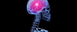
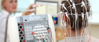
The spinal cord and brain are independent structures, but in order for them to interact together, one formation is required - the pons. This element of the central nervous system acts as a collector, a connecting structure that links the brain and spinal cord together. Therefore, the formation is called a bridge , because it connects two key organs of the central and peripheral nervous system. The pons is part of the structure of the hindbrain, to which the cerebellum is also attached.
- Structure
- Functions
- Symptoms of the lesion
Structure
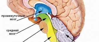
The division is part of the hindbrain. The structure and function of a bridge are very closely related, as in any other structure. It is located in front of the cerebellum, being a section between the midbrain and medulla oblongata.
It is separated from the first by the beginning of the 4th pair of cranial nerves, and from the second by a transverse groove. Outwardly, it resembles a roller with a groove, with nerves passing through it; they are responsible for the sensory abilities of the facial skin. In the groove there was also a place for the basilar arteries; their features include the fact that they supply blood to the back of the brain.
This section has a special diamond-shaped fossa located in the posterior part of the pons. The fossa is bordered at the top by the brain stripes, and above them are the facial colliculi.
Above them there is a median eminence, and next to it is the locus coeruleus, which is responsible for the feeling of anxiety; it includes many nerve endings of the norepinephrine type. The pathways look like thick fibers of nervous tissue that run from the pons to the cerebellum. Thus, they form the arms of the pons and the peduncles of the cerebellum.
Among other things, the structure of the bridge has a “tire”, which is an accumulation of gray matter. This gray matter is the centers of the cranial nerves and the parts that contain the pathways. That is, the upper part of the brain is reserved for the centers that have a connection with the cranial nerves (fifth, sixth, seventh and eighth pairs).
Useful to know: Midbrain: structure, functions, development
Speaking of pathways, the medial lemniscus and lateral lemniscus pass through this part. The same tegmentum contains the reticular formation; it is part of six nuclei and contains structures that are responsible for auditory analyzers.
At the base there are paths that pass from the cerebral cortex to different parts:
- pons brain;
- medulla;
- spinal cord;
- cerebellum.
And the blood supply occurs through arteries that belong to the vertebrobasilar region.
to contents ^
Diseases leading to the development of syndromes
The structure of the Varoliev bridge suggests many possible lesions and an equally large number of manifestations. However, there is a group of diseases that become the foundation for the above syndromes.
This may include:
- Stroke. Acute disruption of cerebral blood flow in one area or another with death of nerve tissue and loss of some functions of cerebral structures. If the brain stem itself suffers, in the most favorable case it will end in a violation of higher activity.
- Ischemic transient attacks. Incorrectly called microstrokes. The same thing is observed, but there is no significant tissue death.
- Atherosclerosis of cerebral vessels. Impaired arterial patency as a result of blockage of the arteries by cholesterol plaques or spontaneous narrowing due to, for example, long-term smoking, hypertension (increased pressure).
- Infectious processes. Especially those that affect cerebral tissue. Encephalitis, meningitis.
- Demyelination. Multiple sclerosis.
The Varoliev bridge is responsible for a lot of important functions and has a systematic structure. Treatment of pathological conditions when the activity of this structure is already impaired is an extremely complex and sometimes impossible process.
Therefore, it makes sense to have a preventive effect on all diseases that may become a source of problems in the future. This is an important preventative measure.
Motor and sensory functions
Speaking in more detail about motor and sensory function, let's talk about cranial nerves. When mentioning cranial nerves, it should be noted the ternary or mixed nerve (V pair). This pair of nerves is responsible for the movement of the masticatory muscles, as well as the muscles that are responsible for the tension of the eardrum and the palatine curtain.
To the sensory part of the trigeminal nerve there are afferent connections of nerve cells from receptors that are located in the skin of the human face, nasal mucosa, 60% of the tongue, eyeball and teeth. The sixth pair, or the so-called abducens nerve, is responsible for the movement of the eyeballs, namely for its rotation outward.
The 7th pair has one of the most important functions for human interaction; it is responsible for the innervation of muscles that allow the production of facial expressions. In addition, the facial nerve controls three glands: salivary, sublingual and submandibular. These glands provide reflexes such as salivation and swallowing.
The bridge also has a connection with the vestibulocochlear nerve. It is clear from the name that the cochlear part reaches the cochlear nuclei, but the vestibular part ends in the triangular nucleus. The eighth pair of nerves is responsible for analyzing vestibular stimuli; it determines the degree of their severity and where they are directed.
Useful to know: Brain stem: features and functions
to contents ^
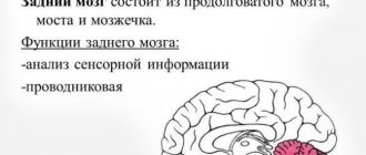
2. Provides various autonomic reflexes, some of them (respiratory, cardiovascular) are vital.
3. Conductive functions: numerous ascending and descending pathways pass through the trunk, connecting the cerebral cortex with the spinal cord.
4. Associative functions: primary analysis of the strength and quality of the sensory stimulus, as well as the interaction of the structures that make up the brain stem.
Medulla
The medulla oblongata is a continuation of the spinal cord, has a length of about 25 mm, it lacks a segmental structure, the gray matter forms separate clusters of neurons - nuclei. It performs its own, vegetative and conductive functions.
Own functions of the medulla oblongata
The medulla oblongata contains the nuclei of the VIII, IX, X, XI and XII pairs of cranial nerves.
VIII pair - vestibular-cochlear nerve (n.vestibulocochlearis). Located on the border between the medulla oblongata and the pons. The fibers of the cochlea or the auditory nerve itself are the beginning of the auditory pathways coming from the spiral ganglion of the cochlea (auditory functions).
IX pair – glossopharyngeal nerve (n.glossopharyngeus). Contains motor (innervation of the muscles of the pharynx and oral cavity), sensory (from the taste receptors of the posterior third of the tongue) and autonomic fibers (innervation of the salivary glands).
X pair - the vagus nerve (n.vagus) has three nuclei. The autonomic nucleus is responsible for the parasympathetic innervation of the larynx, esophagus, heart, stomach, small intestine, and digestive glands. The nerve fibers of the sensory nucleus form the solitary tract in the medulla oblongata. The nucleus receives information from receptors in the alveoli of the lungs and other internal organs. The motor core is responsible for contracting the muscles of the pharynx and larynx during swallowing and breathing.
XI pair - accessory nerve (n.accessorius) - a motor nerve that innervates the sternocleidomastoid and trapezius muscles on its side. When a nerve is damaged, the tone of the innervated muscles decreases and the shoulder on the affected side drops, and turning the head in the direction opposite to the lesion is difficult.
XII pair - hypoglossal nerve (n.hypoglossus) innervates the muscles of the tongue on its side. Unilateral nerve damage is accompanied by dysfunction of the tongue muscles.
Autonomic functions of the medulla oblongata
The medulla oblongata contains a respiratory center (inhalation center - inspiratory and exhalation center - expiratory), a vasomotor center - regulates vascular tone and blood pressure levels, the main center of cardiac activity - a group of neurons in the vagus nerve nucleus (inhibitory) and a group of neurons associated with spinal centers ( stimulating). The vagus nerve regulates (strengthens) the motility of the gastrointestinal tract and its secretory activity. In the medulla oblongata there is a center of salivation, the parasympathetic part of which ensures the release of a large amount of liquid saliva, rich in inorganic substances, and the sympathetic part - a small amount of thick protein secretion.
The medulla oblongata is responsible for some protective reflexes: vomiting, coughing, sneezing, lacrimation, closing the eyelids, as well as reflexes of eating behavior (sucking, chewing, swallowing).
The medulla oblongata implements somatic reflexes: reflexes of maintaining body posture due to static reflexes that regulate the tone of the muscles that maintain the position of the body in space, as well as statokinetic reflexes that ensure the redistribution of the tone of the trunk muscles to organize the posture at the moment of rectilinear and rotational movement. At the bottom of the IV ventricle of the medulla oblongata there are neurons of the “blue spot”, which secrete the neurotransmitter norepinephrine. Through the reticulospinal tract, these neurons inhibit spinal reflexes and reduce muscle tone during the REM sleep phase.
The medulla oblongata regulates some analytical functions, carrying out the primary analysis of sensory stimuli (skin, gustatory, auditory, vestibular).
Conducting functions of the medulla oblongata
Descending pathways begin in the medulla oblongata: vestibulospinal, olivospinal and reticulospinal, providing communication between the vestibular nuclei, olive, reticular formation of the medulla oblongata and spinal cord motor neurons responsible for the tone and coordination of muscle reactions. Irritation of the reticular formation of the medulla oblongata has descending effects on the motor activity of skeletal muscles, as well as ascending, activating effects on the cerebral cortex, causing desynchronization of the electroencephalogram.
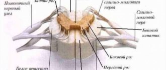
All ascending and descending tracts of the spinal cord pass through the medulla oblongata: spinothalamic, corticospinal, rubrospinal. It ends in the pathways from the cerebral cortex - the corticoreticular, as well as the ascending pathways of proprioceptive sensitivity - the thin fasciculus of Gaulle and the wedge-shaped fasciculus of Burdach. Therefore, when one of the halves of the medulla oblongata is damaged above the intersection of the ascending pathways of proprioceptive sensitivity, the sensitivity and functioning of the muscles of the face and head on the side of the injury occurs, while on the uninjured side, disorders of skin sensitivity and motor paralysis of the trunk and limbs occur. This is due to the fact that all ascending and descending paths intersect.
Pons
The pons Varolii is located above the medulla oblongata and performs motor, sensory, integrative and conductive functions.
Own functions of the pons
The pons includes the nuclei of the VIII pair of cranial nerves, the vestibular nucleus (lateral - Deiters and superior - Bekhterev), which is responsible for the primary analysis of vestibular stimuli.
VII pair - facial nerve (p.facialis), innervates the facial muscles, sublingual and submandibular salivary glands, transmits information from the taste buds of the anterior part of the tongue.
VI pair – abducens nerve (n.abducens), innervates the rectus externus muscle, which abducts the eyeball outward.
V pair – trigeminal nerve (n. trigeminus). The motor nucleus of the nerve innervates the muscles of mastication, the muscles of the velum palatine, and the tensor tympani muscles. The sensitive nucleus receives afferent axons from receptors on the skin of the face, nasal mucosa, teeth, periosteum of the skull bones, and conjunctiva of the eyeball.
The pons contains a pneumotaxic center that triggers the exhalation center of the medulla oblongata, as well as a group of neurons that activate the inhalation center.
The reticular formation of the pons is a continuation of the reticular formation of the medulla oblongata. It affects the cerebral cortex, activating it and causing awakening. The axons of the pontine reticular formation go to the cerebellum and spinal cord.
Conductive function of the pons
All ascending and descending pathways pass through the bridge, connecting the bridge with the cerebellum and spinal cord, the cerebral cortex and other structures of the central nervous system.
Midbrain
The main structural formations of the midbrain are: nucleus of the trochlear nerve - IV pair of cranial nerves (n.trochlearis), nucleus of the oculomotor nerve (n.oculomotorius) - III pair of cranial nerves, red nucleus (n.ruber), substantia nigra ( substantia nigra), quadrigeminalis, cerebral peduncles and nuclei of the reticular formation.
The trochlear nerve innervates the superior oblique muscle of the eye and ensures upward-outward rotation of the eye.
The oculomotor nerve is responsible for raising the upper eyelid, regulating the movements of the eye up, down, towards the nose and down towards the corner of the nose. Neurons of the accessory nucleus of the oculomotor nerve (Yakubovich's nucleus) regulate the lumen of the pupil and the curvature of the lens, ensuring the process of accommodation.
Integrating function
These pons functions connect parts of the brain called the cerebral hemispheres. Also, all other paths, both ascending and descending, pass along the bridge, connecting it with many departments of the central nervous system. Among them are the spinal cord, cerebellum and cerebral cortex.
Impulses passing through the pontocerebellar pathways of the cerebral cortex have an impact on the functioning of the cerebellum. The cortex cannot exert influence directly, so it uses the bridge as an intermediary for these purposes. The pons regulates the medulla oblongata, influencing the centers that are responsible for the respiratory process and its intensity.
to contents ^
Pathologies that impair the functions of the bridge and their symptoms
There is a group of diseases that are typically characterized by disruption of the normal functioning of the body as a result of the destruction of the tissues in question.
Among them:
Brissot-Sicard syndrome
Accompanied by a disorder of cranial nerve activity. It is determined by unilateral paresis or complete paralysis of half the body.
The ability to control the muscles of the facial area is also lost, and ptosis (drooping of the eyelid) with impaired visual function is possible.
This disorder occurs against the background of an infectious, autoimmune or tumor lesion. Less commonly, it results from cerebral ischemia. After a transient attack or a full-fledged stroke.
Bonnier syndrome
Characterized by damage to a group of cranial nerves. In this case, the auditory and vestibular nuclei ultimately suffer.
Symptoms are nonspecific. Problems arise with the perception of sound stimuli. Patients constantly experience dizziness, nausea, and weakness.
Insomnia is also part of the clinic. The patient becomes irritable, and emotional instability is noted. Up to sudden changes in phases, as, for example, with bipolar affective psychosis.
Grenet's syndrome
A typical feature of this pathological process is a violation of the sensitivity of facial muscles, which ultimately leads to problems with the manifestation of non-verbal signals and emotions.
There is partial paresis of the masticatory muscles on one side. On the other hand, controllability impairment is also present, but to a much lesser extent.
Ventral syndrome
An extremely difficult condition. It is characterized by at least loss of speech function. This is the easiest case.
The classic situation is determined by the complete loss of the ability to move. The man cannot move. Communication is possible only through the eyes.
This disorder persists for a long time. Quickly leads to stagnation and death of the patient. Restoration is not possible.
Raymond-Sestan syndrome
Characterized by a key manifestation of the oculomotor nerves. A person loses the ability to voluntarily focus his gaze and move it from one object to another.
Spontaneous relief of the condition and its subsequent return for unclear reasons is possible.
Hübler's syndrome
There are no specific manifestations of blood paralysis of facial muscles. The facial expression is characterized as a mask.
The patient is unable to adequately non-verbally express emotions and respond to surrounding stimuli.
Skin sensitivity also decreases, which is detected by the results of functional tests and physical examination.
Foville syndrome
There is paralysis of facial muscles and strabismus with visual impairment.
Gasperini disease
Combined pathological process. Characterized by mixed symptoms.
Causes and symptoms of gliomas
The nature of the appearance of the neoplasm remains unknown. Experts have only assumptions regarding the reasons for the growth of this type of formation. They may be associated with the influence of unfavorable internal and external factors, and hereditary predisposition.
Glioma is predominantly a primary intracerebral tumor of the hemispheres. It has fuzzy outlines. Experts disagree and cannot say for sure from which cells these neoplasms primarily appear. Many believe that tumor activity is provoked by genetic disorders. The trigger can be unfavorable external and internal factors, including intoxication of the body and radiation exposure.
Symptoms of gliomas
The clinical picture of the disease depends on the severity of the pathological process. Signs of pathology increase gradually. They can be mistaken for a general deterioration in well-being due to overwork and stress.
Symptoms of brain glioma in adults can be focal and cerebral. Dislocation manifestations also occur remotely, which appear as a result of displacement of brain structures and the appearance of local edema.
General cerebral symptoms are characterized by the appearance of headache, nausea and vomiting, seizures, and visual disturbances. They are the first signs of growth of a brain tumor. A headache appears after stress, physical exertion, or a sudden change in body position. At first, the pain syndrome occurs periodically, but as the tumor grows it becomes permanent. Nausea and vomiting are usually not associated with food intake.
Convulsions and visual disturbances occur much later. They appear when the tumor is large enough. Patients note the presence of a constant veil before their eyes. In this case, ophthalmological treatment does not give the expected effect.
How does the bridge affect reflexes?
Due to the fact that the pons is an integral part of the quadrigeminal region, it is related to the development of the auditory and statistical reflex. Thanks to the latter, we are able to hold the body in a certain position. In addition, interacting with the midbrain, it closes a significant part of muscle reflexes.
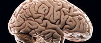
The tissues of the brain are represented by a wide range of formations. In terms of its structure, it is perhaps the most complex part of the human body, which determines the wide nature of the activity of the central nervous system. When assessing the structure, several areas of the central nervous system can be identified in this localization.
At the base of the cerebral structures is the so-called brain stem. It provides a group of vital functions: from breathing and cardiac activity to thermoregulation. Any damage or malfunction results in severe disability or death.
The pons is a component of the brainstem located between the midbrain and medulla oblongata, which ensures normal conduction of nerve impulses and makes it possible to perform a number of voluntary actions.
Responsible for some functions of higher activity. Its damage, for example, due to injury or stroke, leads to critical disruptions in the functioning of the entire body.
Brain structure
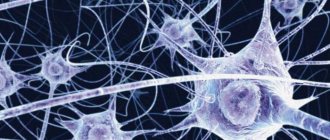
The structural unit of the central nervous system is the neuron. It is this cell that is responsible for receiving, processing and storing information. The entire human brain is a collection of neurons and their processes - axons and dendrites. They ensure the transmission of signals entering the central nervous system and back to the organs. The brain consists of gray and white matter. The first is formed by the neurons themselves, the second by their axons. The main structures of the brain are the hemispheres (left and right), the cerebellum and the brainstem. The first are responsible for a person’s mental abilities, his memory, thinking, imagination. The cerebellum is necessary for coordination of movements, in particular, it provides the ability to stand upright, walk, and grasp objects. Below it is the pons. It is the connecting link between the medulla oblongata and the cerebellum.
What functions does the bridge perform?
The pons is responsible for several important forms of activity.
- Reflexive automatic and voluntary movements of the eyes and eardrum in response to loud sounds, as well as tissues of the oral cavity (palate). Any violation ends in problems.
- The ability for purposeful motor activity. Since the pons in the brain ensures the functioning of the cerebellum, any damage causes problems with the ability to control the body.
- Perception of vestibular stimuli. In this case, we are talking about the ability to perceive one’s body as a whole, its orientation and location in space, to respond to any changes in environmental conditions, and also to dampen unnecessary movements (for example, during sudden braking in public transport, stumbling, etc.). When affected, there is a lack of coordination. Ability to navigate in space.
- Providing olfactory function. The bridge has this ability partially. Other subcortical clusters are also responsible for it.
- Normal innervation of the skin and mucous membranes of the face.
- The pons is involved in the formation of sleep. This is a complex and coordinated work of several cerebral formations at once. Any violations immediately lead to problems with night rest. The patient becomes lethargic, asthenic processes appear.
- The functions of the pons include the acts of chewing and swallowing. Vital for nutrition and breathing.
- In fact, the body’s ability to perform normal gas exchange depends on the functioning of this structure. In the absence of adequate conduction of impulses, problems begin, even lethal disorders.
Reflex function of the bridge
The ability of the central nervous system to respond to external stimuli is called a reflex. An example is the appearance of salivation at the sight of food, the desire to sleep at the sound of soothing music, etc. Reflexes of the brain can be conditioned and unconditioned. A person acquires the first ones in the course of life; they can be developed or adjusted depending on our desire. The latter are not amenable to consciousness, they are laid down at birth, and it is impossible to change them. These include chewing, swallowing, grasping and other reflexes.
Recovery period and life prognosis
The prognosis for life with benign glioma depends on the timeliness of treatment and the degree of malignancy of the tumor. The necessary information about the tumor can only be obtained through a comprehensive and thorough diagnosis.
Low-grade gliomas are attempted to be completely removed surgically. Usually this does not cause any difficulties due to the localized nature of the pathological focus. Surgical treatment is the gold standard of neurosurgery.
It is impossible to indicate the exact life expectancy after surgery for brain glioma. If the patient seeks help on time, and the neurosurgeon manages to completely remove the tumor, the prognosis for glioma will be favorable. Life expectancy can be increased with subsequent radiation therapy. It allows you to increase the five-year survival rate after surgery to 60%.
It is also important to follow all doctor’s recommendations during the rehabilitation period. During recovery after removal of a brain glioma, the patient is under the supervision of specialists of a narrow and broad profile. They monitor important indicators of the body’s functioning, help normalize overall well-being and restore impaired functions of the nervous system.
Reticular formation of the bridge
The reticular formation is a branched network located in the brain and consisting of nerve cells and nuclei. It is present in almost all formations of the central nervous system and smoothly passes from one department to another. The reticular formation of the pons is located between the medulla oblongata and midbrain. Its long processes, called axons, form white matter and pass into the cerebellum. In addition, signals can be transferred from the head to the back along the fibers of the nerve cells of the bridge. In addition, the reticular formation transmits signals to the cerebral cortex, due to which a person awakens or sleeps. The nuclei located in this part of the pons belong to the respiratory center located in the medulla oblongata.