Today, ultrasound diagnostics is rightfully considered one of the safest and most informative. It is applicable in various fields of medicine, and is also used to determine abnormalities in the vascular system. If there is a need to study blood vessels, TCDG is used, which stands for transcranial Dopplerography of cerebral vessels.
Transcranial Dopplerography of cerebral vessels: what is it?
TCD is based on ultrasonic waves and the Doppler effect. Ultrasound waves are reflected from moving red blood cells and vessel walls, and with the help of a special sensor and software, the pulses are converted into an image that is displayed on a computer screen.
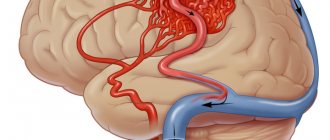
TCD of cerebral vessels will determine the condition of the main arteries, which are responsible for supplying the brain with oxygen, as well as other large arteries and vessels. Thanks to this, it will be possible to assess the condition of the vessels and determine the speed and intensity of blood flow in them.
Description of the TCD method
At clinic No. 1 Viterra Belyaevo, transcranial Dopplerography is carried out using the latest generation devices. The study is carried out using digital technologies in M-mode. It allows you to scan the condition of blood vessels at a depth of up to 90 mm. The examination result is recorded and displayed continuously during the procedure. During its implementation, there is no need to change the depth to locate a vessel with pathologies.
It is possible to display up to 8 windows with Doppler spectra on the device screen, while measurements are taken at different depths. This can be done either sequentially or simultaneously. The device processes the received data and generates the final result, which has a graphical form. It contains digital symbols. Based on this, the Viterra clinic specialist forms his conclusion, which indicates the diagnosis.
Indications for transcranial Doppler ultrasound
There are many reasons why you may need to undergo Doppler ultrasound. Transcranial Dopplerography of cerebral vessels
may show a large number of deviations from the norm.
Vascular malformation
It is a congenital disease that affects the development of the vascular system. Manifests itself in the form of plexuses of abnormal vessels of various sizes. It is dangerous because it can cause subarachnoid hemorrhage of a non-traumatic nature. Among other things, steal syndrome may develop, and malformations may begin to compress the brain tissue.
Cerebral circulatory disorders
TCD is used quite often to examine cerebral vessels. It may be necessary to identify the cause of problems with cerebral circulation. They may consist of the formation of blood clots, loops and kinks in blood vessels. Often the cause is: the formation of narrowings, aneurysms or embolism.
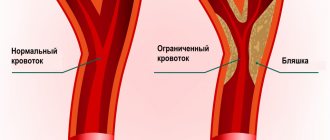
Atherosclerosis of the arteries of the neck and head
It consists of damage to areas of blood vessels by atherosclerotic plaques, which either significantly narrow the lumen of the vessel or clog it completely. In advanced cases, treatment is performed surgically. TCD is used to examine vessels because it allows one to determine not only the presence of plaques, but also their size and location.
Encephalopathy
Occurs in chronic cerebral vascular diseases that slowly progress. This disease combines cognitive impairment, disruptions in the emotional and motor spheres.
If you are worried about dizziness, tinnitus, ringing in the head, headaches, heaviness, weather dependence, etc. - this may be a sign of impaired, insufficient blood circulation to the brain.
In such a situation, it is necessary to study the blood flow in the vessels of the brain. The most accessible and informative method for assessing the state of cerebral circulation is ultrasound examination of blood vessels, which is aimed at identifying obstacles to blood flow to the brain as the cause of vascular diseases of the brain.
Previously, ultrasound equipment made it possible to study vessels only “blindly” - without visualizing the vessel itself, only by the nature of the blood flow in it (using the Doppler effect, the speed and direction of blood flow was determined. This method is called “USDG” - “Ultrasound Dopplerography of Vessels”) .
Currently, the Doppler ultrasound method is not used independently; it is part of a more modern ultrasound examination of blood vessels, which is called “Vascular Duplex Scanning” (vascular DS). Some doctors continue to call this method in the old way: “USDG”. Therefore, there is confusion in the name of this method of ultrasound examination of blood vessels.
The term “ Duplex scanning of vessels ” means “double scanning of vessels” and allows not only to determine the nature of blood movement through the vessels using the Doppler effect (this is ultrasound scanning), but also to examine the vessels in detail: determine their size, the structure of the vascular wall (which becomes denser and thickens with atherosclerosis, thickens with inflammation of the vessel, etc.); determine the course of blood vessels (which can be smooth or tortuous, up to “loop-shaped”), identify intravascular formations - atherosclerotic plaques, blood clots that create an obstacle to blood flow.
Modern ultrasound machines add to these two components of the study the ability to color vessels depending on the direction of blood flow . Therefore, the term “ Triplex vascular scanning ” (that is, triple scanning) appeared.
The terms “Duplex scanning of vessels”, “DS of vessels”, “Triplex scanning of vessels”, “USDG of vessels” mean one thing - ultrasound examination of blood vessels, which is carried out in triplex mode on modern ultrasound machines.
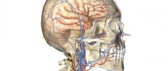
When performing an ultrasound examination of the vessels carrying blood from the heart to the brain, it is customary to distinguish two levels: the vessels of the neck and the vessels of the brain.
For more reliable information about the state of cerebral circulation, it is necessary to examine both levels of blood flow to the brain, since the “neck and head” are one whole: blood flow in the arteries of the neck is an intermediate stage in the movement of blood from the heart to the brain, and blood flow in the cerebral vessels - this is the final result. Blood flow disturbances can occur at any of these levels.
The first stage of ultrasound diagnostics is the examination of the arteries and veins of the neck :
- common carotid arteries and their branches (internal carotid arteries - carry blood to the brain, external carotid arteries - supply blood to the face)
- vertebral arteries, which at the level of the neck pass through the transverse processes of the cervical vertebrae and carry blood to the posterior parts of the brain.
Obstacles to the flow of blood to the brain at the level of the neck can be tortuosity of blood vessels - congenital (up to the formation of a “loop” by the vessel), or acquired (for example, an uneven tortuous course of the vertebral arteries, formed against the background of spinal osteochondrosis); or there may be narrowing of the arteries - congenital (narrow diameter of the artery), or acquired (narrowing of the lumen of the arteries due to inflammation of the vascular wall, atherosclerotic plaques, blood clots, etc., up to complete blockage of the lumen of the artery). The veins of the neck - jugular and vertebral - are also examined to identify disturbances in the outflow of venous blood from the brain to the heart.
The second stage of ultrasound diagnostics is a study of the arteries and veins of the brain , which is performed through the bones of the skull (transcranial). The arteries of the brain are a continuation of the arteries of the neck on the surface of the brain inside the skull, and form two pools of blood circulation in the brain: “Carotid pool” - from the terminal branches of the internal carotid artery (ultrasound examination is carried out through the temporal bone); and the “Vertebro-basilar basin” - consisting of the terminal segments of the vertebral arteries, which, after entering the cranial cavity, merge with each other into the main (or basilar) artery of the brain (ultrasound examination is carried out through the foramen magnum).
Ultrasound examination of cerebral vessels reveals the resulting indicators of cerebral blood flow: either the blood supply to the brain is not impaired, or there are disturbances in arterial blood flow in one or another circulatory system of the brain (arterial spasm, insufficient blood flow to the brain, etc.) or there are signs of obstructed outflow venous blood from the brain (which leads to increased intracranial pressure). This study is called “Transcranial ultrasound examination of cerebral vessels”, its synonyms: “TCDG” “Transcranial Dopplerography”.
Therefore, it is impossible to study, for example, only transcranial blood flow through the vessels of the brain, without knowing the characteristics of blood movement through the arteries of the neck, where there may be various obstacles to the blood flow to the brain.
And vice versa - the study of only the vessels of the neck will be incomplete in its information content without taking into account the resulting indicators of blood flow in the cerebral vessels.
Carrying out an ultrasound examination of the neck arteries separately is only possible to track the growth dynamics of atherosclerotic plaques identified earlier in them (for timely referral of the patient to vascular surgeons).
There are cases when doctors prescribe a blood flow study in only one of the levels of blood flow to the brain: either in the vessels of the neck or in the vessels of the brain. It is not right.
This may be due to terminological confusion in the name of the method of ultrasound examination of blood vessels (out of habit, they continue to call it “USDG of the neck”).
Or it may be due to mistrust in the results of transcranial examination of cerebral blood flow, which developed in the era of “blind” Doppler examination of cerebral vessels. But those days are already gone. Modern ultrasound equipment allows you to clearly locate cerebral vessels and the movement of blood through them.
The method of ultrasound examination of blood vessels is fast, simple, informative and safe for the patient’s health.
To clarify the state of cerebral blood flow, it is necessary to conduct an ultrasound examination of the vessels of the neck and brain for such complaints as:
- headache
- dizziness
- tinnitus, ringing in the head
- decreased vision and hearing
- memory loss
- sleep disorders
- emotional disorders, psycho-emotional overload, etc.
To prevent the development of stroke, thanks to the identification of atherosclerotic plaques in the arteries, ultrasound examination of the vessels of the neck and brain is indicated for all patients:
- over 40 years of age
- with obliterating atherosclerosis of the arteries of the lower extremities
- for coronary heart disease, heart rhythm disturbances
- for arterial hypertension
- diabetes mellitus
- kidney diseases
- patients who have had a stroke or myocardial infarction.
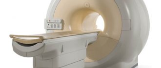
No other studies other than Duplex Vascular Scanning provide information about the functional state of the vessels of the head and neck and the movement of blood through them. Including this study is not replaced by MRI or CT scan of the brain (since they characterize the structure and condition of the brain tissue itself, and do not characterize cerebral blood flow). Ultrasound examinations of cerebral vessels, MRI or CT scans of the brain complement each other in their information content.
In our department, ultrasound diagnostics of blood vessels is carried out by very experienced and responsible specialists using modern ultrasound equipment. In addition to studies of the vessels of the neck and brain, studies are carried out of the arteries and veins of the upper and lower extremities, arteries of the kidneys, and vessels of the abdominal cavity.
| Name of service | Price |
| Functional diagnostics | |
| Code: A04.12.001.002 (5.067) Duplex scanning of the arteries and veins of the kidneys, | 1050.00 |
| Code: A04.12.003 (5.069) Duplex scanning of the abdominal aorta | 1050.00 |
| Code: A04.12.003.002 (5.070) Duplex scanning of arteries and veins of the iliac segment | 1600.00 |
| Code: A04.12.005.002 (5.042) Duplex scanning of the arteries of the upper extremities | 1500.00 |
| Code: A04.12.005.003 (5.003) DS of brachiocephalic arteries and veins | 1700.00 |
| Code: A04.12.005.004 (5.040) Duplex scanning of the veins of the upper extremities | 1500.00 |
| Code: A04.12.006.001 (5.043) Duplex scanning of the arteries of the lower extremities | 1500.00 |
| Code: A04.12.006.002 (5.041) Duplex scanning of the veins of the lower extremities | 1500.00 |
| Code: A04.12.018 (5.038) DS transcranial arteries and veins | 1100.00 |
Research methodology
It is important not only to understand what it is - TCD of cerebral vessels, but also to know how such a procedure is carried out. During this procedure, the patient will need to lie on his back, at which time the doctor will apply a special gel to the area being examined, after which he will apply a sensor and move it along the course of the arteries. You may need to breathe more frequently and also turn your head in certain ways. Unpleasant sensations during TCD to examine the cerebral vessels are excluded; the patient only feels the pressure of the sensor.
What vessels can be studied using TCD?
The position of the acoustic window will determine the list of blood vessels that can be examined. There are only three windows:
- temporal;
- orbital;
- suboccipital.
Examination of the brain using the TCD method using a temporal window will allow you to examine the internal carotid artery, middle, anterior and posterior cerebral arteries. Involves examination through the temporal region.
Orbital window - scanning the arteries through the orbits of the eyeballs. Allows you to assess the condition of the ophthalmic artery and the internal carotid siphon.
The suboccipital window involves ultrasound examination through the junction of the occiput with the spine. The basilar artery is examined, as well as the intracranial segments of the vertebral blood vessels.
Neurosonography and TCD
If you do not know what kind of examination TCD and neurosonography
, then you should understand that it applies to infants. In fact, this is an ultrasound of the child’s brain, which is completely safe and allows you to see the early manifestations of various abnormalities.
Indications
The main indications for transcranial ultrasound Dopplerography of the vessels of the neck and head are:
- frequent severe headaches, migraines, dizziness
- pathologies of the cervical spine
- loss of consciousness
- fatigue and severe weakness
- high blood sugar
- cramps and numbness in the legs and arms
- obesity
- cardiac ischemia
- heart attack
- stroke
In children, Doppler ultrasound is used for:
- delayed speech development
- restlessness, disinhibited behavior
- manifestation of asthenic conditions with increased fatigue, decreased memory and attention
What does transcranial Doppler sonography show?
After you have learned what TCD is, it is worth familiarizing yourself with the data that will become known as a result of the procedure. The following will be taken into account:
- vein or artery wall thickness, diameter;
- minimum and maximum blood flow speed, its nature;
- resistive and ripple indices.
Each of these indicators may indicate the presence or absence of disease.
The speed of blood flow through the arteries
A TCD examination may show an abnormality, which is a deterioration in blood flow. The cause may be the presence of blood clots or plaques, as well as other problems that can be identified through duplex testing.
Venous drainage from the cranial cavity
There can be many reasons for this phenomenon; they may include severe traumatic brain injuries, the occurrence of tumors that compress blood vessels and/or brain tissue. Often, outflow occurs against the background of a stroke and its consequences, with a decrease or underdevelopment of the network of blood vessels.
Degrees of development of the collateral network of brain vessels
TCD examination can reveal underdevelopment of the blood vessel network. It often causes problems with blood supply to the brain. All detected violations are recorded in the conclusion.
Why is TKDG prescribed?
Transcranial Doppler ultrasound reveals the following:
- if you have constant headaches, you can find out the cause of their origin - vasospasm or high intracranial pressure;
- what condition are the vertebral arteries in?
- examination reveals vascular lesions in the initial stages;
- the actual state of the venous blood flow of the head and neck vessels is determined;
- The presence of narrowing of the arteries and the degree of this change are determined.
Why do we have
- Having carefully “examined” the vessels of the brain, specialists at the Broad Hearts MC can prescribe correct, effective treatment, as well as identify and prevent the likelihood of a stroke recurrence. If necessary, related consultations and all necessary related studies will be carried out: ECG; Holter ECG; stress test - bicycle ergometry; Vascular ultrasound.
- The technical equipment of the “Broad Hearts” MC allows transcranial Doppler sonography of the brain to be performed on both the youngest patients (children from the first month of life) and adult patients.
- A conclusion in electronic form is issued immediately after TCD, saved in the center’s electronic database and, if necessary, the attending physician can compare the dynamics of the study with previous data.
- The results (ultrasound, examination, ECG, Holter-ECG, ABPM, bicycle ergometry, tests) are printed and given immediately to the patient in a convenient folder. In addition, they are stored electronically in the database (electronic medical record of the patient) of the Broad Hearts MC and allow the doctor to assess the dynamics of treatment and observation. Also on our website there is a service: Personal account, where all results are stored electronically and can be opened by patients anywhere and at any time independently. The personal account login is a phone number, and the password is sent via SMS to the specified phone number after payment for services. If problems arise, you can contact the center administrators by phone.
Find out more about studies of cerebral vessels from the administrator of the “Broad Hearts” MC by phone: (473) 280-20-30.
Operating principle of TKDS
The essence of TCD is to visualize the veins and arteries located inside the skull, as well as the vessels of the cervical spine using color mapping of blood flow. The method allows you to determine the presence of vascular damage.
The technique used for diagnostics connects the obtained drawings and displays an overall picture, according to which a specialist can make an accurate analysis. Procedures can be carried out in two modes:
Description of the procedure
The TCD procedure lasts no more than half an hour, is absolutely painless, has no contraindications, except for the general serious condition of the patient, in which the person is unable to remain motionless. We also strongly do not recommend taking pharmaceuticals that affect the functioning of the heart, the speed and nature of blood flow the day before the study, as this may distort the data obtained.
For the same reason, it is not advisable to drink alcohol or smoke on the day of the study.
Extracranial Dopplerography
Ultrasound using grayscale imaging, spectral Doppler analysis, and color Doppler imaging (CDI) is a validated method for assessing the extracranial cerebrovascular system. Although it is not possible to detect every anomaly, adherence to practical parameters will maximize the likelihood of detecting the majority of extracranial cerebrovascular anomalies. Sometimes additional and/or specialized examination may be required.
Transcranial Dopplerography
(TCD) provides rapid, non-invasive, real-time measurement of cerebrovascular function. TCD can be used to measure blood flow velocity in the basal arteries of the brain to assess relative changes in blood flow, diagnose focal vascular stenosis, or detect embolic signals in these arteries. TCD can also be used to assess the physiological state of a specific vascular territory by measuring the response of blood flow to changes in blood pressure, changes in final CO2 concentration (cerebral vasoreactivity), or cognitive and motor activation. TCD is widely used in the clinical diagnosis of a number of cerebrovascular disorders such as acute ischemic stroke, vasospasm, subarachnoid hemorrhage, sickle cell disease, as well as other conditions such as brain death. Clinical indications and research applications for this imaging modality continue to expand.
Where is ultrasound done?
You can undergo Doppler ultrasound for a fee in our medical centers, conveniently located in the central regions of Odintsovo and Zvenigorod. A professional team of doctors, modern reliable methods of diagnosis and treatment, high-precision equipment, the best service at affordable prices - all this is VERAMED. Registration for ultrasound examination is carried out through a special form on the website or call center. All services are provided for a fee. You can leave reviews about the work of the clinics on the website in the appropriate section.
How is an ultrasound scan of the brain performed?
There are no contraindications to the study. No special preparation is required, but the day before you should refrain from taking cardiovascular drugs and tranquilizers, drinking alcohol, and smoking.
The duration of the examination usually does not exceed 15 minutes. The patient is asked to lie on the couch and may need to turn onto his stomach or side. During Dopplerography of the brain, it is forbidden to talk, move or rotate your head unless the doctor asks for it.
The procedure does not cause discomfort - the doctor will simply move a special sensor along the neck, back of the head, forehead, and temporal region. The elasticity, density and thickness of the walls of arteries and veins, vasomotor activity, features of the circulatory network, the presence and description of hemodynamic anomalies, and intraluminal formations are recorded.