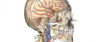
MRI of the head and cervical spine is a method for diagnosing diseases of the brain and neck. The procedure can be performed in standard mode, to study soft tissue, or in angio mode, to study the blood vessels supplying the central nervous system. The method is based on the use of magnetic fields and radio waves to obtain layer-by-layer images of the area of interest with excellent clarity and detail.
Why is virtual angiography needed?
With the help of such an examination it is possible to establish:
- Structural anomalies;
- Aneurysms;
- Dissection of vascular walls;
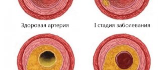
- Atherosclerosis;
- Neoplasia of various origins;
- Secondary foci of cancer;
- Degree of stenosis;
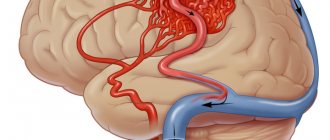
- Ischemic and hemorrhagic stroke;
- Hemorrhage;
- Pathological vascularization;
- Hemodynamic disturbances leading to decreased cerebral circulation;
- Vertebrobasilar insufficiency;
- Thrombosis of the cerebral sinuses.
How to sign up for an examination in Moscow
To do this, just use the services of our portal. It is designed for the convenience of patients and works for their benefit free of charge. Our staff will tell you which medical institutions operate in the area you are interested in. Operators will contact the selected clinic and make an appointment at a convenient time. The website contains information about the licenses of all medical centers with which we cooperate. There are price lists with the cost of their services, benefits and current promotions for brain MRI and MR angiography.
We tried to place important and useful information on the site. They talked about different types of examinations. There is information on how to prepare for them, what to take with you to the diagnosis. The portal notes which contraindications hinder hardware research.
Here is information about:
- Addresses, telephone numbers, opening hours of medical centers;
- Free recording time;
- Models and technical characteristics of devices, type of tunnel, weight restrictions;
- Qualifications and work experience of medical personnel;
- Availability of support services for visitors with limited mobility and an accessible environment: ramps, gurneys, freight elevators;
- Location of parking nearby;
- Acceptable payment options.
There is a list of banks whose cards you can make payments with. The service contains verified patient reviews. Based on them, a rating is formed. It allows you to select the most client-oriented institutions.
This article is posted for educational purposes only and does not constitute scientific material or professional medical advice.
How to do an MRI of the head and neck
MRI of the head and neck is performed without special preparation, at any time of the day. The study does not require adherence to a special diet, refusal of water or food. At the same time, great attention is paid to the safety of the procedure - it is strictly forbidden to undergo MRI with metal objects (keys, jewelry, piercings, tools) or electronic devices (cell phones, batteries, smart watches and other gadgets) on the body or in the pockets. Magnetic fields operating during operation of MRI equipment can accelerate metal objects, turning them into projectiles, and damage electronics, causing injuries and burns to the patient.
The study is carried out using a special device - a tomograph. It looks like a cylinder lying on its side, with a through hole 60 cm long and wide, forming a tunnel. This tunnel is the working part of the apparatus, in which the patient lies on his back during the procedure. For this purpose, there is a movable table that moves the patient.
After installation, the patient is asked to put on headphones - the tomograph makes loud sounds. Headphones help protect your hearing and make the examination more comfortable. They also transmit operator commands and calm music to help you relax.
The duration of the procedure is 30 minutes. Immediately after completing the examination, you must wait for the images to be decrypted - this usually takes no more than 30 minutes. The images on a free CD or on film/flash drive (for a fee), together with the radiologist’s report, must be transferred to the attending physician. If the patient comes without a referral, our radiologist will consult the patient and/or his accompanying person about the results of the study free of charge.
How the research works
During screening, no intervention in the body is expected, so you should not expect pain. The patient is prepared for the scan with a brief instruction. Main rules:
- Warn in advance about previous allergic reactions to medications, implanted implants, or electronic devices.
- Remove foreign objects from the body and clothing, especially those made of metal.
- Do not worry or move while the equipment is operating, otherwise the pictures will have motion artifacts, blurred and not suitable for interpretation.
After checking the patient for readiness, he is escorted to the room with the equipment, helped to sit on the retractable couch of the device, and fastened with clamps so that there is no accidental displacement of the head during the examination. Wear headphones or earplugs to protect against discomfort from the noise of the operating scanner. Next, the couch is moved inside the installation tunnel, where layer-by-layer filming of the head takes place using remote sensors. Some tomographs are designed differently. In this case, instead of a tunnel, scanning is carried out by the ring part of the machine, which slowly moves along the area of interest and reads the data.
If enhanced scanning with intravenous dyes is expected, an injection step is added. MRI of the head vessels with contrast is performed to obtain clearer images of the vascular network, as well as to search for tumor formations at the earliest stages of their inception. The patient is given an injection with dye into a vein, after which filming continues.
After the session, the patient is helped to get up and escorted to another room, where he can clean up, change clothes, or simply take a break from prolonged immobility (the session takes from 30 minutes to an hour and a half). Rehabilitation after screening is not needed; you can go on other matters. The results take some time to prepare, so you can pick them up later, or leave contacts to send digital documents to an email address.
Preparing for the brain MRI procedure - MEDSI
Table of contents
- What is an MRI of the brain?
- When is a brain MRI necessary for an adult?
- When is a brain MRI necessary for a child?
- Preliminary preparation
- Preparation in the MRI room
- Features of preparation for the procedure with the introduction of contrast
- In what cases is anesthesia required?
- Strict contraindications
- Relative contraindications
- Who is not recommended to undergo an MRI of the brain?
- Are there possible complications with an MRI of the brain?
- We invite you to undergo an MRI of the brain at MEDSI!
What is an MRI of the brain?
Magnetic resonance imaging (MRI) is a diagnostic method that is designed to search for and identify damage in various organs.
To carry it out, special equipment is used - a tomograph. The doctor orders an MRI of the brain to determine whether the patient has any disease or damage to this organ. The research is based on the phenomenon of magnetic resonance. Each human organ consists of matter of varying density and structure. During the diagnostic process, signals are recorded that, under the influence of a magnetic field, emanate from each point of the organs being examined. Due to the dissimilarity of their properties, organs are displayed differently on a tomogram. Each section has its own meaning. The data is recorded on a digital medium. As a result of the examination, the doctor receives projections of brain slices from different sides. This allows you to most accurately diagnose the problem and select treatment.
When is a brain MRI necessary for an adult?
The doctor orders an MRI to find out if there are pathologies or abnormalities in the patient's brain, diagnose the disease at an early stage, and then choose and monitor treatment. For MRI of the brain, indications may be as follows:
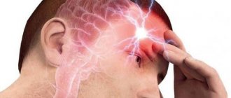
- Severe and frequent headaches
- Stroke
- Epileptic seizures
- Head injuries
- Pain and discomfort in the eyes and ears
- Dizziness
MRI of the brain is used to identify or confirm a number of diseases and disorders associated with the functioning of the brain, circulatory system, and nervous system, including chronic ones. These are diseases such as:
- Epilepsy
- Multiple sclerosis
- Oncology
- Hemorrhage
- Aneurysm
- Eye and/or ear diseases
- Infection
- Cyst
- Arterial obstruction or venous thrombosis
- Congenital defects in the structure of the brain
- Hydrocephalus
- Chronic diseases of the nervous system
- Intracranial hypertension
The doctor may order an MRI before surgery to determine the condition of the tissues and cerebral cortex and localize the lesion due to injury or disease. A tomogram helps to identify not only tumors or recently emerging brain problems, but also chronic ailments.
The procedure is completely safe, so not only adults, but also children are prescribed MRI of the brain.
When is a brain MRI necessary for a child?
Indications for examining a child are as follows:
- Tumor recognition
- The need to identify pathologies and disorders of the brain structure
- Study of the cerebral cortex and blood vessels
- Examination of his sinuses and ventricles
It must be remembered that a doctor should prescribe an MRI of the brain, since he is the one who knows whether the procedure is necessary and what nuances of the examination should be taken into account. Therefore, if the patient independently wants to obtain such a study, the results may be less accurate than if the process is supervised by an experienced physician.
Preliminary preparation
MRI of the brain requires preparation. First of all, it is necessary to inform the doctor about the presence of implants, because with some of them it is impossible to undergo an MRI of the brain, despite the indications for the procedure. The doctor should also be warned if the patient has claustrophobia (fear of closed spaces). Then the study can be carried out under sedation (putting the patient into medicated sleep). A pregnant woman needs to warn doctors about this, as there are some restrictions.
Before the MRI, you can take medications that the patient usually uses. No special diet is required, except in individual cases determined by the doctor.
Preparation in the MRI room
If there are no contraindications, immediately before the brain MRI procedure, preparation is minimal. You should:
- Remove all metal and magnetic objects, including jewelry, hairpins, etc.
- Remove plastic cards, phones, keys, etc. from your pockets
- Remove clothing with metal elements
- Remove those prostheses that are possible: hearing aids, dentures
Features of preparation for the procedure with the introduction of contrast
Separately, preparation for MRI of the brain is required if the patient is undergoing an MRI with the introduction of a contrast agent. This is required if it is necessary to obtain even more accurate analysis results. A special gadolinium-based substance is injected into the patient through a catheter. It makes the human body more sensitive to magnetic resonance.
In this case, it is necessary to consult a doctor - an allergist-immunologist, in order to exclude possible allergic reactions to the administered medicine.
In what cases is anesthesia required?
Young children cannot remain still and control themselves, so an MRI of the child's brain may be performed under sedation. An older child who is not afraid and understands the importance of the procedure will be able to undergo it independently.
Thus, examination under sedation can be carried out in the following cases:
- the patient has claustrophobia
- a small child is being examined
- possible panic attacks
- severe pain does not allow lying still
- have mental disorders
- antisocial behavior of the patient
If you require a study under sedation, you must notify the administrator when registering. He will provide up-to-date information about the centers where this service is available on the day of registration.
When the basic preparations are completed, the patient is placed on a sliding table and then placed in the tomograph chamber. The tomograph is a cylinder around which there is a magnet. The latest machines allow you to scan the brain in 15 minutes. After the examination, the patient will receive a disk or image with a transcript.
Strict contraindications
The magnetic field is harmless to humans, unlike X-ray radiation. Therefore, most of the contraindications for MRI of the brain apply to patients who have any metal implants. This is due to the nature of the procedure: it is carried out using a magnet, which can attract or distort the operation of the mechanism. Dangerous items include:
- pacemakers
- defibrillators
- ear implants
- metal prostheses
- clips on vessels
Relative contraindications
Other items are not prohibited during the procedure, but may distort the results. Here they are:
- nerve stimulants
- non-metallic prostheses
- staples, plates, pins
- artificial heart valves
- dental implants
- braces
Who is not recommended to undergo an MRI of the brain?
It is not recommended to undergo MRI if the patient
- has a body diameter greater than 150 cm
- overstimulated and unable to remain still
It must be remembered that MRI is used with caution for pregnant women and is not recommended in the first trimester.
Are there possible complications with an MRI of the brain?
As a rule, there are no complications when undergoing MRI of the brain. But strange sensations may arise. If the study is carried out using a contrast agent, then during its administration such unpleasant reactions of the body as:
- pain at the injection site
- nausea
- rush of blood
- allergic reaction
The first three symptoms usually pass quickly, and in the latter case it is necessary to immediately inform the doctor about the deviations so that he can take the necessary measures.
You may feel warm during the scan. If this bothers the patient greatly, he needs to tell the doctor about it.
In rare cases, after undergoing an MRI of the brain, the patient may feel unwell. Therefore, if the following symptoms occur, you should immediately report them to your doctor:
- headache
- dizziness
- vision problems
We invite you to undergo an MRI of the brain at MEDSI!
At MEDSI clinical diagnostic centers you can undergo an MRI of the brain quickly and painlessly.
MRI at MEDSI is:
- Urgent examinations in case of injury or sudden illness
- Performing tomography under sedation (for those suffering from claustrophobia and panic attacks)
- Equipment from well-known manufacturers (Siemens, Philips)
- Research for adults and children
- Interpretation of results exclusively by qualified radiologists
Who needs to get scanned?
Numerous signs indicate a violation of cerebral circulation, and if they appear, you should immediately go to the doctor. You can go for an MRI without his referral, but you will have to consult with a radiologist to rule out possible contraindications. You may be sent for a scan if you have the following symptoms:
- recurring headaches;
- fainting and dizziness that cannot be explained by other reasons;
- signs of vascular stenosis or aneurysm formation;
- post-stroke condition;
- inflammatory diseases of the central nervous system (encephalopathy, meningitis, etc.);
- loss of vision or hearing not related to the functioning of the organs themselves;
- Previous head injuries and their long-term consequences;
- Suspicions of the presence of tumors, germination of metastases from other areas with previously diagnosed oncology.
A study with repeated procedures is prescribed for monitoring Alzheimer's disease, Parkinson's disease, monitoring inoperable benign neoplasms, delayed psychodevelopment, stuttering, development of hydrocephalus, as well as inappropriate behavior, if the patient has not previously observed brain pathologies. The appearance of sudden seizures is also a direct indication for tomography. If, despite persistent symptoms, no brain disease is detected, it is recommended to simultaneously undergo an MRI of the neck vessels.
When is an MRI of the head and neck prescribed?
Magnetic resonance imaging of the head and neck is used in the diagnosis of the following diseases:
- Primary headaches (cluster headache, tension headache, migraine) as a method to exclude organic brain damage;
- Inflammatory diseases of the central nervous system (meningitis, encephalitis);
- Volumetric formations of the brain (cysts, abscesses, tumors);
- Metastases to the brain, cervical vertebrae;
- Vascular diseases (atherosclerosis, aneurysms, malformations, vertebral artery syndrome);
- Strokes;
- Demyelinating diseases (multiple sclerosis), other autoimmune pathologies of the central nervous system;
- Degenerative-dystrophic processes in the cervical spine;
- Herniated intervertebral discs in the cervical spine;
- Neck injuries;
- Diseases of the thyroid gland (cysts, tumors, nodes);
- Other pathologies of the head and neck.
Symptoms for which it is recommended not to delay an MRI examination of the head and neck:
- Headache;
- Pain in the neck;
- Dizziness, tinnitus;
- Unsteadiness of gait;
- Decreased hearing, vision;
- Deterioration of memory, ability to concentrate;
- High blood pressure;
- Convulsive syndrome;
- Feeling of crawling, numbness and burning of the skin;
- Impaired tactile sensitivity;
- Impaired or limited mobility of the spine in the cervical region, crunching when rotating, tilting the head;
- Enlarged lymph nodes, foreign tumors, lumps in the neck (thyroid gland) and head, which are determined visually or by touch.
Restrictions
Screening using magnetic resonance equipment may be refused for the following reasons:
- The presence of metal implants built into living tissues that respond to a powerful magnetic field.
- The initial stages of pregnancy are up to the 13th week, when any external influence on the actively developing fetus is undesirable.
- An immune reaction to contrast agents has been previously documented.
- There are kidney diseases that affect urinary retention.
- Exacerbation of chronic diseases of the respiratory system.
- Mental abnormalities and claustrophobia, which does not allow one to remain motionless in a confined space for a long time.
Many of the listed contraindications can be overcome if you undergo the study without contrast, use open equipment, or consider general anesthesia during the recording of the tomogram. If the restrictions are temporary, it is better to postpone the procedure to a later date (as during pregnancy). If this cannot be done, it is better to choose an alternative, the best of which is CT angiography of cerebral vessels.
Contraindications for MR imaging of the head and neck
Magnetic resonance imaging has a minimum of contraindications - this is one of the significant advantages of the method. Almost all of them are related to the fact that during the procedure the equipment generates high-intensity magnetic fields.
MR imaging is contraindicated:
- Patients with electrically active implants - defibrillators, neurostimulators, heart pacemakers, cochlear implants, insulin pumps (due to the risk of breakdown and failure, causing harm to health);
- Patients with metal foreign bodies - metal particles in the eyeballs (in people involved in metalworking), stents and vascular clips made of stainless steel, fragments, shrapnel and shot (due to the risk of displacement and bleeding);
- Pregnant women during the 1st trimester (due to the fact that the effect of magnetic fields on the developing fetus has not been studied for ethical reasons, but indirect evidence suggests that the method is absolutely safe during pregnancy);
- Children under 5 years of age (young patients cannot lie still during the examination, which is a prerequisite for obtaining clear images);
- Persons with excess body weight of more than 130 kg and a torso circumference of more than 150 cm (due to the fact that MRI equipment has limited dimensions and is not suitable for examination in obese patients).
Diagnostic cost
Every year, prices for magnetic resonance scanning become more affordable, as the number of clinics offering this service grows. Our service contains all verified tomography centers, distributed by region, ratings, reviews and other parameters. Starting prices for the examination are posted in the table; more accurate data is available via links to price lists of specific organizations. Go to the main page of the portal, indicate the name of the procedure and choose the best clinics based on your own request.
| Name of procedure | Prices, rub. |
| MRI of the brain | 2500 |
| MR angiography of cerebral vessels | 2500 |
| MR angiography of the vessels of the head with contrast | 2500 |
| MR angiography of neck vessels | 3600 |
| CT angiography of cerebral vessels | 3600 |
What does an MRI of the head and neck show?
Each MRI image of the head and cervical spine is a layer-by-layer image of the area under study, taken in several planes. All anatomical structures are clearly visible on it - the brain and spinal cord, nerves, blood vessels, muscles, joints, etc. The method allows you to take intravital photographs of the human body with high clarity and the smallest details.
When interpreting the images, the radiologist pays attention to the slightest signs of the disease. MRI perfectly “sees” inflammation, swelling, circulatory disorders, tumors and other typical pathological processes. This is the only method today that allows you to diagnose almost all diseases of the head and cervical spine at one time.
Registration for examination
To book the type of study you need, contact the service advisory service at the number at the top of the page. Operators will select the required options, answer remaining questions, provide information on addresses and costs, and reserve an MRI or CT session with special discounts for portal users. Consultation and other services of the recording service are absolutely free.
Sources used:
- Belova, L.A. Venous cerebral discirculation in chronic cerebral ischemia: clinical picture, diagnosis, treatment / L.A. Belova // Neurological Bulletin named after. V.M. Bekhtereva – .2010. – T. 42, No. 2. – P. 62–67.
- Bozhko, O.V. The role of neuroimaging methods in the early detection of Alzheimer's disease / O.V. Bozhko, S.I. Gavrilova, Ya.B. Fedorova // “Psychiatry”. – 2006. – No. 2 – P.54–59.
- Alzheimer's disease: textbook. allowance / A.Yu. Emelin et al. – St. Petersburg, 2016. – 76 p.
- Varakin, Yu.Ya. Prevention of cerebrovascular accidents / in the book. Essays on angioneurology // Yu.Ya. Varakin; edited by BEHIND. Suslina. – M., 2005 – 368 p.
- Vasenina, E.E. Primary progressive aphasia / E.E. Vasenina, O.S. Levin // Journal of Neurology and Psychiatry. – 2014. –T. 6, No. 2. – P. 3–12.
- Venous dysgemia and cognitive deficit in patients with dyscirculatory encephalopathy / S.V. Lobzin et al. // Vestn. Northwestern State Medical. University named after I.I. Mechnikov – .2013. – T.5, No. 2. – pp. 12–18.
List of references on the topic:
- Modern methods of diagnosis, treatment and prevention of diseases: collection. instructional method. doc.: in 7 volumes / Ministry of Health Rep. Belarus. - Vol. 9. - Minsk: RNMB. – 2008.T. 7: Oncology. Radiology and medical radiology. — 160 s.
- Gusev E.I., Nikiforov A.S., Konovalov A.N. Nervous diseases, neurosurgery. M., 2001.
- Shtulman D.R., Levin O.S. handbook of a practical physician on neurology. - M., 1999.
- Diseases of the nervous system. Guide for doctors / Ed. N.N. Yakhno.
The text was checked by the doctor Mikhail Mikhailovich Motov
Therapist