Not to be confused with red blood cell nucleus.
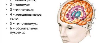
Structure in the human brain
| Red core | |
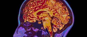
| Cross section through the midbrain, showing the location of the red nuclei. In the improved colliculus, the quadrigeminals are at the top of the image and the cerebral peduncles are at the bottom of the image - as in section. | |
| Details | |
| Part | Midbrain |
| Identifiers | |
| Latin | ruber core |
| MeSH | D012012 |
| NeuroNames | 505 |
| NeuroLex ID | birnlex_1478 |
| TA98 | A14.1.06.323 |
| TA2 | 5898 |
| F.M.A. | 62407 |
| Anatomical terms of neuroanatomy [edit in Wikidata] | |
Red core
or
the nucleus of the nucleus
is a structure in the rostral brain involved in the coordination of movements. [1] The red core is pale pink in color, which is believed to be due to the presence of iron in at least two different forms: hemoglobin and ferritin. [2] [3] This structure is located in the covering of the midbrain next to the substantia nigra and includes the caudal magnocellular and rostral parvocellular components. [1] The red nucleus and substantia nigra are subcortical centers of the extrapyramidal motor system.
Function [edit]
In a vertebrate without a significant corticointestinal tract, gait is controlled primarily by the red nucleus. [4] However, in primates, in which the corticospinal tract is dominant, the rubrospinal tract may be considered vestigial in terms of motor function. Therefore, the red nucleus is less important in primates than in many other mammals. [1] [5] However, the crawling of infants is controlled by the red nucleus, just as the arm swings in a typical walk. [6] The red nucleus may play an additional role in controlling the muscles of the shoulder and shoulder through projections of its intercellular part. [7] [8] In humans, the red nucleus also has limited control over the arms, as the rubrospinal tract is more involved in the movement of large muscles such as the arms (but not the legs, as the tract ends in the upper thoracic spinal cord). Fine control of the fingers does not depend on the functioning of the red nucleus, but depends on the corticospinal tract. [9] Most axons of the red nucleus do not project to the spinal cord, but through its parvocellular part transmit information from the motor cortex to the cerebellum through the inferior olivary complex, an important relay center in the medulla. [1]
Pathologies in violation
It all started when science received a description of severe muscle tension in animals. Tension was created by breaking the connections of the red core. This break is called decerebrate rigidity. Based on this observation, a conclusion was made, the essence of which is that when the connection between the red and vestibular nuclei is lost, severe tension occurs in the skeletal muscles, muscles of the limbs, as well as the muscles of the neck and back.
The above muscles are distinguished by their ability to counteract gravity, so it was concluded that this development of events is associated with the vestibular system. As it turned out later, the vestibular nucleus of Deiters is capable of triggering the work of extensor motor neurons. The activity of these neurons is significantly slowed down under the influence of the red nuclei and Deiters nucleus.
It turns out that active muscle work is the result of the joint work of the entire complex. In humans, decerebrate rigidity occurs as a result of traumatic brain injury. You can also encounter this phenomenon after a stroke. It should be understood that such a condition is a bad sign. You can find out about its presence by the following signs:
- arms straight, spread in different directions;
- hands lie palms up;
- all fingers are clenched except the thumbs;
- legs extended and folded together;
- feet extended;
- toes curled;
- the jaws press tightly against each other.
In case of injuries, severe infectious diseases, all kinds of internal damage to organs, including the brain, as well as tumor processes and aggression of the immune system - all this leads to disruption of the brain. Thus, if connections with the red nuclei are disrupted, decerebrate rigidity may occur, as well as disruption of the eyeball and eyelid muscles, the latter being an easier reaction of the body to the rupture of connections.
Input and output [edit]
The red nucleus receives many inputs from the cerebellum (nucleus intermedius and lateral cerebellar nucleus) opposite
side and input signals from the motor cortex of the
same
side. [10]
The red nucleus has two sets of efferents: [10]
- In humans, most of the output goes into a bundle of fibers that passes through the medial tegmental area to the inferior olive on the same side
, forming part of a pathway that ultimately affects the cerebellum. - The other exit (the rubrospinal projection) goes to the rhombencephalic reticular formation and the spinal cord on the opposite
side, constituting the rubrospinal tract, which runs ventrally to the lateral corticospinal tract. As stated earlier, the rubrospinal tract is more important in non-primates: in primates, due to the well-developed cerebral cortex, the corticospinal tract has taken over the role of the rubrospinal tract.
Age characteristics and prevention
The brain is a complex structure. It operates with close interaction between all segments. The center that controls the middle section is the cerebral cortex. With age, connections become weaker, and reflex activity weakens. Since the area is responsible for motor function, even minor disruptions in this tiny segment lead to the loss of this important ability. It is more difficult for a person to move, and serious disorders lead to diseases of the nervous system and complete paralysis. How to prevent disorders in the functioning of the brain in order to remain healthy until old age?
First of all, you should avoid hitting your head. If this happens, it is necessary to begin treatment immediately after the injury. It is possible to preserve the functions of the midbrain and the entire organ until old age if you train it with regular exercises:
- The lifestyle a person leads is important for physical and mental health. Drinking alcohol and smoking destroy neurons, which gradually leads to a decrease in mental and reflex activity. Therefore, you should give up bad habits, and the sooner you do this, the better.
- Moderate physical activity and walks in nature supply the brain with oxygen, which has a beneficial effect on its activity.
- You shouldn’t give up reading, solving charades and puzzles: intellectual activity keeps the brain active.
- An important aspect of the functioning of brain structures is nutrition: fiber, protein, and greens must be present in the diet. The midbrain responds positively to intake of antioxidants and vitamin C.
- It is necessary to control blood pressure: the health of the vascular system affects the general condition of a person.
The brain is a flexible system that can be successfully developed. Therefore, by constantly improving your mind and body, you can maintain clarity of thoughts and motor activity until old age.
Additional images[edit]
- Schematic representation of the main ganglion categories (I to V).
- Deep dissection of the brain stem. View from below.
- Cross section of the midbrain.
- Cross section of the midbrain at the level of the superior colliculi.
- The coronal region of the brain is immediately in front of the pons.
- Frontal (coronal) section of the human brain
- Red core
- Brain stem, optic fissure, cerebral aqueduct. The lowest kind. Deep dissection.
- Cerebrum. Bottom view. Deep dissection
Midbrain nuclei
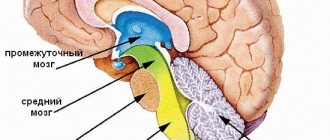
In the midbrain, gray matter is located in the form of a concentration of nerve cells, forming the nerve nuclei of the skull:
- The nuclei of the oculomotor nerve are located in the tegmentum, closer to the middle, ventral to the aqueduct. They form a layered structure and participate in the occurrence of reflexes and visual reactions in response to signals. Also, during the formation of visual stimuli, the nuclei control the movement of the eyes, body, head and facial expressions. The system complex includes the main nucleus, consisting of large cells, and small cell nuclei (central and outer).
- The nucleus of the trochlear nerve consists of paired elements and is located in the tegmental segment in the region of the inferior colliculi directly under the aqueduct. It is represented by a homogeneous mass of large isodiametric cells. Neurons are responsible for hearing and complex reflexes; with their help, a person reacts to sound stimuli.
- The reticular formation is represented by a cluster of reticular nuclei and a network of neurons, located in the thickness of the gray matter. In addition to the middle center, it includes the diencephalon and medulla oblongata; the formation is connected with all parts of the central nervous system. It affects motor activity, endocrine processes, affects behavior, attention, memory, inhibition.
Links[edit]
- ^ abcd Cacciola, Alberto; Milardi, Demetrio; Basile, Gianpaolo Antonio; Bertino, Salvatore; Calamuneri, Alessandro; Chillemi, Gaetana; Paladina, Giuseppe; Impellizzeri, Federica; Trimarchi, Fabio; Anastasi, Giuseppe; Bramanti, Alessia; Rizzo, Giuseppina (08/20/2019). "The corticorubral and cerebellar-rural tracts are topographically organized in the human red nucleus". Scientific reports
.
9
(1):12117. DOI:10.1038/s41598-019-48164-7. ISSN 2045-2322. PMC 6702172. PMID 31431648. - Wikipedia Red Nucleus revision https://sites.google.com/site/childrenoftheamphioxus/table-of-contents/wikipedia-red-nucleus-revision
- Drayer, B.; Burger, P.; Darwin, R.; Riederer, S.; Herfkens, R.; Johnson, Georgia (July 1986). "MRI of the brain gland." A.J.R. American Journal of Radiology
.
147
(1):103–110. DOI: 10.2214/ajr.147.1.103. ISSN 0361-803X. PMID 3487201. - ten Donkelaar, H. J. (1988-04-01). "Evolution of the red nucleus and rubrospinal tract". Behavioral Brain Research
.
28
(1): 9–20. DOI: 10.1016/0166-4328 (88) 90072-1. ISSN 0166-4328. PMID 3289562. S2CID 54367413. - Onodera, Satoru; Hicks, T. Philip (13 August 2009). "A comparative neuroanatomical study of the red nucleus of cat, macaque and human". PLOS ONE
.
4
(8): –6623. DOI: 10.1371/journal.pone.0006623. ISSN 1932-6203. PMID 19675676. S2CID 18760268. - Being and Perception. Manupod Press. 2011. p. 49. ISBN 978-0-9569621-0-2.
- Gibson, AR; Houk, J. C.; Colerman, N. J. (1985). "Magnocellular activity of the red nucleus during different types of limb movements in the macaque monkey". Journal of Physiology
.
358
(1):527–549. DOI: 10.1113/jphysiol.1985.sp015565. ISSN 1469-7793. PMC 1193356. PMID 3981472. - Van Kan, Peter L.; McCurdy, Martha L. (01/01/2002). "Discharge of primate magnocellular red nucleus neurons during grasping at different spatial positions." Experimental Brain Research
.
142
(1):151–157. DOI: 10.1007/s00221-001-0924-5. ISSN 1432-1106. PMID 11797092. S2CID 42113292. - Bunday, Karen L.; Tazoe, Toshiki; Rothwell, John S.; Perez, Monica A. (May 21, 2014). "Subcortical control of precision grip after human spinal cord injury". Journal of Neuroscience
.
34
(21):7341–7350. DOI: 10.1523/JNEUROSCI.0390-14.2014. ISSN 1529-2401. PMC 4028504. PMID 24849366. Retrieved August 30, 2020. - ^ ab Milardi, Demetrio; Cacciola, Alberto; Cutroneo, Giuseppina; Marino, Sylvia; Irrera, Mariangela; Cacciola, Giorgio; Santoro, Giuseppe; Ciolli, Pietro; Anastasi, Giuseppe; Calabro, Rocco Salvatore; Quartarone, Angelo (28 July 2016). "Red core connectivity as revealed by constrained spherical deconvolution tractography". Neuroscience Letters
.
626
: 68–73. DOI: 10.1016/j.neulet.2016.05.009. ISSN 0304-3940. PMID 27181514. S2CID 46756208. Retrieved August 30, 2021.
Red core in Latin
Midbrain, mesencephalon,
develops in the process of phylogenesis under the predominant influence of the visual receptor, therefore its most important formations are related to the innervation of the eye. Hearing centers were also formed here, which, together with the vision centers, later grew in the form of four mounds of the roof of the midbrain.
With the appearance in higher animals and humans of the cortical end of the auditory and visual analyzers in the forebrain cortex, the auditory and visual centers of the midbrain themselves fell into a subordinate position and became intermediate, subcortical. With the development of the forebrain in higher mammals and humans, pathways began to pass through the midbrain, connecting the telencephalon cortex with the spinal cord (cerebral peduncles).
As a result, the human midbrain contains:
1) subcortical centers of vision and nuclei of nerves innervating the muscles of the eye; 2) subcortical auditory centers; 3) all ascending and descending pathways connecting the cerebral cortex with the spinal cord and passing through the midbrain; 4) bundles of white matter connecting the midbrain with other parts of the central nervous system.
Accordingly, the midbrain
, which is the smallest and most simply structured part of the brain in humans, has two main parts: the roof, where the subcortical centers of hearing and vision are located, and the cerebral peduncles, where the pathways predominantly pass.
Dorsal part, roof of the midbrain, tectum mesencephali.
It is hidden under the posterior end of the corpus callosum and is divided by two criss-crossing grooves - longitudinal and transverse - into four colliculi, arranged in pairs
.
The upper two colliculi, colliculi superiores
, are subcortical centers of vision, both lower,
colliculi inferiores
, are subcortical centers of hearing.
The pineal body lies in a flat groove between the superior tubercles. Each colliculus
passes into the so-called
handle of the colliculi, brachium colliculi
, which goes laterally, anteriorly and upward, to the diencephalon.
The handle of the superior colliculus, brachium colliculi superioris
, goes under
the pillow, pulvinar
, of the thalamus to
the lateral geniculate body, corpus geniculatum laterale.
Handle of the inferior colliculus, brachium colliculi inferioris
, passing along the upper edge of the trigonum lemnisci to the sulcus lateralis mesencephali, disappears under
the medial geniculate body, corpus geniculatum mediale
. The named geniculate bodies already belong to the diencephalon.
Ventral part, cerebral peduncles, pedunculi cerebri
, contains all the pathways to the forebrain.
Brain stems
They look like two thick semi-cylindrical white cords that diverge from the edge of the bridge at an angle and plunge into the thickness of the cerebral hemispheres.
Midbrain cavity
which is a remnant of the primary cavity of the middle cerebral bladder, looks
like a narrow canal
and is called
the cerebral aqueduct, aqueductus cerebri
.
It is a narrow, ependymal-lined canal 1.5 - 2.0 cm long, connecting the IV ventricle with the III. Dorsally, the aqueduct
is limited by the roof of the midbrain,
ventrally
by the tegmentum of the cerebral peduncles.
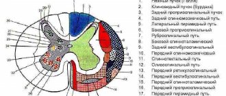
In a cross section of the midbrain, three main parts are distinguished:
1) roof plate, lamina tecti; 2) tire, tegmentum, representing the upper section of the pedunculi cerebri; 3) the ventral section of the pedunculi cerebri, or the base of the cerebral peduncle, basis pedunculi cerebralis.
According to the development of the midbrain under the influence of the visual receptor, it contains various nuclei related to the innervation of the eye.
In lower vertebrates, the superior colliculus
serves as the main ending point of the optic nerve and is the main visual center.
In mammals and in humans, with the transfer of visual centers to the forebrain, the remaining connection of the optic nerve with the superior colliculus is important only for reflexes. of the auditory loop (lemniscus lateralis)
end in the nucleus of the inferior colliculus, as well as in the medial geniculate body .
The roof of the midbrain has a bilateral connection with the spinal cord - tractus spinotectalis and tractus tectobulbaris et tectospinalis
. The latter, after crossing in the tegmentum, go to the muscle nuclei in the medulla oblongata and spinal cord. This is the so-called visual-sound reflex pathway, which was mentioned when describing the spinal cord. Thus, the plate of the roof of the midbrain can be considered as a reflex center for various types of movements that arise mainly under the influence of visual and auditory stimuli.
Brain plumbing
surrounded by central gray matter, which in its function is related to the autonomic system.
In it, under the ventral wall of the aqueduct, in the tegmentum of the cerebral peduncle, the nuclei of two motor cranial nerves are located - n.
oculomotorius (III pair) at the level of the superior colliculus and
n.
trochlearis (IV pair) at the level of the inferior colliculus. The nucleus of the oculomotor nerve consists of several sections, corresponding to the innervation of several muscles of the eyeball.
Medial and posterior to it is another small, also paired, vegetative accessory nucleus, nucleus accessories.
, and
unpaired median nucleus
.
The accessory nucleus and the unpaired median nucleus innervate the involuntary muscles of the eye, m.
ciliaris and m. sphincter pupillae . This part of the oculomotor nerve belongs to the parasympathetic system. Above (rostral) the nucleus of the oculomotor nerve in the tegmentum of the cerebral peduncle is the nucleus of the medial longitudinal fasciculus.
Lateral to the cerebral aqueduct is the nucleus of the midbrain tract of the trigeminal nerve, nucleus mesencephalicus n. trigemini
.
The cerebral peduncles divide
, as already noted, on the ventral part, or
base of the cerebral peduncle, basis pedunculi cerebralis
, and
tegmentum, tegmentum
.
The border between them is the black substance, substantia nigra
, which owes its color to
the black pigment contained in its constituent nerve cells - melanin
.
Midbrain tegmentum, tegmentum mesencephali,
- part of the midbrain located between its roof and the black substance (substantia nigra) of the cerebral peduncles.
The tractus tegmentalis centralis - the central tegmental tract - departs from it
- projection descending nerve pathway located in the central part of the midbrain tegmentum. It contains fibers coming from the thalamus, globus pallidus, red nucleus and reticular formation of the midbrain to the reticular formation and olive of the medulla oblongata; belongs to the extrapyramidal system.
Substantia nigra
extends along the entire length of the cerebral peduncle from the pons to the diencephalon; according to its function it belongs to the extrapyramidal system.
Located ventrally from the substantia nigra
the base of the cerebral peduncle contains longitudinal nerve fibers descending from the cerebral cortex to all underlying parts of the central nervous system (tractus corticopontmus, corticonuclearis, corticospinalis, etc.).
Tegmentum
, located dorsal to
the substantia nigra
, contains predominantly ascending fibers, including the medial and lateral lemniscus. As part of these loops, all sensory pathways ascend to the cerebrum, with the exception of the visual and olfactory ones.
Among the nuclei of gray matter
the most significant is
the red nucleus, nucleus ruber
.
This elongated sausage-shaped formation extends in the tegmentum of the cerebral peduncle from the hypothalamus of the diencephalon to the inferior colliculus, where it begins an important descending tract, tractus rubrospinal
, connecting
the red nucleus
with the anterior horns of the spinal cord.
This bundle, after leaving the red nucleus
, intersects with a similar bundle of the opposite side in the ventral part of the median suture - the ventral decussation of the tegmentum.
Nucleus ruber
is a very important coordination center of the extrapyramidal system, connected with its other parts.
Fibers pass to it from the cerebellum as part of the upper peduncles of the latter after their decussation under the roof of the midbrain, ventrally from the aqueductus cerebri
, as well as from
the pallidum
- the lowest and most ancient of the subcortical nodes of the brain that are part of the extrapyramidal system. Thanks to these connections, the cerebellum and the extrapyramidal system, through the red nucleus and the tractus rubrospinal extending from it, influence the entire skeletal muscle in the sense of regulating unconscious automatic movements.
The reticular formation, formatio reticularis, also continues into the tegmentum of the midbrain.
, and
fasciculus longitudindlis medialis
. The latter originates in various places. One of its parts begins from the vestibular nuclei, passes on both sides along the sides of the midline, directly under the gray matter of the bottom of the aqueduct and the IV ventricle, and consists of ascending and descending fibers going to the nuclei of the III, IV, VI and XI cranial nerves .
Medial longitudinal fasciculus
is an important associative pathway connecting the various nuclei of the nerves of the eye muscles with each other, which determines the combined movements of the eyes when they are deviated in one direction or another. Its function is also associated with eye and head movements that occur when the balance apparatus is stimulated.
Basic information
The central nervous system contains neurons with processes and glia. The brain has only five sections. First
– oblong – continuation of the dorsal.
It transmits information to and from other departments. Performs a regulatory function in relation to the coordination of movements. The second
is the bridge - here are the midbrain centers responsible for the assimilation of audio and video information.
This department stands for coordination of movements. The third
- the cerebellum - connects the posterior and anterior sections.
The fourth
- middle - is responsible for facial expressions, movements of the eyeballs, and the auditory pathways pass through it.
This is exactly what we will consider. The fifth
- the front - normalizes mental activity.
This is interesting. There is no connection between brain size and mental abilities in humans. The number of nerve connections is much more important.
What role does it play?
It is the middle section that regulates muscle tone. His role corresponds to his intermediate position. Due to the fact that the midbrain has a special structure, its functions include the transmission of information. It has a lot of different purposes:
- sensory
– to convey tactile sensations; - motor
– coordination depends on this part of the midbrain; - reflex
- for example, oculomotor, reaction to light and sound.
Due to the work of the middle section, a person can stand and walk. Without it, a person would not be able to fully move in space. Also, the work of the vestibular apparatus is controlled at the level of the midbrain.
How many parts does the middle section have?
There are three parts in total.
Dorsal - the roof of the middle section. It is divided into 4 mounds using grooves intersecting in pairs. The two upper hills are the subcortical centers for regulating vision, and the remaining lower ones are auditory. The ventral one is the so-called cerebral peduncle. The conductive channels to the anterior section are based here. The internal space of the brain looks like a hollow canal.
Helpful information.
If a person does not breathe oxygen for more than five minutes, the brain will be permanently damaged, resulting in death.