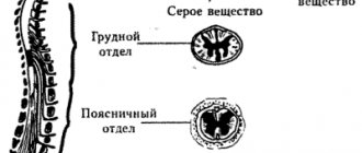
The spine is a complex system consisting not only of bone tissue, but also of the delicate spinal cord trunk. It is thanks to him that a person is able to live a full life, feel the touch of objects, and distinguish them from each other. It consists of a huge number of nerve fibers and unique pathways along which impulses move. The anatomy of the spinal cord is distinguished by its high organization, since millions of signals coming from receptors in the periphery constantly pass through this organ.
Brief Definition
The spinal cord pathways or tracts are collections of nerve fibers located inside the spine that carry impulses from the brain to all parts of the body and back. The nerve endings, the combination of which form the pathways, are distinguished by a similar structure, development and general functions. They are divided among themselves according to the tasks assigned to them. The paths are classified as follows:
- Associative. Their main purpose is to unite gray matter cells from different segments to form their own anterior, lateral or posterior bundles.
- Commissural. These fibers connect the gray matter from the two hemispheres. With their help, the coordinated work of individual areas, nerve centers, and both hemispheres occurs.
- Projection. With the help of such pathways, the work of the overlying and underlying parts of the brain is combined. They are the ones who provide the projection of pictures of the surrounding world, as on a monitor screen.
Projection pathways, in turn, are efferent and afferent. They form the basis of the central nervous system, and are divided into ascending (centripetal or sensitive) and descending (centrifugal, motor).
Important! Nerve fibers provide a constant, unbreakable connection to the brain located in the skull and spine. It is thanks to them that the impulse is quickly transmitted, all body movements are coordinated with each other.
The pathways of the brain and spinal cord differ from each other, but they always act harmoniously, ensuring the passage of an incredibly large number of nerve signals from receptors to the central nervous system. Paths are formed from long axons, special fibers that are capable of creating connections among themselves, thus connecting individual segments of the spinal trunk, providing control of effector organs.
Spinal cord pathways
The spinal cord pathways or tracts are collections of nerve fibers located inside the spine that carry impulses from the brain to all parts of the body and back. The nerve endings, the combination of which form the pathways, are distinguished by a similar structure, development and general functions. They are divided among themselves according to the tasks assigned to them. The paths are classified as follows:
· Associative . Their main purpose is to unite gray matter cells from different segments to form their own anterior, lateral or posterior bundles.
· Commissural . These fibers connect the gray matter from the two hemispheres. With their help, the coordinated work of individual areas, nerve centers, and both hemispheres occurs.
· Projection . With the help of such pathways, the work of the overlying and underlying parts of the brain is combined. They are the ones who provide the projection of pictures of the surrounding world, as on a monitor screen.
Projection pathways, in turn, are efferent and afferent. They form the basis of the central nervous system, and are divided into ascending (centripetal or sensitive) and descending (centrifugal, motor).
Important! Nerve fibers provide a constant, unbreakable connection to the brain located in the skull and spine. It is thanks to them that the impulse is quickly transmitted, all body movements are coordinated with each other.
The pathways of the brain and spinal cord differ from each other, but they always act harmoniously, ensuring the passage of an incredibly large number of nerve signals from receptors to the central nervous system. Paths are formed from long axons, special fibers that are capable of creating connections among themselves, thus connecting individual segments of the spinal trunk, providing control of effector organs.
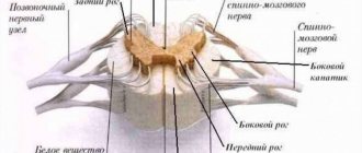
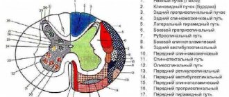
All tracts of the spinal cord are located in the white matter, which is divided into the anterior, lateral and anterior cord. Their main volume consists of supraspinal tracts, which provide two-way communication between the spinal region and the head organ. These stripes occupy little space around the gray matter and are called propriospinal.
The pathways of the spinal and head sections are divided conditionally, depending on the characteristics of their structure and functionality. They are an integral part of the spine as a whole, and allow you to control not only the motor activity of the body, but also the functioning of the internal organs. They are located outside the main brain bundles. They develop in parallel with the formation of the head section.
Important! When the neurons carrying the impulses begin to die, conduction may stop completely, leading to loss of sensation in the limbs or paralysis.
RISING PATHS
The ascending tracts of the spinal cord are responsible for transporting pain impulses, tactile sensations, information about body temperature, and sensitivity from receptors to the cerebellum. That is, their main feature is the movement of flow from the periphery to the center. It is thanks to them that a person understands what is happening to his body at a given second of time, processes constantly incoming information from the outside world, and makes decisions in a timely manner based on the impulses received. The table will tell you more about the varieties of this type of path and their main tasks.
| Name of paths | Location | Their main tasks |
| Thin beam (Gaull beam) | Rear pillar | This is the basis of the ascending tracts, as they run along the entire spinal trunk. Impulses from it are directed to the cerebral cortex. With their help, conscious impulses are transmitted from muscle receptors to the “center”. |
| Wedge-shaped fasciculus (Burdach's path) | Rear pillar | Nerve currents are directed to the cortex. The pathways are responsible for transmitting impulses from the musculoskeletal system. |
| Posterior spinocerebellar tract (Flexig's tract) | Dorsal | Responsible for the transmission of unconscious nerve currents from proprioceptors of muscle fibers, ligaments, tendons to the cerebellum. |
| Anterior spinocerebellar fasciculus (Gowers' tract) | More ventral | As in the previous case, it is responsible for transporting currents from muscles, ligaments and tendons to the cerebellum. Impulses are transmitted unconsciously. |
| Lateral spinothalamic tract | They are responsible for the sensation of temperature changes and pain, since impulses are carried out precisely according to them. | |
| Anterior spinothalamic tract | Responsible for transmitting nerve currents about tactile sensations, pressure, touch and other things. |
The ascending tracts of the spinal trunk are generally responsible for transmitting any incoming information to the articular receptors of the body. Thanks to them, a person understands the position of his body, is aware of tactile sensations, passive movements performed, and feels vibration.
DESCENT ROUTES
Descending pathways are responsible for the movement of currents from underlying sections to working systems. In general, they are divided into pyramidal and extrapyramidal. The first ones are responsible for transmitting impulses of voluntary motor reactions, namely the control of conscious movements, the second ones control involuntary movements (maintaining balance in the event of a fall). Through these nerve bundles, formed from the axons of cells, they are responsible for distributing “instructions” from the brain to the main motor departments. Through them, the spinal cord performs leading executive tasks.
The following structure diagram will help you understand the structure of descending paths:
Symptoms and treatment of spinal cord inflammation
· Pyramidal or cortinospinal tracts. They pass through the medulla oblongata, located in the anterior and lateral cords of the spinal cord. Its main task is to transport nerve currents from the head region, namely: from the motor centers and departments located in it that are responsible for motor functions to similar areas in the spinal organ. With its help, a person is able to perform voluntary actions with the musculoskeletal system.
· Rubrospinal tract. Another main path, related to the descending ones. It originates in the red nucleus and gradually, as part of the white matter, descends to the segments of the spinal cord. The path ends in the intermediate part of the gray matter. Responsible for the transmission of nerve currents that provide support for the tone of the skeletal muscle corset necessary for normal motor activity.
· Reticulospinal tract. It is located in the anterior part of the column, starting from the reticular formation of the medulla oblongata. The main task is the transportation of impulses, as well as maintaining the tone of the skeletal muscles with the help of inhibitory and exciting effects on motor neurons. Thanks to it, the state of the spinal autonomic center is monitored and regulated.
· Vestibulospinal tract. Passes in front of the column, starting from the Deiters cores. With its help, impulses are transmitted that maintain a certain posture and are responsible for the balance of the body.
· Tectospinal tract. Impulses move along it, which provide motor reflexes of the organs of vision and hearing.
The descending pathways allow impulses to move freely from the head to the underlying motor nuclei in the spinal canal, thereby maintaining normal motor activity. With their help, the work of the higher motor center, namely the cerebral cortex, is carried out.
Damage to central or peripheral motor neurons leads to the development of paralysis and paresis. These disorders are accompanied by the complete disappearance of reflexes, usually due to loss of the efferent part of the reflex arc, and a complete decrease in muscle tone. If it is necessary to determine the affected area, individual areas are stimulated, causing wave-like contractions and small twitches. Where they are not observed, the problem is localized.
The treatment most often prescribed is surgery, which helps restore patency in the spinal canal. But sometimes doctors resort to hirudotherapy or apitherapy. Bee stings, which involve injecting bee venom, help increase blood flow and repair damage. But this is not always permissible and is carried out only under the supervision of a medical professional.
Source: https://spina.guru/anatomiya/provodyashchie-puti-golovnogo-spinnogo-mozga
Structure of pathways
All tracts of the spinal cord are located in the white matter, which is divided into the anterior, lateral and anterior cord. Their main volume consists of supraspinal tracts, which provide two-way communication between the spinal region and the head organ. These stripes occupy little space around the gray matter and are called propriospinal.
The pathways of the spinal and head sections are divided conditionally, depending on the characteristics of their structure and functionality. They are an integral part of the spine as a whole, and allow you to control not only the motor activity of the body, but also the functioning of the internal organs. They are located outside the main brain bundles. They develop in parallel with the formation of the head section.
Important! When the neurons carrying the impulses begin to die, conduction may stop completely, leading to loss of sensation in the limbs or paralysis.
No ads 1
Ascending Paths
The ascending tracts of the spinal cord are responsible for transporting pain impulses, tactile sensations, information about body temperature, and sensitivity from receptors to the cerebellum. That is, their main feature is the movement of flow from the periphery to the center. It is thanks to them that a person understands what is happening to his body at a given second of time, processes constantly incoming information from the outside world, and makes decisions in a timely manner based on the impulses received. The table will tell you more about the varieties of this type of path and their main tasks.
| Name of paths | Location | Their main tasks |
| Thin beam (Gaull beam) | Rear pillar | This is the basis of the ascending tracts, as they run along the entire spinal trunk. Impulses from it are directed to the cerebral cortex. With their help, conscious impulses are transmitted from muscle receptors to the “center”. |
| Wedge-shaped fasciculus (Burdach's path) | Rear pillar | Nerve currents are directed to the cortex. The pathways are responsible for transmitting impulses from the musculoskeletal system. |
| Posterior spinocerebellar tract (Flexig's tract) | Dorsal | Responsible for the transmission of unconscious nerve currents from proprioceptors of muscle fibers, ligaments, tendons to the cerebellum. |
| Anterior spinocerebellar fasciculus (Gowers' tract) | More ventral | As in the previous case, it is responsible for transporting currents from muscles, ligaments and tendons to the cerebellum. Impulses are transmitted unconsciously. |
| Lateral spinothalamic tract | They are responsible for the sensation of temperature changes and pain, since impulses are carried out precisely according to them. | |
| Anterior spinothalamic tract | Responsible for transmitting nerve currents about tactile sensations, pressure, touch and other things. |
The ascending tracts of the spinal trunk are generally responsible for transmitting any incoming information to the articular receptors of the body. Thanks to them, a person understands the position of his body, is aware of tactile sensations, passive movements performed, and feels vibration.
Descending Paths
Descending pathways are responsible for the movement of currents from underlying sections to working systems. In general, they are divided into pyramidal and extrapyramidal. The first ones are responsible for transmitting impulses of voluntary motor reactions, namely the control of conscious movements, the second ones control involuntary movements (maintaining balance in the event of a fall). Through these nerve bundles, formed from the axons of cells, they are responsible for distributing “instructions” from the brain to the main motor departments. Through them, the spinal cord performs leading executive tasks.
The following structure diagram will help you understand the structure of descending paths:
Symptoms and treatment of spinal cord inflammation
- Pyramidal or cortinospinal tracts. They pass through the medulla oblongata, located in the anterior and lateral cords of the spinal cord. Its main task is to transport nerve currents from the head region, namely: from the motor centers and departments located in it that are responsible for motor functions to similar areas in the spinal organ. With its help, a person is able to perform voluntary actions with the musculoskeletal system.
- Rubrospinal tract. Another main path, related to the descending ones. It originates in the red nucleus and gradually, as part of the white matter, descends to the segments of the spinal cord. The path ends in the intermediate part of the gray matter. Responsible for the transmission of nerve currents that provide support for the tone of the skeletal muscle corset necessary for normal motor activity.
- Reticulospinal tract. It is located in the anterior part of the column, starting from the reticular formation of the medulla oblongata. The main task is the transportation of impulses, as well as maintaining the tone of the skeletal muscles with the help of inhibitory and exciting effects on motor neurons. Thanks to it, the state of the spinal autonomic center is monitored and regulated.
- Vestibulospinal tract. Passes in front of the column, starting from the Deiters cores. With its help, impulses are transmitted that maintain a certain posture and are responsible for the balance of the body.
- Tectospinal tract. Impulses move along it, which provide motor reflexes of the organs of vision and hearing.
The descending pathways allow impulses to move freely from the head to the underlying motor nuclei in the spinal canal, thereby maintaining normal motor activity. With their help, the work of the higher motor center, namely the cerebral cortex, is carried out.
Damage to central or peripheral motor neurons leads to the development of paralysis and paresis. These disorders are accompanied by the complete disappearance of reflexes, usually due to loss of the efferent part of the reflex arc, and a complete decrease in muscle tone. If it is necessary to determine the affected area, individual areas are stimulated, causing wave-like contractions and small twitches. Where they are not observed, the problem is localized.
The treatment most often prescribed is surgery, which helps restore patency in the spinal canal. But sometimes doctors resort to hirudotherapy or apitherapy. Bee stings, which involve injecting bee venom, help increase blood flow and repair damage. But this is not always permissible and is carried out only under the supervision of a medical professional.
No ads 2
DESCENDING PROJECTION PATHWAYS
General characteristics of motor descending pathways:
1.2-neuron structure scheme;
2.the fibers of 1 neuron cross over;
3.2 neuron - in the anterior horns of the spinal cord.
Descending projection pathways (effector, efferent) conduct impulses from the cortex, subcortical centers to the underlying sections, to the nuclei of the brain stem and the motor nuclei of the anterior horns of the spinal cord. These pathways can be divided into two groups: 1) the main motor, or pyramidal, pathway (cortical-nuclear and corticospinal tracts) carries impulses of voluntary movements from the cerebral cortex to the skeletal muscles of the head, neck, trunk, and limbs through the corresponding motor nuclei of the brain and spinal cord; 2) extrapyramidal motor pathways transmit impulses from the subcortical centers to the motor nuclei of the cranial and spinal nerves, and then to the muscles.
The pyramidal tract includes a system of fibers along which motor impulses from the cerebral cortex, from the precentral gyrus, from gigantopyramidal neurons (Betz cells) are sent to the motor nuclei of the cranial nerves and the anterior horns of the spinal cord, and from them to the skeletal muscles. Taking into account the direction of the fibers, as well as the location of the bundles in the brain stem and cords of the spinal cord, the pyramidal tract is divided into three parts: 1) corticonuclear - to the nuclei of the cranial nerves; 2) lateral corticospinal (pyramidal ) - to the nuclei of the anterior horns of the spinal cord; 3) anterior corticospinal (pyramidal) - also to the anterior horns of the spinal cord.
The corticonuclear tract is a bundle of processes of gigantopyramidal neurons, which from the cortex of the lower third of the precentral gyrus descend to the internal capsule and pass through its genu. Next, the fibers of the cortical-nuclear tract go to the base of the cerebral peduncle. Starting from the midbrain and further, in the pons and medulla oblongata, the fibers of the cortical-nuclear tract pass to the opposite side to the motor nuclei of cranial nerves III and IV - in the midbrain, V, VI, VII - in the pons, IX, X, XI, XII - in the medulla oblongata. In these nuclei the corticonuclear (pyramidal) pathway ends. Its constituent fibers form synapses with the motor cells of these nuclei. The processes of the mentioned motor cells leave the brain as part of the corresponding cranial nerves and are directed to the skeletal muscles of the head and neck and innervate them.
The lateral and anterior corticospinal (pyramidal) tracts also begin from the giant pyramidal neurons of the precentral gyrus, its upper 2/3 . The axons of these cells are directed to the internal capsule, pass through the anterior part of its posterior peduncle (immediately behind the fibers of the corticonuclear tract), and descend to the base of the cerebral peduncle. Further, the corticospinal fibers descending, pierce the bundles of pons fibers running in the transverse direction and exit into the medulla oblongata , where on its anterior (lower) surface they form protruding ridges - pyramids . In the lower part of the medulla oblongata, part of the fibers passes to the opposite side and continues in the lateral funiculus of the spinal cord, gradually ending in the anterior horns of the spinal cord with synapses on the motor cells of its nuclei.
This part of the pyramidal tracts, involved in the formation of the pyramidal decussation (motor decussation), is called the lateral corticospinal (pyramidal) tract. Those fibers of the corticospinal tract that do not participate in the formation of the pyramidal decussation and do not pass to the opposite side continue their journey down as part of the anterior cord of the spinal cord. These fibers make up the anterior corticospinal (pyramidal) tract.
Then these fibers also pass to the opposite side, but through the white commissure of the spinal cord and end on the motor cells of the anterior horn of the opposite side of the spinal cord. It should be noted that all pyramidal paths are crossed, i.e. their fibers, on the way to the next neuron, sooner or later move to the opposite side. The second neuron of the descending voluntary motor pathway (corticospinal cord) are the cells of the anterior horns of the spinal cord, the long processes of which emerge from the spinal cord as part of the anterior roots and are sent as part of the spinal nerves to innervate skeletal muscles.
Extrapyramidal pathways , combined into one group, unlike the pyramidal pathways, have extensive connections in the brain stem and with the cerebral cortex, which has taken over the functions of monitoring and managing the extrapyramidal system. The cerebral cortex, which receives impulses both through direct (cortical direction) ascending sensory pathways and from subcortical centers, controls the motor functions of the body through extrapyramidal and pyramidal pathways.
The cerebral cortex influences the motor functions of the spinal cord through the cerebellum-red nuclei system, through the reticular formation, which has connections with the thalamus and striatum, and through the vestibular nuclei.
Thus, the centers of the extrapyramidal system include the red nuclei, one of the functions of which is to maintain muscle tone necessary to keep the body in a state of balance without effort of will. The red nucleus, which also belongs to the reticular formation, receives impulses from the cerebral cortex, the cerebellum (from the cerebellar proprioceptive pathways) and itself has connections with the motor nuclei of the anterior horns of the spinal cord. The red nucleus-spinal tract is part of the reflex arc, the afferent part of which is the spinal-cerebellar proprioceptive pathways. This pathway originates from the red nucleus (Monakov's bundle), passes to the opposite side ( Forel's decussation) and descends in the lateral funiculus of the spinal cord, ending on the motor cells of the spinal cord. The fibers of this pathway pass in the posterior part (tegmentum) of the pons and the lateral parts of the medulla oblongata.
An important link in the coordination of motor functions of the human body is the vestibulospinal tract . It connects the nuclei of the vestibular apparatus with the anterior horns of the spinal cord and provides adjustment reactions of the body in case of imbalance. of the vestibular nuclei take part in the formation of the vestibular-spinal tract . These fibers descend in the anterior funiculus of the spinal cord and end on the motor cells of the anterior horns of the spinal cord.
The nuclei that form the vestibulospinal tract are in direct connection with the cerebellum, as well as with the posterior longitudinal fasciculus, which in turn is connected with the nuclei of the oculomotor nerves. The presence of a connection with the nuclei of the oculomotor nerves ensures the preservation of the position of the eyeball (direction of the visual axis) when turning the head and neck.
In the formation of the posterior longitudinal fasciculus and those fibers that reach the anterior horns of the spinal cord (reticular-spinal tract), of the reticular formation take part , mainly the intermediate nucleus (nucleus of Cajal), the nucleus of the epithalamic (posterior) commissure (nucleus of Darkshevich ), to which fibers come from the basal ganglia of the cerebral hemispheres.
The functions of the cerebellum, which is involved in coordinating movements of the head, trunk and limbs and is in turn connected with the red nuclei and the vestibular apparatus, are controlled from the cerebral cortex through the bridge along the cortico-pontocerebellar pathway . This pathway consists of two neurons . The cell bodies of the first neuron lie in the cortex of the frontal, temporal, parietal and occipital lobes. Their processes, the cortico-pontine fibers, are directed to the internal capsule and pass through it. Fibers from the frontal lobe, which can be called frontopontine fibers, pass through the anterior limb of the internal capsule, nerve fibers from the temporal, parietal and occipital lobes pass through the posterior limb. Next, the fibers of the cortical-pontine tracts go through the base of the cerebral peduncle. In the anterior part (at the base) of the pons, the fibers of the corticopontine tracts end with synapses on the cells of the pons nuclei of the same side of the brain. The cells of the pontine nuclei with their processes constitute the second neuron of the corticocerebellar tract. The axons of the cells of the pons nuclei are folded into bundles - the transverse fibers of the pons, which pass to the opposite side, cross the descending bundles of fibers of the pyramidal tracts in the transverse direction and are directed through the middle cerebellar peduncle to the cerebellar hemisphere of the opposite side.
Thus, the pathways of the brain and spinal cord establish connections between afferent and efferent (effector) centers and participate in the formation of complex reflex arcs in the human body. Some pathways (fiber systems) begin or end in the brain stem and nuclei, providing functions that have a certain automaticity. These functions (for example, muscle tone, automatic reflex movements) are carried out without the participation of consciousness, although under the control of the cerebral cortex. Other pathways transmit impulses to the cerebral cortex, to the higher parts of the central nervous system, or from the cortex to the subcortical centers (to the basal ganglia, nuclei of the brain stem and spinal cord). The pathways functionally unite the body into one whole and ensure the consistency of its actions.
Test questions for the lecture:
1. General characteristics of motor pathways.
2. Structural and functional elements of the cortical-nuclear pathway.
3. Structural and functional elements of the cortical-nuclear pathway.
4. Structural and functional elements of the extrapyramidal system.
5. The role of subcortical structures in the formation of muscle tone.
6. The role of the structural elements of the midbrain in the regulation of muscle tone and the regulation of the quadrigeminal reflex.
Conductive function of the spinal cord
One of the key functions of the spinal cord is conduction, as ascending and descending pathways pass through it. That is, the organ serves as a certain “conductor” through which all systems in the body communicate with the head section. It is thanks to it that the brain receives all the necessary information about what is happening to the body and transmits impulses to all parts and organs. Ascending nerve signals come from the skin as a result of muscle contractions and the work of internal systems. From the head, descending impulses also pass through the spinal cord and are capable of changing the state of skeletal muscles and influencing the functioning of all vital departments.
The ability to perform assigned tasks is provided by the white matter, nerve fibers and neurons that make up the spinal cord. Its pathways are a collection of nerve endings that provide the movement of impulses from different segments and connect the spinal cord and brain. Their special structure provides “two-way communication,” that is, the ability to move impulses in one direction and the other.
Reflex function
An equally significant task facing the spinal cord is the implementation of autonomic and motor reflexes. Impulses coming from the brain along descending pathways are responsible for the movements of the entire torso and limbs. It is thanks to the passage of impulses that motor, food and vasomotor reflexes are performed.
Basic reflex activity of the spinal cord:
- Regulation of muscle tone.
- Formation of normal walking.
- Contraction of the anterior and abdominal muscle wall.
- Reflex movement of the limbs: rhythmic, extension, flexion, posture.
The reflex function of the spinal cord is based on communication with the brain. When a signal is received, the flexion and extension reflexes of the spinal cord are activated. They themselves are quite simple in nature. With repeated stimulation, the strength and duration of the reflex increases significantly. The reflex and conduction function of the spinal cord is controlled by the overlying parts of the central nervous system.
The pathways of the brain and spinal cord are a single system that always works harmoniously. This is what ensures the consistency of all body actions and its normal reaction to a given situation. For example, the receipt of a signal along the ascending pathways from receptors that the street is slippery allows, during the sliding process, impulses to be transmitted along the ascending pathways to ensure balance is maintained.