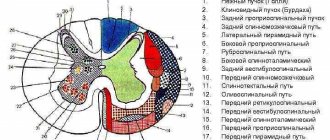
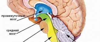
Descending pathways
All descending tracts of the spinal cord with their detailed characteristics and course of movement are clearly demonstrated in Table No. 2.
| No. | Descending Path View | Characteristics | |
| 1 | Lateral corticospinal, also called lateral corticospinal or main crossed pyramidal. | This pathway includes a considerable proportion of fibers of the pyramidal system. The lateral tract is localized in the lateral funiculus. As they travel, the fibers gradually become thinner. Lateral fibers conduct signals that cause conscious actions in humans. | Lateral fibers conduct signals that cause conscious actions in humans. |
| 2 | Anterior corticospinal, otherwise called corticospinal, and also straight or uncrossed pyramidal. | This path lies in the anterior spinal cord. Like the lateral pyramidal tract, the direct pyramidal tract includes cellular axons of the motor hemisphere, although they are located ipsilaterally. Initially, these axons descend towards their “own” segment. After this, as part of the anterior spinal commissure, they are transported to the opposite side, ending in the mononeurons of the anterior horn. | |
| 3 | Red-spinal or rubrospinal. | Beginning in the red nucleus of the spinal cord, this tract subsequently descends to the motor nerve cells of the anterior horns. This pathway is responsible for transmitting unconscious motor signals. | |
| 4 | Tectospinal, otherwise called tectospinal. | It is localized in the anterior funiculus near the anterior pyramidal tract. This tract starts on the roof of the midbrain. The mononeurons of the anterior horns are its final point. The tectospinal tract provides reflex protective actions in response to visual and auditory stimuli. | |
| 5 | vestibulospinal, otherwise called vestibulospinal. | This path is localized in the anterior spinal cord. The vestibular nuclei of the pons are its beginning, and the anterior spinal horns are its end. The balance of the human body is ensured precisely through the transmission of impulses from the vestibulospinal tract. | |
| 6 | Reticulospinal or reticulospinal. | This pathway ensures the transmission of excitatory signals from the reticular formation to the spinal nerve cells. |
To understand the neurophysiology of the human spinal cord pathways, you will need to briefly familiarize yourself with the structure of the spine. In its structure, the spinal cord is a bit like a cylinder, covered with muscle tissue on all sides. The pathways control the functioning of the internal organs, as well as all organ systems and functions performed by the body. Injuries, various injuries, and other ailments of the spinal cord can somehow reduce conductivity. By the way, conduction may even stop completely due to the death of neurons. Complete loss of conduction of spinal signals is characterized by paralysis, manifested in a complete lack of sensation in the limbs. This is very fraught with problems with internal organs that are responsible for damage to the communication of nerve cells. Thus, injuries and other ailments of the lower spinal cord are often characterized by urinary incontinence and even spontaneous defecation.
Drug treatment will consist of prescribing medications to prevent the death of brain cells, as well as to further increase blood flow to the damaged spinal areas. Electrical impulses may be prescribed as an additional treatment to stimulate the functioning of neurons and also help maintain muscle tone.
Also, if necessary, the attending physician may prescribe the use of the following folk remedies.
Apitherapy
- Apitherapy. Bee stings effectively restore the conductivity of efferent tracts. Thus, the poisons of these insects, penetrating into damaged areas, provide them with additional blood flow. If the cause of spinal pathology is radiculitis, a growing hernia and other similar ailments, apitherapy will be an excellent addition to traditional treatment.
- Herbal medicine. Medications are prescribed to normalize blood circulation and improve metabolism.
- Hirudotherapy. Thanks to treatment with leeches, it becomes possible to eliminate congestion - inevitable attributes of vertebral pathologies.
Spinal cord pathways
The spinal cord pathways or tracts are collections of nerve fibers located inside the spine that carry impulses from the brain to all parts of the body and back. The nerve endings, the combination of which form the pathways, are distinguished by a similar structure, development and general functions. They are divided among themselves according to the tasks assigned to them. The paths are classified as follows:
· Associative . Their main purpose is to unite gray matter cells from different segments to form their own anterior, lateral or posterior bundles.
· Commissural . These fibers connect the gray matter from the two hemispheres. With their help, the coordinated work of individual areas, nerve centers, and both hemispheres occurs.
· Projection . With the help of such pathways, the work of the overlying and underlying parts of the brain is combined. They are the ones who provide the projection of pictures of the surrounding world, as on a monitor screen.
Projection pathways, in turn, are efferent and afferent. They form the basis of the central nervous system, and are divided into ascending (centripetal or sensitive) and descending (centrifugal, motor).
Important! Nerve fibers provide a constant, unbreakable connection to the brain located in the skull and spine. It is thanks to them that the impulse is quickly transmitted, all body movements are coordinated with each other.
The pathways of the brain and spinal cord differ from each other, but they always act harmoniously, ensuring the passage of an incredibly large number of nerve signals from receptors to the central nervous system. Paths are formed from long axons, special fibers that are capable of creating connections among themselves, thus connecting individual segments of the spinal trunk, providing control of effector organs.
All tracts of the spinal cord are located in the white matter, which is divided into the anterior, lateral and anterior cord. Their main volume consists of supraspinal tracts, which provide two-way communication between the spinal region and the head organ. These stripes occupy little space around the gray matter and are called propriospinal.
The pathways of the spinal and head sections are divided conditionally, depending on the characteristics of their structure and functionality. They are an integral part of the spine as a whole, and allow you to control not only the motor activity of the body, but also the functioning of the internal organs. They are located outside the main brain bundles. They develop in parallel with the formation of the head section.
Important! When the neurons carrying the impulses begin to die, conduction may stop completely, leading to loss of sensation in the limbs or paralysis.
RISING PATHS
The ascending tracts of the spinal cord are responsible for transporting pain impulses, tactile sensations, information about body temperature, and sensitivity from receptors to the cerebellum. That is, their main feature is the movement of flow from the periphery to the center. It is thanks to them that a person understands what is happening to his body at a given second of time, processes constantly incoming information from the outside world, and makes decisions in a timely manner based on the impulses received. The table will tell you more about the varieties of this type of path and their main tasks.
| Name of paths | Location | Their main tasks |
| Thin beam (Gaull beam) | Rear pillar | This is the basis of the ascending tracts, as they run along the entire spinal trunk. Impulses from it are directed to the cerebral cortex. With their help, conscious impulses are transmitted from muscle receptors to the “center”. |
| Wedge-shaped fasciculus (Burdach's path) | Rear pillar | Nerve currents are directed to the cortex. The pathways are responsible for transmitting impulses from the musculoskeletal system. |
| Posterior spinocerebellar tract (Flexig's tract) | Dorsal | Responsible for the transmission of unconscious nerve currents from proprioceptors of muscle fibers, ligaments, tendons to the cerebellum. |
| Anterior spinocerebellar fasciculus (Gowers' tract) | More ventral | As in the previous case, it is responsible for transporting currents from muscles, ligaments and tendons to the cerebellum. Impulses are transmitted unconsciously. |
| Lateral spinothalamic tract | They are responsible for the sensation of temperature changes and pain, since impulses are carried out precisely according to them. | |
| Anterior spinothalamic tract | Responsible for transmitting nerve currents about tactile sensations, pressure, touch and other things. |
The ascending tracts of the spinal trunk are generally responsible for transmitting any incoming information to the articular receptors of the body. Thanks to them, a person understands the position of his body, is aware of tactile sensations, passive movements performed, and feels vibration.
DESCENT ROUTES
Descending pathways are responsible for the movement of currents from underlying sections to working systems. In general, they are divided into pyramidal and extrapyramidal. The first ones are responsible for transmitting impulses of voluntary motor reactions, namely the control of conscious movements, the second ones control involuntary movements (maintaining balance in the event of a fall). Through these nerve bundles, formed from the axons of cells, they are responsible for distributing “instructions” from the brain to the main motor departments. Through them, the spinal cord performs leading executive tasks.
The following structure diagram will help you understand the structure of descending paths:
Symptoms and treatment of spinal cord inflammation
· Pyramidal or cortinospinal tracts. They pass through the medulla oblongata, located in the anterior and lateral cords of the spinal cord. Its main task is to transport nerve currents from the head region, namely: from the motor centers and departments located in it that are responsible for motor functions to similar areas in the spinal organ. With its help, a person is able to perform voluntary actions with the musculoskeletal system.
· Rubrospinal tract. Another main path, related to the descending ones. It originates in the red nucleus and gradually, as part of the white matter, descends to the segments of the spinal cord. The path ends in the intermediate part of the gray matter. Responsible for the transmission of nerve currents that provide support for the tone of the skeletal muscle corset necessary for normal motor activity.
· Reticulospinal tract. It is located in the anterior part of the column, starting from the reticular formation of the medulla oblongata. The main task is the transportation of impulses, as well as maintaining the tone of the skeletal muscles with the help of inhibitory and exciting effects on motor neurons. Thanks to it, the state of the spinal autonomic center is monitored and regulated.
· Vestibulospinal tract. Passes in front of the column, starting from the Deiters cores. With its help, impulses are transmitted that maintain a certain posture and are responsible for the balance of the body.
· Tectospinal tract. Impulses move along it, which provide motor reflexes of the organs of vision and hearing.
The descending pathways allow impulses to move freely from the head to the underlying motor nuclei in the spinal canal, thereby maintaining normal motor activity. With their help, the work of the higher motor center, namely the cerebral cortex, is carried out.
Damage to central or peripheral motor neurons leads to the development of paralysis and paresis. These disorders are accompanied by the complete disappearance of reflexes, usually due to loss of the efferent part of the reflex arc, and a complete decrease in muscle tone. If it is necessary to determine the affected area, individual areas are stimulated, causing wave-like contractions and small twitches. Where they are not observed, the problem is localized.
The treatment most often prescribed is surgery, which helps restore patency in the spinal canal. But sometimes doctors resort to hirudotherapy or apitherapy. Bee stings, which involve injecting bee venom, help increase blood flow and repair damage. But this is not always permissible and is carried out only under the supervision of a medical professional.
Source: https://spina.guru/anatomiya/provodyashchie-puti-golovnogo-spinnogo-mozga
Ascending afferent pathways originating in the brainstem
The medial lemniscus, trigeminal lemniscus, ascending tract of the auditory analyzer, optic radiation, and thalamic radiation begin in the brain stem.
1. The medial lemniscus as a continuation of the thin and wedge-shaped fascicles was described earlier.
2. The trigeminal loop, lemniscus trigeminalis, is formed by processes of nerve cells that make up the sensory nuclei of the trigeminal nerve (V pair), facial nerve (VII pair), glossopharyngeal nerve (IX pair) and vagus nerve (X pair).
The axons of afferent neurons located in the trigeminal ganglion approach the sensory nuclei of the trigeminal nerve. The common sensory nucleus of the other three nerves - the nucleus of the solitary tract - is approached by axons of afferent neurons located in the genu node (VII pair) and in the upper and lower nodes of the IX and X pairs of nerves. The bodies of the first neurons are localized in the listed nodes, and the bodies of the second neurons of the path along which impulses from the head receptors are transmitted are located in the sensitive nuclei.
The fibers of the trigeminal lemniscus pass to the opposite side (some of the fibers follow on their side) and reach the thalamus, where they end in its nuclei.
The nerve cells of the thalamus are the bodies of the third neurons of the ascending tracts of the cranial nerves, the axons of which, as part of the central thalamic radiates, through the internal capsule are directed to the cerebral cortex (postcentral gyrus).
3. The ascending path of the auditory analyzer has, as its first neurons, cells located in the ganglion of the cochlear part of the vestibulocochlear nerve. The axons of these cells approach the cells of the anterior and posterior cochlear nuclei (second neurons). The processes of the second neurons, moving to the opposite side, form a trapezoidal body, and then take an ascending direction and are called the lateral loop, lemniscus lateralis. These fibers end on the bodies of the third neurons of the auditory pathway, located in the lateral geniculate body. The processes of the third neurons form the auditory radiation, radiatio acustica, which runs from the medial geniculate body through the posterior limb of the internal capsule to the middle part of the superior temporal gyrus.
Editor's Note: What is Attention Deficit Disorder?4. Visual radiance, radiatio optica (see Fig.), connects the subcortical centers of vision with the cortex of the calcarine sulcus.
The optic radiance includes two systems of ascending fibers:
- the geniculate-cortical optic tract, which originates from the cells of the lateral geniculate body;
- the cushion-cortical tract, starting from the cells of the nucleus located in the cushion of the thalamus; in humans it is poorly developed.
The set of these fibers is designated as posterior thalamic radiation, radiationes thalamicae posteriores.
Ascending to the cerebral cortex, both systems pass through the posterior limb of the internal capsule.
5. Thalamic radiations, radiationes thalamicae (see Fig.), are formed by processes of thalamic cells and constitute the final sections of the ascending tracts of the cortical direction.
The thalamic radiations include:
- anterior thalamic radiations, radiationes thalamicae anteriores, are radial fibers of the white matter of the cerebral hemispheres. They begin from the superior medial nucleus of the thalamus and are directed through the anterior limb of the internal capsule to the cortex of the lateral and inferior surfaces of the frontal lobe. Some of the fibers of the anterior thalamic radiates connect the anterior group of thalamic nuclei with the cortex of the medial surface of the frontal lobes and the anterior part of the cingulate gyrus;
- central thalamic radiations, radiationes thalamicae centrales, are radial fibers connecting the ventrolateral group of thalamic nuclei with the cortex of the pre- and postcentral gyrus, as well as with the adjacent sections of the cortex of the frontal and parietal lobes. They pass as part of the posterior limb of the internal capsule;
- the lower peduncle of the thalamus, pedunculus thalami inferior, contains radial fibers connecting the thalamic cushion and medial geniculate bodies with areas of the temporal choir;
- posterior thalamic radiates (see earlier).
Associative paths, short and long.
Association pathways are the link between the last neuron of the afferent pathway and the first neuron of the efferent pathway.
Associative pathways are chains of interneurons that connect different areas of the cortex within the same hemisphere. In the SM, associative paths connect neighboring segments.
There are short and long associative paths.
Short association fibers connect areas of the cortex of neighboring gyri, without going beyond the lobe of the cerebral hemisphere. These are arcuate fibers located superficially under the bark - at the bottom of the furrows. This also includes fibers that connect cells of neighboring nuclei of the brain stem.
Long association fibers are located under a layer of short association fibers in the cerebral hemispheres. They connect areas of the cortex of different lobes of one hemisphere.
Long associative fibers include: 1. Belt. This is a group of nerve fibers located deep in the vaulted gyrus. Connects areas of the cortex of the frontal, occipital and temporal lobes in the region of the medial surface of the cerebral hemisphere. In ontogenesis, the belt develops earlier than other associative pathways. 2. Upper longitudinal beam. It is localized under the superolateral surface of the cerebral hemisphere, lateral to the corona radiata.
Its fibers connect areas of the cortex of the lower parts of the frontal lobe, the inferior parietal lobule, the temporal and occipital lobes. The formation of the superior longitudinal fasciculus in ontogenesis is associated with the development of the cortical ends of the cutaneous, motor, auditory and visual analyzers.
3. Lower longitudinal fascicle. It is located under the inferomedial surface of the cerebral hemisphere, along the outer wall of the posterior and inferior horns of the lateral ventricle.
Bundles of fibers connect areas of the cortex of the occipital and temporal lobes. The lower longitudinal fasciculus develops earlier than the upper one and provides connection between the cortical end of the visual analyzer and the autonomic centers, and therefore their combined actions. 4. Hook-shaped bundle. Located on the inferolateral surface of the cerebral hemisphere. Connects areas of the cortex of the frontal, temporal and occipital lobes of the hemisphere. Long associative tracts include fibers passing through the stria terminalis, the medullary stria of the thalamus, and the dorsal and medial longitudinal fasciculi.
In the SM, associative fibers are located around the SV in the form of a narrow strip.
These are their own bundles, which are classified as short associative paths. They are part of local reflex arcs, connecting segments of the SC, as well as sensory neurons of the spinal ganglia with motor neurons of the anterior horns of the SC.
In ontogenesis, associative pathways are formed much later compared to commissural and projection pathways.
However, in the future, associative pathways quickly develop and dominate over other pathways.
Commissural pathways. Projection pathways: a) ascending (afferent) fiber systems.
Commissural nerve tracts are pathways of the central nervous system that connect the symmetrical parts of the cerebral hemispheres or other parts of the central nervous system. Commissural (commissural) pathways connect areas of the cortex of the right and left hemispheres of the brain and ensure the unity of brain activity.
Commissural, that is, commissural tracts connect both symmetrical and asymmetrical areas of the cortex of different hemispheres.
The commissural tracts include: corpus callosum, anterior commissure of the brain, posterior commissure of the brain, commissure of the fornix.
Corpus callosum. Among other commissures, it is phylogenetically the youngest formation.
It consists of transversely directed nerve fibers connecting similar areas of the neocortex of the right and left hemispheres of the cerebrum with each other. Part of the fibers that form the beak of the corpus callosum connects both thalami and the heads of the caudate nuclei, ensuring synchronous activity not only of the cortex of the right and left hemispheres, but also of the subcortex. Anterior commissure of the brain. Represented by fibers belonging to the pathways of the olfactory brain.
The anterior commissure of the brain is located in front of the columns of the medullary vault and consists of anterior and posterior parts. The formation of the anterior part of the commissure is associated with the formation of the ancient cortex, and the posterior part is associated with the formation of the new cortex. It is suggested that the anterior commissure is important for the paired activity of not only the olfactory, but also the auditory and visual analyzers. The anterior commissure provides interhemispheric integration of impulses.
Posterior commissure of the brain. Located in the posterior wall of the third ventricle. The fibers that make up the commissure connect the thalami cushions to each other. The commissure of the fornix (hippocampal commissure). This is an ancient formation related to the pathways of the olfactory brain. The commissure fibers of the fornix connect the structures of the hippocampus of the right and left hemispheres and ensure the synchronous functioning of the hippocampus.
The commissure of the fornix is located between the lower surface of the splenium of the corpus callosum and the crura of the fornix.
Projection nerve tracts are pathways of the central nervous system that connect the cerebral cortex with the periphery and pass through various parts of the central nervous system. Projection nerve pathways are divided into afferent (ascending) and efferent (descending).
The ascending pathways of the lateral column include the following: spinothalamic, or tractus spinothalamicus - fibers of the second neurons of the pain and temperature, partly tactile sense, which passed into the opposite lateral column after crossing in the anterior gray commissure.
In addition to the simplest reflex arcs that arise within one segment of the SC, there are also intersegmental reflex ascending and descending pathways.
Let's look at the ascending paths. When performing the knee reflex, the extensor muscle contracts and the leg involuntarily extends. Signals from the sensory neuron, indicating that the leg has changed position, travel up the collateral of the sensory neuron.
This collateral passes in the spinal cord to the thalamus. This is where incoming signals are filtered. For example, if the signal is single and weak, then it simply does not pass through.
Such a system allows you to regulate the passage of signals to the cortex and makes it possible to respond to more important signals with the greatest speed. After passing through the thalamus, the nerve impulse arrives at the neurons of the somatosensory cortex of the cerebral hemispheres of the telencephalon. In this case, the person has the feeling that the leg has straightened.
In order to return it to its original position, the nerve impulse is transmitted to the motor cortex of the cerebral hemispheres, where movement programs are built. Intersegmental ascending pathways allow us to control voluntary movements triggered at different levels of the SC.
What types of pathways are divided into according to: direction, function, length, localization and significance?
- Towards:
- Rising,
- Descending;
- By function:
- Sensitive - impulses are formed in receptors. These pathways are formed by sensory and interneurons.
- Motor – impulses go to the executive organs (muscles).
- By lenght:
- Short - localized within one department of the central nervous system, or between adjacent departments.
- Long - connect distant parts of the central nervous system.
- By localization:
- Associative - in one hemisphere.
- Commissural - connects the two hemispheres.
- Projection - connect the hemispheres with other parts of the brain.
- By importance:
- The main ones are inside the central nervous system.
- Circuitous - outside the central nervous system. They pass through the membrane of the GM and SM, through the vessels. Conducts feelings of gravity and vibration.
Classification of spinal tracts
The main part of the pathways is formed by neurons, which allows them to be classified according to the functional characteristics of the nerve fibers:
- commissural connection;
- associative pathways;
- projection fibers.
Nerve tissues are located in the white and gray matter of the brain and connect the cerebral cortex and the spinal horns. The morphofunctionality of the descending pathways sharply limits the transmission of impulses in one direction.
Main ascending spinal tracts
The wire function comes with the following features:
- Associative pathways are a kind of “bridge” that connects areas between the nucleus and the cerebral cortex. Associative pathways consist of long ones (signal transmission occurs in 2-3 segments of the medulla) and short ones (located in 1 part of the hemisphere).
- Commissural tracts - consist of the corpus callosum, which connects new sections in the spinal cord and brain, and diverge to the sides in the form of rays.
- Projection fibers can be functionally afferent or descending. The location of these fibers allows the impulse to reach the cerebral cortex as quickly as possible.
The conductive function of the spinal cord is determined by descending and ascending pathways
In addition to this classification, depending on the main functions, the following forms of pathways are distinguished:
- The main system of nerve fibers is the corticospinal tract of impulse transmission, which is responsible for motor activity. Depending on the direction, it is divided into the lateral, corticonuclear and lateral corticospinal systems.
- In the projection-descending nervous system, which begins in the cortex of the middle hemisphere and passes through its cord and trunk, ending in the anterior horns of the spinal column, the presence of a tectospinal impulse transmission pathway is noted.
- Diagnosing the vestibular cord normalizes work in the vestibular apparatus. In this case, the nerve tissues pass in the anterior part of the spinal cord, starting from the lateral nucleus in the region of the vestibulocochlear nerve.
- Conducting nerve impulses from the cerebral hemisphere to the gray matter and improving muscle tone belongs to the reticular-spinal cord development path.
It is important to remember that the pathways are united by the totality of all the nerve endings that provide the signal to various parts of the brain
Characteristic
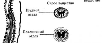
The spinal cord lies in the canal of the spinal column. In an adult, the length of the cord is about 45 cm. The upper segment of the spinal cord borders the medulla of the brain, which ensures the continuity of the pathways. The basis of the cord is gray matter, which when cut looks in the shape of a butterfly. Gray matter is surrounded by white fibers, which form pathways - ascending and descending.
The spinal cord contains tracts of white matter that support sensory functions and complex motor activities. There are ascending (afferent) and descending (efferent) pathways within the spinal cord. In the first case, we are talking about the transmission of impulses from all parts of the body to the head part of the central nervous system, in the second - about the direction of impulses from the cortical structures of the head part of the central nervous system to parts of the body and limbs.
The medulla receives impulses emanating from exteroreceptors (perceive environmental stimuli) located on the surface of the skin, proprioceptors (sensitive receptors), and partially visceroreceptors (located in blood vessels and internal organs). The ascending sensory pathways within the spinal cord perform a conducting function. They convey:
- Proprioceptive sense (receive impulses from elements of the musculoskeletal system - muscles, tendons, joint capsules).
- Extraceptive feeling (receive impulses from skin receptors).
- Intraceptive sense (perceive impulses from elements of the circulatory system and internal organs).
The fibers that carry the proprioceptive sense are divided into conscious and unconscious sensory pathways. In the first case, impulses arrive in the cortical parts of the hemispheres, in the second - in the cerebellum, where balance reactions and complex motor activity that require fine coordination are modulated.
The nerve endings of the spinal cord innervate the skeletal muscles with the exception of the muscles of the head, which are innervated by the cranial nerves. The pathways pass in the posterior, lateral, and anterior cords, formed from white matter, where nerve fibers are grouped within the spinal cord, transmitting impulses of various types to certain parts of the central nervous system.
Ascending and descending tracts of the spinal cord
The white matter of the spinal cord consists primarily of ascending (afferent) and descending (efferent) myelinated nerve fibers. They are grouped according to their functions into relatively clearly defined bundles (fasciculi) or into more diffuse tracts (tractus), which are usually named according to their point of exit and destination. Together, the tracts and bundles form funiculi. Intersegmental fibers of the spinal cord run directly adjacent to the gray matter. They do not leave the spinal cord and serve primarily spinal reflexes. Outside the own bundles there are posterior, anterior and lateral cords, although the last two are often combined into the anterolateral cord.
Ascending tracts: 1. Tracts of the anterolateral funiculus: afferent tracts to the thalamus, providing gross perception of touch, tactile sensations, as well as pressure, pain, temperature sensations from the limbs and trunk (anterior and lateral spinothalamic tracts). 2. Posterior funiculus tracts (posterior columns): afferent tracts to the thalamus, providing deep sensation from muscles, tendons and joints (pronrioception), sensation of vibration, light touch and fine tactile sensations on the limbs and trunk - the thin fasciculus gracilis and wedge-shaped bundle (fasciculus cuneatus). The gracilis and cuneate fasciculi project to the nuclei of the same name in the medulla oblongata, which in turn project to the thalamus through the median lemniscus medialis, an important fibrous tract of the brainstem. 3. Spinocerebellar tracts: afferent tracts to the cerebellum, providing unconscious deep sensitivity from muscles, tendons and joints (iroprioception) - ventral (tractus spinocerebellaris centralis) and dorsal spinocerebellaris tracts (tractus spinocerebellaris dorsalis).
From the editor: Symptoms, causes and treatment of dysarthria in children
Descending tracts: 1. Pyramidal tract: efferent tracts from the motor cortex to the motor neurons of the anterior horn, providing fine movements of the limbs and trunk (corticosinial tracts). 2. Extrapyramidal tracts: efferent tracts from the brain stem to the motor neurons of the anterior horn, responsible for involuntary movements, for example, those associated with maintaining posture and balance, reflex movements, conjugal movements (reticulosiinal tract, etc.).
Massage courses in St. Petersburg: become a professional!
How to find out about the pain threshold and temperature differences?
To determine the level of pain, doctors use the pricking method. In the most unexpected places for the patient, the doctor applies several light injections with a pin. The patient's eyes should be closed, because He shouldn't see what's happening.
The temperature sensitivity threshold is easy to determine. In a normal state, a person experiences different sensations at temperatures, the difference of which was about 1-2°. To identify a pathological defect in the form of impaired skin sensitivity, doctors use a special device - a thermoesthesiometer. If it is not there, you can test for warm and hot water.
Functions of pathways
The generation of reflexes occurs with the participation of the spinal cord and brain
The ascending tracts of the spinal cord are formed from sensory neurons responsible for transmitting electrical impulses characterizing various sensations to the brain from the periphery of the body. These neurons carry out four main types of sensations:
- tactile;
- temperature;
- pain;
- proprioceptive, helping to control and change the position of the body and limbs.
Their axons run throughout the spinal cord and are directed to its upper parts. From here the impulse goes directly to the cerebral cortex.
Descending nerve pathways are divided into two main groups. Those that originate from convex, wedge-shaped areas of the brain are called the corticospinal or pyramidal tract. In evolutionary terms, this tract is the youngest. The neurons of this pathway lie in the motor zone of the cerebral hemispheres, where there are centers that direct purposeful, finely coordinated movements of the limbs, which are especially important for humans. Therefore, they received significant development only among Homo sapiens. Animals also have pyramidal tracts, but are much less developed.
Extrapyramidal pathways are responsible for involuntary reflex motor reactions. They are formed by neurons in the gray matter of the brain. The main structural unit of this substance is the basal ganglion. The extrapyramidal system of the brain also includes the thalamus, cerebellum and association centers of the cerebral cortex.
If we list the functions of the extrapyramidal system, we get the following list:
- regulation of automatic motor acts of congenital and acquired nature;
- maintaining balance;
- regulation of muscle tone;
- involuntary contraction of facial muscles;
- regulation of movements that serve as accompanying movements (for example, rapid movement of the arms when running).
Classification
A brief description of the main function is the conduction of nerve impulses. The classification of pathways located within the brain and spinal cord involves the identification of afferent, efferent, and associative tracts. Afferent - conduct impulses from receptors to the controlling (integration, coordinating activities, ensuring interaction) head parts of the central nervous system.
Efferent - conduct impulses from the controlling head parts of the central nervous system to the working (executive) organs. Association pathways, consisting of bundles of nerve fibers within the spinal cord, maintain communication between different control (integration) departments that are located in the head structures of the central nervous system.
Ascending are the pathways that transmit impulses from the spinal cord (from sensory organs, blood vessels, internal organs, elements of the musculoskeletal system) to the brain. The ascending (sensitive) tracts are formed by afferent (the bodies are located in the spinal nuclei) and associative (the bodies are located in the dorsal horns within the spinal cord) neurons.
Descending pathways are located within the pyramidal and extrapyramidal systems and conduct impulses from the brain to the spinal cord. Descending pathways of the projection type conduct impulses coming from the cortical, subcortical sections to the nuclei of the trunk and to the region of the anterior horns (to the motor nuclei) within the spinal tract.
The pathways within the spinal cord, functionally divided into tracts of conscious and unconscious sensitivity, include vestibular, auditory, visual, gustatory and other sections. The tracts of conscious sensitivity are associated with analyzers that are located in the cortical head structures of the central nervous system.
Tracts of conscious sensitivity
In anatomy, the pathways of conscious and unconscious sensitivity within the brain and spinal cord are a chain of neurons that transmit impulses of the same type in one direction. Nerve fibers of conscious sensitivity play the role of informants of cortical structures.
As a result, the cortical sections receive information regarding muscle tone, the position of the torso in space, the sensation of pressure and vibration. The pathways of conscious sensitivity lie in the posterior cords of the spinal cord and consist of neuronal structures of 3 types.
The bodies of the first neurons are located in the spinal ganglia; nerve cells act as an intermediate link, receiving impulses from proprioceptors and redirecting them to the spinal tract. The bodies of the second neurons are located in the nuclei - thin (Gaull) and cuneate (Burdach), from which axons extend, forming the bulbar-thalamic fascicle. Its branches are directed to the thalamus region.
The bodies of third neurons are located in the nuclei of the thalamus. The axons of these nerve cells form thalamocortical fibers, which extend to the cortex of the cerebral hemispheres. Fibers of conscious sensitivity pass through the brain stem, their collaterals (bypass, lateral branches) are directed to the reticular formation within the brain stem, to the cerebellum and limbic system.
Tracts of unconscious sensitivity
Nerve impulses coming from proprioceptors are redirected to the cerebellum, where a general picture of the state of the elements of the musculoskeletal system is formed. The information is analyzed, after which the cerebellum regulates indicators such as muscle tone, which leads to coordination of movements and maintenance of balance. The system of nerve fibers is represented by the spinal-cerebellar tracts (anterior - Gowers and posterior - Flexig).
Receptors are located in muscle tissue, tendons, periosteum, joint capsules, and bone structures that form the musculoskeletal system. Both pathways are composed of two types of neuronal structures. The bodies of neurons of the 1st type are located in the spinal ganglia, which are also called sensitive, where the pathways from the receptors to the spinal cord pass.
The axons of these neurons penetrate into the gray matter, where they end in the region of the nuclei of neuronal structures of the 2nd type. The bodies of neurons of the 2nd type are located in the nuclei - thoracic and intermedial. The axons of neurons of the 2nd type enter the lateral funiculi, forming the spinocerebellar tracts leading to the cerebellum and having similar characteristics:
- Conducts impulses of unconscious sensitivity from proprioceptors.
- Their receptors are located in the elements of the musculoskeletal system.
- Transmits nerve impulses to the cerebellum.
- The pathways within the spinal cord are located in the lateral cords, where they continue the tract coming from the spinal nodes.
At the same time, a number of differences are revealed between the posterior and anterior tracts, including the location of type 2 neurons in different nuclei (thoracic and intermedial) suggesting the formation of nerve fibers from different axons - the posterior tract is formed by axons emanating from the thoracic nucleus, the anterior - axons extending from the intermedial nucleus.
The anterior tract is crossed and long, the posterior tract is straight and short. The lateral cords contain afferent and efferent fibers, including the spinocerebellar tracts - posterior (Flexiga) and anterior (Goversa). The descending systems of nerve fibers of the lateral cords form the pyramidal (cortical sections - spinal tract) and extrapyramidal (red nucleus - spinal tract) pathways.
Motor tracts
In short, from the motor centers located in the brain, impulses depart along the motor tracts to the skeletal muscles, determining the nature, speed, amplitude and other parameters of movements of the body and limbs. There are pyramidal and extrapyramidal motor tracts. The pyramidal ones originate in the cortical parts of the brain and control conscious movements. Types of pyramidal tracts:
- Corticospinal. Conducts motor stimuli directed by the will of a person, his consciousness, which leads to the execution of differentiated, precise movements by the muscles. Conducts inhibitory stimuli from the cortical regions to the anterior horns located in the spinal tract.
- Corticonuclear. Conducts motor stimuli directed by the will of a person, his consciousness, which causes voluntary mobility of the neck and head, supports the performance of differentiated, precise movements by these parts of the body.
- Corticopontine-cerebellar. It transmits impulses from the cortical sections to the pontine nuclei and then to the cerebellum, due to which the cortex controls the regulatory activity of the cerebellum, which ensures balance and maintaining a given posture.
The extrapyramidal system includes the subcortical regions and nuclei of the brain stem. The extrapyramidal tracts conduct impulses that regulate complex movements of the automatic (reflex) type by controlling muscle tone and motor coordination. Types of extrapyramidal tracts:
- Reticulospinal. Supports the execution of complex automatic motor acts that require the participation of many muscle groups. An example is the breathing and swallowing reflexes.
- Red-nucleus spinal. The main tract of the extrapyramidal system runs through the red nucleus.
- Roof-spinal. Provides reflexive, involuntary motor reactions in response to sharp, sudden stimuli - visual, auditory, tactile, olfactory.
- vestibulospinal. Supports involuntary motor activity in case of imbalance.
- Olivospinal. Conducts impulses that regulate muscle tone, ensuring the stability of the body's balance.
The thalamus and reticular nuclei of the sensory type are afferent centers within the pathways of the extrapyramidal system. Olive, hypothalamic, red, vestibular nuclei are efferent centers within the extrapyramidal system.
What organs does the spinal cord control?
It is also important to understand which internal organs are connected to the spinal cord and may suffer if a particular area of the spine is damaged. Specific spinal segments control specific parts of the body by transmitting nerve impulses and transmitting responses along motor neurons
What each vertebra is responsible for can be clearly seen in the table.
| Back segment | Vertebra serial number | Controlled internal organs |
| Cervical | 3-5 | Diaphragm |
| Cervical | 6-8 | Articular tissue of the upper limbs |
| Chest | 1,2, 5-8 | Muscle tissue and epidermis of the hands, elbows and forearms |
| Chest | 2-12 | Muscles, skin of the body |
| Chest | 1-11 | Intercostal muscles |
| Chest | 1-5 | Heads, heart |
| Chest | 5-6 | Lower esophagus |
| Chest | 6-10 | Gastrointestinal tract |
| Lumbar | 1-2 | Prostate, groin area, adrenal glands, bladder, uterus. |
| Lumbar | 3-5 | Muscles and skin of the legs |
| Sacral | 1-2 | Muscle tissue and epidermis of the lower extremities |
| Sacral | 3-5 | External genitalia, reflex centers, erectile dysfunction and defecation |
Damage to the spinal cord in a specific section negatively affects the functioning of these internal organs. In some cases, dysfunction occurs before vertebral compression or displacement is detected.
Due to the characteristic structure of the brain, it is connected with most systems in the body
The integrity of its structure is extremely important for the correct functioning of the musculoskeletal system and the health of internal organs. Any injury, regardless of severity, can lead to disability
Treatment of minor spinal injuries is carried out on an outpatient basis, using medications, therapeutic exercises and massage. Severe injuries require surgical intervention, especially if compression of the spinal cord is detected. Cells are quickly damaged and die, so any delay can cost a person’s health.
The spinal cord is a key element of the human central nervous system, which is connected in one way or another with almost all internal organs and human muscle tissue. The specific structure allows you to transmit impulses and signals, ensure full motor activity, and perform a number of other functions.
From the editor: The most effective exercises for memory development
How the sense of touch is born
The fibers that provide sensitivity take different paths. For example, from proprioceptors the pathways go to the cerebellum and cortex. They send a signal to this area about the condition of the joints, tendons, and muscles.
This path is made up of axons of sensory type neurons. The afferent neuron processes the received signal and, using an axon, conducts it to the thalamus. After processing in the thalamus, information about the motor system is sent to the postcentral cortex. Here the formation of sensations occurs about how tense the muscles are, in what position the limbs are, at what angle the joints are bent, whether there is vibration, passive movements.
The thin bundle also contains fibers that are associated with skin receptors. They conduct a signal that generates information about tactile sensitivity during vibration, pressure, and touch.
The axons of the second interneurons form other sensory pathways. The area where the cell bodies of these neurons are located is the dorsal horn (spinal cord). In their segments, these axons create a cross, then they go on the opposite side to the thalamus.
This pathway contains fibers that provide temperature and pain sensitivity. Also here are fibers that are involved in tactile sensitivity. Neurons located in the spinal cord receive information from brain structures.
Extrapyramidal neurons participate in the formation of the rubrospinal, reticulospinal, vestibulospinal, and tectospinal tracts. Nerve efferent impulses pass along all of these paths. They are responsible for maintaining muscle tone, performing various involuntary movements, and posture. Acquired or innate reflexes participate in these processes. In these pathways, the conditions for performing all voluntary movements controlled by the cerebral cortex are formed.
The spinal cord conducts all signals that come from the centers of the ANS to the neurons that make up the sympathetic nervous system. These neurons are located in the lateral horns of the spinal cord.
Also involved in the process are neurons from the parasympathetic nervous system, which are also localized in the spinal cord (sacral region). These pathways are entrusted with the function of maintaining the tone of the sympathetic nervous system.
Methods for restoring spinal cord patency
All therapeutic measures are primarily aimed at stopping cellular necrosis and eliminating the factors that were catalysts for this condition.
Drug therapy involves the use of drugs that prevent the death of brain cells and ensure sufficient blood supply to damaged areas in the spinal cord. In this case, it is necessary to take into account the age category of the patient and the severity of the lesion. In addition, in order to provide additional stimulation to nerve cells, it is recommended to use electrical impulses that maintain muscle tone.
If necessary, surgery is performed to restore conduction, which affects 2 areas: removing the catalyst and stimulating the spinal cord to ensure restoration of lost function.
The operation to restore conductivity is performed by experienced neurosurgeons using the most modern methods of monitoring the process
Before the operation begins, a thorough diagnostic examination of the patient is performed, which makes it possible to identify the localization of the degenerative process, after which neurosurgeons narrow the surgical field. In case of severe symptoms, the doctor’s action is primarily aimed at eliminating the compression that provoked spinal cord syndrome.
In addition to surgical and therapeutic treatment, apitherapy, herbal medicine and hirudotherapy are often used, which have a positive effect on the structural pathways of the spinal column and brain. However, it should be borne in mind that in all cases a mandatory medical consultation is required.
It must be taken into account that restoration of neural connections after various kinds of negative influences requires long-term treatment. In this case, early seeking of highly qualified help is of great importance. Otherwise, the chances of restoring the functionality of the spinal cord are significantly reduced. This indicates that the pathways in the brain and spinal cord interact closely with each other, uniting the entire body, which ensures unity of action.
Recovery methods
Damage to the substance of the spinal cord of traumatic or other origin is accompanied by a disruption of the conductive function of the nerve tracts. Axon regeneration is regulated by therapeutic methods. To stimulate axon regeneration, transplantation of embryonic tissue, as well as artificially grown neuroblasts (germ cells), is used.
The transplanted cells take root, after which differentiation and further growth occur, which in some cases leads to the restoration of lost functions. In parallel, drugs with a neuroprotective effect are prescribed for the rehabilitation and stabilization of surviving nerve structures. Stimulation of axon growth is achieved through the infusion of neurotrophic (providing nutrition to nervous tissue) factors.
The pathways within the spinal cord transmit impulses from the executive organs to the brain and vice versa, supporting the sensory and motor functions of the body.