The dendrite of a neuron (dendrum - branch) is a process of the neuron body through which it receives a signal from other cells. The dendrite receives a signal from the axon of another neuron or a receptor protein that responds to the environment.
Answering the question of what dendrites are, we can say that traditionally dendrites are considered as antennas of a neuron. Information exchange occurs in one direction: from axon to dendrite. The more dendrites a neuron has, the more information channels, the more complex decisions the neuron makes.
Synaptic cleft
You may be interested in: Semantic barriers and ways to eliminate them
The signal from other cells reaches the body of the neuron along one of its dendrites. The dendrite in the human nervous system usually receives a chemical signal (neurotransmitter) from the axon. The junction of a dendrite and an axon is called a synapse.
Synapses allow precise messages to be passed from neuron to neuron. Thanks to synapses, there is neuroplasticity and the ability to fine-tune the functions and behavior of the body.
The dendrite contains receptors that receive the neurotransmitter. Receptors are specialized proteins that capture a neurotransmitter molecule and, depending on their type, trigger further reactions in the cell.
Dendritic malfunction
Dendrites play a very important role in transmitting information between neurons. Thus, it is not surprising that dendritic malfunctions are associated with various disorders of the nervous system. Malfunctions vary in type and severity and range from abnormal morphology to abnormal dendritic branching, abnormalities in dendritic development, and abnormal loss of dendritic branching and dendritic genesis. All this is associated with disorders such as schizophrenia, autism, depression, anxiety, Alzheimer's disease and Down syndrome and others.
Dendritic spines
Small growths called spines form on the dendrites. The latter can take many forms, but the most persistent is that of a fungus.
The number of dendritic spines ranges from 20 to 50 per 10 µm of dendrite length. The spines are very variable in shape and volume.
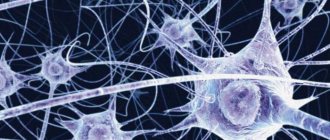
There are 86 billion neurons in the brain. Axons, dendrites and cell bodies of neurons form huge neural networks.
Dendrites are responsible for learning and memory, and also control the balance of the system. When there is a local strengthening of connections between certain neurons, it is in the dendrites that the production of a protein increases, regulating the decrease in the activity of other synapses.
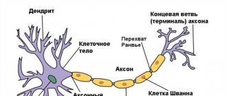
What is a dendrite - functions and morphology
Dendrites are numerous thin tubular or round protrusions of the cell body (perikarya) of a nerve cell.
The term itself speaks of the extreme branching of these areas of neurons (from the Greek δένδρον (dendron) - tree). The surface structure of neurocytes can contain from zero to many dendrites. The axon is most often single. The surface of dendrites does not have a myelin sheath, unlike axon processes.
The cytoplasm contains the same cellular components as the nerve cell body itself:
- endoplasmic granular reticulum;
- accumulations of ribosomes - polysomes (protein-synthesizing organelles);
- mitochondria (energy “stations” of the cell, which, using glucose and oxygen, synthesize the necessary high-energy molecules);
- Golgi apparatus (responsible for the delivery of internal secretions to the outer layer of the cell);
- neurotubules (microtubules) and neurofilaments are the main components of the cytoplasm, thin supporting structures that ensure the preservation of a certain shape.
The structure of dendritic endings is directly related to their physiological functions - receiving information from axons, dendrites, and perikarya of neighboring nerve cells through numerous interneuron contacts based on selective sensitivity to certain signals.
Training and Spikes
Dendritic spines are responsible for the possibility of learning and memory formation. Thanks to the spines and their plasticity, a neuron can easily connect to one or another neighbor and quickly disconnect from them, controlling the possibility of receiving a signal.
It would be logical to assume that if synaptic connections are responsible for memories, then their plasticity is a problem for maintaining memories of the past. In 2009, Nature published a publication in which the authors examined how learning experience affects synaptic connections in mice.
The work showed that a large number of new spines formed from a new experience disappeared over time if the experience was not repeated periodically. But those that were preserved were most likely responsible for the acquired skills.
Moreover, if the training was repeated for a long time, spines were removed; apparently, the removed spines were responsible for incorrect actions. Learning and daily sensory experience leave permanent marks in the form of a small group of spines formed at different stages of learning.
What are dendrites if not a huge library of memories? But the main problem of dendritic spines is that they are very sensitive to any mechanical and chemical influences. Therefore, brain injuries, even if localized in one place, usually affect the entire neural network.
Conduction of nerve impulses
The receptor membrane of the surface of the dendrites (as well as the body of the nerve cell) is covered with numerous synaptic plaques that transmit excitation to the receptive area of the surface membrane of the neuron, where a bioelectric potential is generated.
Information encoded in the form of electrical impulses is transmitted to the electrically excitable conductive membrane of the axon. In this way, the body's neural networks are formed.
Sleep and learning
A 2014 study by ZG Yang showed how, after 24 hours of training and sleep, new dendritic spines appeared in mice and some of the existing ones disappeared. The authors note that the rate of formation of new spines in mice trained to learn a new behavior was significantly higher within 6 hours of training compared to untrained mice.
In addition, the authors showed that spines form much more slowly when mice are deprived of sleep. And neither new skill training nor late sleep can correct the situation.
Structure of a neuron and synapse
Neurons are the main building blocks of the nervous system.
Their main function is to transmit signals through small connections (synapses). Although neurons come in different types, their structure and functions are generally similar. Our brain is approximately one and a half kilograms of tissue that contains about 1.1 trillion cells, including 100 billion neurons. On average, each neuron forms about five thousand connections with other neurons - so-called synapses.
The cell body of a neuron produces processes called dendrites. They receive neurotransmitters from other neurons (some neurons communicate with each other through electrical impulses.)
Let's imagine one typical neuron. It releases a neurotransmitter called serotonin.
This small neuron is part of the nervous system, but it itself is a complex system that requires the interaction of several subsystems to operate. When a neuron fires, tendrils at the tips of its axons fire molecules at synapses—the connections our neuron has made with other neurons.
If these are receiver synapses, then through them the neuron receives signals from other neurons (usually in the form of chemical elements called neurotransmitters). The signal tells the neuron whether to fire or not.
The excitation of a neuron depends mainly on the combination of signals received at any given moment. When a neuron is excited, it begins to send signals through synapses to other neurons, conveying to them a message: to be excited or not.
A typical neuron fires 5–50 times per second. In the time it takes you to read this text, there will be literally quadrillions of signals transmitted in your head.
On each neuron there are about two hundred small bubbles, vesicles, full of serotonin. Every time a neuron fires, five to ten vesicles open. If on average a neuron fires ten times per second, each antennae is emptied every few seconds.
As a consequence, the little molecular machines must either produce new serotonin or recycle the free serotonin that floats around the neuron. After this, they need to build vesicles, fill them with serotonin, and bring them to the site of action at the tip of each process.
Agree, this cannot be done without maintaining a balance between many processes, which means that many processes can go wrong. And let's not forget that serotonin metabolism is just one system out of thousands that function in your body.
To greatly simplify, we can say that the total excitation of a neuron depends on the sum of all the excitatory and inhibitory signals that it receives in each millisecond.
When a neuron is excited, an electrochemical wave travels along its axon (the process through which signals are transmitted). It releases neurotransmitters that travel to the synapses of the receiving neurons and cause either inhibition or excitation.
The speed of transmission of nerve signals is increased by myelin, a fatty substance that insulates axons.
The gray matter of the brain is largely composed of the cell bodies of neurons. The white matter contains axons and glial cells.
Glial cells provide metabolic support - they promote the formation of the myelin sheath on axons and recycle unused neurotransmitters.
Functionally, the cell bodies of neurons are like a hundred billion On/Off switches connected by wires called axons into a complex network right inside your head.
Each nerve signal is a bit of information. The nervous system moves information throughout your body the same way the heart pumps blood.
The number of possible combinations of excitation or inhibition of hundreds of billions of neurons is 10 to the millionth power (that is, one followed by a million zeros). Essentially, this is the number of possible states of your brain. To better understand the meaning of this number, let's compare it to the number of atoms in the Universe - "only" ten to the eightieth power.
Conscious mental events are based on temporary combinations of synapses that form and disintegrate (usually within a couple of seconds) like eddies in a stream. Neurons are also capable of creating long-term neural circuits, the connections in which are strengthened every time a certain mental activity occurs.
Richard Mendius, Rick Hanson. The Practical Neuroscience of Happiness, Love & Wisdom. 2010
Dendrite as an independent unit
What dendrites are is still being figured out. The fact is that it is difficult to study the behavior and functions of dendrites in living objects.
If the size of a neuron is about ten microns, then the length of a dendrite can reach up to a thousand. Dendrites are usually understood as not very active participants in the process.
In 2021, a study was published in the journal Science that reconsiders the classic view of dendrites. It turned out that dendrites generate signals several times more often than the neuron body does, which suggests that information is encoded at the dendritic level as well.
It was previously discovered that if during an experience the cell bodies of neurons were activated and the dendrites were silent, then long-term memory was not formed regarding this experience. It was suggested that the activity of neurons is associated more with real time, with current experiences, and dendrites - with what will remain in memory.
What are dendrites, given the new data? These are amazing structures that make up 90% of the nervous tissue and perhaps take on most of the work of storing and transforming experience.
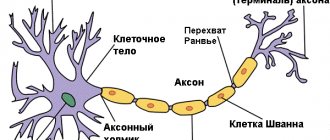
Long axon process
As mentioned above, the axon, body and dendrites are the main parts of a neuron. An adult nerve cell always has one axon. As a rule, it starts from the soma, but in some cases it can grow from one of the dendrites. The axon has a cone shape, that is, it gradually narrows towards its end. Along the entire process there are constrictions called nodes of Ranvier. Inside the process there is cytoplasm, which has a set of organelles different from the soma.
Speaking about axon length, it should be noted that most neurons have processes only a few millimeters long, however, spinal cord axons can reach a meter in length. Its main function is the transmission of nerve impulses, which it performs at a speed of more than 27 m/s.
Development
The formation of nervous tissue occurs in the third week of the embryonic period. At this time, a plate is formed. From it develop:
- Oligodendrocytes.
- Astrocytes.
- Ependymocytes.
- Macroglia.
During further embryogenesis, the neural plate turns into a tube. In the inner layer of its wall there are stem ventricular elements. They proliferate and move outward. In this area, some cells continue to divide. As a result, they are divided into spongioblasts (components of microglia), glioblasts and neuroblasts. From the latter, nerve cells are formed. There are 3 layers in the tube wall:
- Internal (ependymal).
- Medium (raincoat).
- External (marginal) - represented by the white medulla.
At 20-24 weeks, the formation of bubbles begins in the cranial segment of the tube, which are the source of the formation of the brain. The remaining sections serve for the development of the spinal cord. Cells involved in the formation of the crest extend from the edges of the neural groove. It is located between the ectoderm and the tube. From these same cells, ganglion plates are formed, which serve as the basis for myelocytes (pigmented skin elements), peripheral nerve ganglia, melanocytes of the integument, and components of the APUD system.
Cell membrane
This element provides a barrier function, separating the internal environment from the neuroglia located outside. The thinnest film consists of two layers of protein molecules and phospholipids located between them. The structure of the neuron membrane suggests the presence in its structure of specific receptors responsible for recognizing stimuli. They have selective sensitivity and, if necessary, “turn on” in the presence of a counterparty. The connection between the internal and external environments occurs through tubules that allow calcium or potassium ions to pass through. At the same time, they open or close under the influence of protein receptors.
Thanks to the membrane, the cell has its potential. When it is transmitted along the chain, excitable tissue is innervated. Contact between the membranes of neighboring neurons occurs at synapses. Maintaining a constant internal environment is an important component of the life of any cell. And the membrane subtly regulates the concentration of molecules and charged ions in the cytoplasm. At the same time, they are transported in the required quantities for metabolic reactions to occur at an optimal level.
Fibers
Glial sheaths are located independently around the nerve processes. Together they form nerve fibers. The branches in them are called axial cylinders. There are unmyelinated and myelinated fibers. They differ in the structure of the glial membrane. Unmyelinated fibers have a fairly simple structure. The axial cylinder approaching the glial cell bends its cytolemma. The cytoplasm closes over it and forms a mesaxon - a double fold. One glial cell can contain several axial cylinders. These are “cable” fibers. Their branches can pass into neighboring glial cells. The pulse travels at a speed of 1-5 m/s. Fibers of this type are found during embryogenesis and in the postganglionic areas of the autonomic system. The myelin segments are thick. They are located in the somatic system, innervating the skeletal muscles. Lemmocytes (glial cells) pass sequentially, in a chain. They form a strand. An axial cylinder runs through the center. The glial membrane contains:
- The inner layer of nerve cells (myelin). It is considered the main one. In some areas between the layers of the cytolemma there are extensions that form myelin notches.
- Peripheral layer. It contains organelles and a nucleus - the neurilemma.
- Thick basement membrane.
What is a synapse
To put it too simply, a synapse is the junction of two nerve cells. It would seem, what's special here? But in fact, a synapse is a rather complex device, thanks to which the entire mechanism for collecting and processing information can work properly. A synapse is what allows you to transform the simplest signals and unconditioned reflexes into the most complex patterns of mental activity: ideas, ideas, images, works of art, scientific theories. What is the structure of a synapse?
Components
There are 5-10 times more gliocytes in the system than nerve cells. They perform different functions: supporting, protective, trophic, stromal, excretory, suction. In addition, gliocytes have the ability to proliferate. Ependymocytes are distinguished by their prismatic shape. They make up the first layer, lining the brain cavities and the central spinal cord. The cells are involved in the production of cerebrospinal fluid and have the ability to absorb it. The basal part of ependymocytes has a conical truncated shape. It passes into a long thin process that penetrates the medulla. On its surface it forms a glial delimiting membrane. Astrocytes are represented by multi-processed cells. They are:
- Protoplasmic. They are located in the gray matter of the brain. These elements are distinguished by the presence of numerous short branches and wide endings. Some of the latter surround blood capillary vessels and participate in the formation of the blood-brain barrier. Other processes are directed to the neural bodies and carry nutrients from the blood through them. They also provide protection and insulate synapses.
- Fibrous (fibrous). These cells are found in the white matter. Their endings are weakly branched, long and thin. They have branches at their ends and form demarcation membranes.
Oliodendrocytes are small elements with extending short tails located around neurons and their endings. They form the glial membrane. Through it, impulses are transmitted. At the periphery, these cells are called mantle cells (lemmocytes). Microglia are part of the macrophage system. It is presented in the form of small mobile cells with poorly branched short processes. The elements contain a light core. They can be formed from blood monocytes. Microglia restore the structure of a nerve cell that has been damaged.
Literature
- Polyakov G.I., On the principles of neural organization of the brain, M: Moscow State University, 1965
- Kositsyn N. S. Microstructure of dendrites and axodendritic connections in the central nervous system. M.: Nauka, 1976, 197 p.
- Nemechek S. et al. Introduction to neurobiology, Avicennum: Prague, 1978, 400 p.
- Brain (collection of articles: D. Hubel, C. Stevens, E. Kandel, etc. - issue of Scientific American (September 1979)). M.:Mir, 1980
- Savelyeva-Novoselova N. A., Savelyev A. V. Device for modeling a neuron. A. s. No. 1436720, 1988
- Savelyev A.V.
Sources of variations in the dynamic properties of the nervous system at the synaptic level // Journal “Artificial Intelligence”, NAS of Ukraine. - Donetsk, Ukraine, 2006. - No. 4. - P. 323-338.