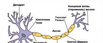
The nervous system consists of neurons (specific cells that have processes) and neuroglia (it fills the space between nerve cells in the central nervous system). The main difference between them is the direction of transmission of the nerve impulse. Dendrites are the receiving branches, along which the signal travels to the body of the neuron. Transmitting cells - axons - conduct a signal from the soma to the receiving cells. These can be not only neuron processes, but also muscles.
Structure of a neuron and synapse
Neurons are the main building blocks of the nervous system.
Their main function is to transmit signals through small connections (synapses). Although neurons come in different types, their structure and functions are generally similar. Our brain is approximately one and a half kilograms of tissue that contains about 1.1 trillion cells, including 100 billion neurons. On average, each neuron forms about five thousand connections with other neurons - so-called synapses.
The cell body of a neuron produces processes called dendrites. They receive neurotransmitters from other neurons (some neurons communicate with each other through electrical impulses.)
Let's imagine one typical neuron. It releases a neurotransmitter called serotonin.
This small neuron is part of the nervous system, but it itself is a complex system that requires the interaction of several subsystems to operate. When a neuron fires, tendrils at the tips of its axons fire molecules at synapses—the connections our neuron has made with other neurons.
If these are receiver synapses, then through them the neuron receives signals from other neurons (usually in the form of chemical elements called neurotransmitters). The signal tells the neuron whether to fire or not.
The excitation of a neuron depends mainly on the combination of signals received at any given moment. When a neuron is excited, it begins to send signals through synapses to other neurons, conveying to them a message: to be excited or not.
A typical neuron fires 5–50 times per second. In the time it takes you to read this text, there will be literally quadrillions of signals transmitted in your head.
On each neuron there are about two hundred small bubbles, vesicles, full of serotonin. Every time a neuron fires, five to ten vesicles open. If on average a neuron fires ten times per second, each antennae is emptied every few seconds.
As a consequence, the little molecular machines must either produce new serotonin or recycle the free serotonin that floats around the neuron. After this, they need to build vesicles, fill them with serotonin, and bring them to the site of action at the tip of each process.
Agree, this cannot be done without maintaining a balance between many processes, which means that many processes can go wrong. And let's not forget that serotonin metabolism is just one system out of thousands that function in your body.
To greatly simplify, we can say that the total excitation of a neuron depends on the sum of all the excitatory and inhibitory signals that it receives in each millisecond.
When a neuron is excited, an electrochemical wave travels along its axon (the process through which signals are transmitted). It releases neurotransmitters that travel to the synapses of the receiving neurons and cause either inhibition or excitation.
The speed of transmission of nerve signals is increased by myelin, a fatty substance that insulates axons.
The gray matter of the brain is largely composed of the cell bodies of neurons. The white matter contains axons and glial cells.
Glial cells provide metabolic support - they promote the formation of the myelin sheath on axons and recycle unused neurotransmitters.
Functionally, the cell bodies of neurons are like a hundred billion On/Off switches connected by wires called axons into a complex network right inside your head.
Each nerve signal is a bit of information. The nervous system moves information throughout your body the same way the heart pumps blood.
The number of possible combinations of excitation or inhibition of hundreds of billions of neurons is 10 to the millionth power (that is, one followed by a million zeros). Essentially, this is the number of possible states of your brain. To better understand the meaning of this number, let's compare it to the number of atoms in the Universe - "only" ten to the eightieth power.
Conscious mental events are based on temporary combinations of synapses that form and disintegrate (usually within a couple of seconds) like eddies in a stream. Neurons are also capable of creating long-term neural circuits, the connections in which are strengthened every time a certain mental activity occurs.
Richard Mendius, Rick Hanson. The Practical Neuroscience of Happiness, Love & Wisdom. 2010
Dendrites and axons in the structure of a nerve cell
Dendrites and axons are integral parts of the structure of a nerve cell. The axon of a neuron is often contained in one number and carries out the transmission of nerve impulses from the cell of which it is a part to another, which receives information through the perception of it by such a part of the cell as the dendrite.
Dendrites and axons, in contact with each other, create nerve fibers in peripheral nerves, the brain, and also the spinal cord.
A dendrite is a short, branched process that serves primarily to transmit electrical (chemical) impulses from one cell to another. It acts as a receiving part and conducts nerve impulses received from a neighboring cell to the body (nucleus) of the neuron, of which it is a structural element.
It got its name from the Greek word, which means tree due to its external resemblance to it.
Cell body
Neuron (from ancient Greek νεῦρον - fiber, nerve) is a structural and functional unit of the nervous system. This cell has a complex structure, is highly specialized and in structure contains a nucleus, a cell body and processes. There are more than one hundred billion neurons in the human body.
The complexity and variety of functions of the nervous system are determined by the interactions between neurons, which, in turn, represent a set of different signals transmitted as part of the interaction of neurons with other neurons or muscles and glands. Signals are emitted and propagated by ions that generate an electrical charge that travels along the neuron.
Structure
Cell body
The body of a nerve cell consists of protoplasm (cytoplasm and nucleus), and is externally bounded by a membrane of a double layer of lipids (bilipid layer).
Lipids consist of hydrophilic heads and hydrophobic tails, arranged with hydrophobic tails facing each other, forming a hydrophobic layer that allows only fat-soluble substances (eg oxygen and carbon dioxide) to pass through.
There are proteins on the membrane: on the surface (in the form of globules), on which growths of polysaccharides (glycocalyx) can be observed, thanks to which the cell perceives external irritation, and integral proteins that penetrate the membrane through, in which ion channels are located.
Typical structure of a neuron
A neuron consists of a body with a diameter of 3 to 130 µm, containing a nucleus (with a large number of nuclear pores) and organelles (including a highly developed rough ER with active ribosomes, the Golgi apparatus), as well as processes. There are two types of processes: dendrites and axons.
The neuron has a developed and complex cytoskeleton that penetrates its processes. The cytoskeleton maintains the shape of the cell; its threads serve as “rails” for the transport of organelles and substances packaged in membrane vesicles (for example, neurotransmitters).
The cytoskeleton of a neuron consists of fibrils of different diameters: Microtubules (D = 20-30 nm) - consist of the protein tubulin and stretch from the neuron along the axon, right up to the nerve endings. Neurofilaments (D = 10 nm) - together with microtubules, provide intracellular transport of substances.
Microfilaments (D = 5 nm) - consist of actin and myosin proteins, especially expressed in growing nerve processes and in neuroglia. A developed synthetic apparatus is revealed in the body of the neuron; the granular ER of the neuron is stained basophilically and is known as the “tigroid”.
The tigroid penetrates the initial sections of the dendrites, but is located at a noticeable distance from the beginning of the axon, which serves as a histological sign of the axon.
There is a distinction between anterograde (away from the body) and retrograde (toward the body) axon transport.
Dendrites and axon
An axon is usually a long process adapted to conduct excitation from the neuron body.
A neuron may have several dendrites and usually only one axon. One neuron can have connections with many (up to 20 thousand) other neurons.
Dendrites divide dichotomously, while axons give off collaterals. Mitochondria are usually concentrated at branching nodes.
Dendrites do not have a myelin sheath, but axons may have one. The place of generation of excitation in most neurons is the axon hillock - a formation at the point where the axon departs from the body. In all neurons, this zone is called the trigger zone.
Neuron structure diagram
Synapse
Synapse (Greek σύναψις, from συνάπτειν - to hug, clasp, shake hands) is the point of contact between two neurons or between a neuron and the effector cell receiving the signal.
It serves to transmit a nerve impulse between two cells, and during synaptic transmission the amplitude and frequency of the signal can be adjusted. Some synapses cause depolarization of the neuron, others hyperpolarization; the former are excitatory, the latter are inhibitory.
Typically, stimulation from several excitatory synapses is necessary to excite a neuron.
The term was introduced in 1897 by the English physiologist Charles Sherrington.
- Classification
- Structural classification
- Based on the number and arrangement of dendrites and axons, neurons are divided into axonless neurons, unipolar neurons, pseudounipolar neurons, bipolar neurons, and multipolar (many dendritic arbors, usually efferent) neurons.
Structure
Together they create a specific nervous tissue system responsible for perceiving the transmission of chemical (electrical) impulses and transmitting them further. They are similar in structure, only the axon is much longer than the dendrite, the latter is the loosest, with the least density.
A nerve cell often contains a fairly large branched network of dendritic branches. This gives her the ability to increase her intelligence collection from the environment around her.
Dendrites are located near the neuron body and form more contacts with other neurons, performing their main function of transmitting a nerve impulse. They can be connected to each other by small processes.
The features of its structure include:
- can reach up to 1 mm long;
- it does not have an electrically insulating shell;
- has a large number of a regular unique system of microtubules (they are clearly visible in sections, they run parallel, without intersecting with each other, often some are longer than others, they are responsible for the movement of substances along the processes of the neuron);
- has active zones of contact (synapses) with the bright electron density of the cytoplasm;
- has extensions from the cell trunk such as spines;
- has ribonucleoproteins (carrying out protein biosynthesis);
- has granular and non-granular endoplasmic reticulum.
Differences between axons and dendrites
What is the difference between them? Let's consider.
- The neuron dendrite is shorter than the transmitting process.
- There is only one axon; there can be many receiving branches.
- The dendrites branch heavily, and the transmitting processes begin to separate towards the end, forming a synapse.
- Dendrites become thinner as they move away from the neuron body; the thickness of the axons remains virtually unchanged along their entire length.
- Axons are covered with a myelin sheath consisting of lipid and protein cells. It acts as an insulator and protects the process.
Because the nerve signal is transmitted as an electrical impulse, cells require insulation. Its functions are performed by the myelin sheath. It has tiny breaks that facilitate faster signal transmission. Dendrites are sheathless processes.
Consequences of dendrite destruction
Although they, after eliminating the conditions that caused disturbances in their construction, are able to recover, completely normalizing metabolism, but only if these factors have a short, insignificant effect on the neuron, otherwise, parts of the dendrites die, and since they do not have the opportunity to leave the body , accumulate in their cytoplasm, provoking negative consequences.
In animals this leads to disturbances in forms of behavior, with the exception of the simplest conditioned reflexes, and in humans it can cause disorders of the nervous system.
In addition, a number of scientists have proven that in cases of dementia in old age and Alzheimer's disease, the processes in neurons are not tracked. The dendrite trunks externally become charred (charred).
No less important is the change in the quantitative equivalent of spines due to pathogenic conditions. Since they are recognized as structural components of interneuronal contacts, disturbances that occur in them can provoke quite serious dysfunctions of brain activity.
Functioning of the nervous system
The normal functioning of the nervous system depends on impulse transmission and chemical processes at the synapse. The creation of neural connections plays an equally important role. The ability to learn is present in humans precisely because of the body's ability to form new connections between neurons.
Any new action at the learning stage requires constant control by the brain. As it is mastered, new neural connections are formed, and over time the action begins to be performed automatically (for example, the ability to walk).
Dendrites are transmission fibers that make up approximately one third of all nervous tissue in the body. Through their interaction with axons, humans have the ability to learn.
Synapse - what is it, their types and functions
The nervous system, as we know, consists of neurons. These special cells are able to receive, store and process information; they are responsible for connecting the body with the outside world and for the operation of all systems of this body. Memory, attention, thinking, imagination, creativity - all these are the results of the work of neurons. However, all this diverse activity could not take place if the neuron did not have such an important element as a synapse. In a certain sense, it is the synapses, and not the neurons themselves, that are the basis of the nervous system.
What is a synapse
To put it too simply, a synapse is the junction of two nerve cells. It would seem, what's special here? But in fact, a synapse is a rather complex device, thanks to which the entire mechanism for collecting and processing information can work properly. A synapse is what allows you to transform the simplest signals and unconditioned reflexes into the most complex patterns of mental activity: ideas, ideas, images, works of art, scientific theories. What is the structure of a synapse?
Synapse structure
Each nerve cell has a large number of processes. All of these processes, except one, are dendrites; these are short and highly branched formations that are designed to receive information from other neurons. The remaining long process is called an axon; it is responsible for transmitting information from a given nerve cell to the next.
Connected by processes, nerve cells form a complex network through which signals travel in different directions. Scattered signals from the peripheral nervous system enter the central one, where from them the body forms a holistic picture of the world, decides what to do in the future, and sends signals to the necessary organs. The axon of a nerve cell can reach an impressive length - up to one and a half meters. And this is only in the human body. In giraffes, axons in the spinal cord can reach up to five meters. Apparently, in larger extinct animals, such as dinosaurs, the axons of nerve cells in the spinal cord were even longer. It turns out that nerve cells are the largest cells in the body.
However, most often a signal cannot pass directly from one nerve cell to another, because the space between the dendrites and the axon is filled with intercellular substance. In order for nervous information to pass from one process to another, it is necessary to build a kind of bridge. Such bridges are called neurotransmitters, or neurotransmitters; They are formed as a result of biochemical reactions and are protein molecules.
The nerve cells themselves are very small - the largest of them usually do not exceed a length of 100 micrometers. The processes of neurons, therefore, are completely microscopic in size. However, even at this microscopic level, the structure of the synapse is quite complex. It consists of three departments. The first is a thickening at the end of the axon called the presynaptic membrane, which is necessary for the formation of neurotransmitters. The second section is a similar thickening at the end of the dendrite, which serves to receive signals from the neurotransmitter. Between them there is a third section - the synaptic cleft itself, in which neurotransmitters are formed.
But the structure of the synapse is not limited to this. On the thickening of the axon there are special formations - synaptic vesicles, which contain either a neurotransmitter or an enzyme that destroys the neurotransmitter. And on the thickening of the dendrite there are receptors that receive signals from a specific neurotransmitter.
Synapse
The place where contact occurs between branches of neurons or between an axon and a receiving cell (such as a muscle cell) is called a synapse. It may involve only one branch from each cell, but most often contact occurs between several processes. Each axon extension can contact a separate dendrite.
The signal at a synapse can be transmitted in two ways:
- Electric. This occurs only when the width of the synaptic cleft does not exceed 2 nm. Thanks to such a small gap, the impulse passes through it without being delayed.
- Chemical. Axons and dendrites come into contact due to the potential difference in the membrane of the transmitting process. On one side the particles have a positive charge, on the other - a negative one. This is due to different concentrations of potassium and sodium ions. The first are located inside the membrane, the second are outside.
When a charge passes through, the permeability of the membrane increases, and sodium enters the axon and potassium leaves it, restoring the potential.
Immediately after contact, the process becomes immune to signals, after 1 ms it is capable of transmitting strong impulses, and after 10 ms it returns to its original state.
Dendrites are the receiving side that transmits impulses from the axon to the body of the nerve cell.
Classification of synapses
There are several classifications of nerve cell connections. We considered the first of them above - this is the division into chemical, electrical and mixed synapses. Synapses can also be divided according to the nature of the transmitted signal: excitatory
and
braking
.
Synapses can also be divided by location: central
, located in the brain, and
peripheral
, located in the peripheral nervous system.
Synapses are also divided depending on the neurotransmitters they produce. Some produce norepinephrine, others produce acetylcholine, serotonin, glutamate and others. In total, there are about sixty types of neurotransmitters, each of which has a specific function. Thus, norepinephrine is an stimulant; it activates all systems of the body and creates a feeling of rage. Dopamine is a happiness hormone that imparts a state of bliss to the body and generates positive emotions; it is also responsible for cognitive processes. Both an excess and a lack of neurotransmitters leads to various disorders in the nervous system and the body as a whole. Thus, a lack of dopamine causes depression, loss of strength, and leads to dementia. An excess of glutamate can lead to the death of nerve cells.
The structure and functioning of the biological nervous system allowed scientists to create its artificial analogue. In an artificial neural network, connections between individual “neurons” are also called synapses, and they contain both “dendrites” and “axons”. In artificial neural networks, it is possible to simulate even individual types of signals - for example, there are excitatory and inhibitory signals. Of course, an artificial neural network is a simplified model of a real, biological one, but as technology develops, the model becomes more detailed. Thus, in 2015, in Sweden, researchers created one of the most advanced artificial analogs of a neuron to date. The device was created based on organic bioelectronics. Such an artificial neuron most fully replicates the work of a natural nerve cell and can even communicate with other neurons.