A common brain lesion is the formation of a cyst, which is a benign neoplasm filled with a liquid substance. It can be located in different parts of the department. Two forms of the disease are quite common: arachnoid and retrocerebellar. Each of these types corresponds to specific features and techniques of therapy. The second form usually appears as fluid accumulation in areas where there has been loss of gray matter. To prevent destruction of the brain structure, it will be necessary to identify the causes of the death of SV. This decision will contribute to the correct choice of treatment method.
Classification of brain cysts
Cysts are divided according to the type of acquisition into the following types:
- Primary or congenital - found in newborns already in the maternity hospital;
- Secondary or acquired - these brain formations are a consequence of various pathological abnormalities or the result of surgical intervention.
The localization of the formations under consideration largely determines their variety. The most common are arachnoid and retrocerebellar cysts.
- If the location of the cyst is determined between the brain and its arachnoid membrane, then the cyst is called arachnoid.
- If the cyst occupies a certain part of the brain, then we are talking about an intracerebral (cerebral) cyst.
What is a retrocerebellar cyst of the brain? In particular, the name of this type of disease comes from its location, where the connection is determined with the cerebellum.
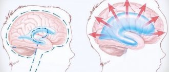
A retrocerebellar or intracerebral cyst develops at the site of death of areas of gray matter, that is, in the thickness of the brain, and not in its shell or on the surface. Speaking about this type of neoplasm, they can be located in any part of the brain, so there is a certain classification of the disease in question depending on their location (for example, superior and inferior retrocerebellar cyst).
A similar clinical picture is observed in the presence of a tumor.
The causes of brain meningioma are hormonal changes after 50 years. Read our article about who is more susceptible to this disease and what preventative measures need to be taken. From birth, babies may develop a malignant tumor, such as neuroblastoma. Signs of the development of neuroblastoma in a child can be seen in this article.
The provoking reasons for the development of this type of cyst are:
- Deficiency of oxygen and the required amount of microelements entering the brain;
- necrosis of nerve cells (strokes);
- previous operations in the cranial area;
- development of inflammation of infectious origin;
- injuries and mechanical damage (concussion).
Retrocerebellar arachnoid cyst
The arachnoid membrane refers to one of the three meninges located between the dura superficial mater and the soft deep mater of the brain.
Thus, a pathology in which a bubble of liquid content (cerebrospinal fluid) is formed, located, as a rule, between the meninges, is called an arachnoid cyst.
Factors for the development of this type of cyst:
- the process of inflammation of the meninges;
- increased liquid filling pressure;
- brain injuries.
Retrocerebellar cerebrospinal fluid cyst
A cyst is a formation that has a wall and some contents. If its filler is liquid, then they talk about a cerebrospinal fluid cyst
, which is a consequence of processes such as:
- the process of inflammation of the membranes of the brain;
- injuries;
- stroke and hemorrhage;
- undergone surgical procedures.
CSF cysts of the brain occur in approximately 4-5% of the population, while only 2 out of 10 patients experience a severe course of the disease. Retrocerebellar arachnoid cerebrospinal fluid cyst of the brain is divided into forms
:
- Congenital
– a consequence of a disturbance in the intrauterine state of the fetus (birth trauma). - Acquired
- the development of a cerebrospinal fluid cyst is caused by inflammation of the superficial membranes of the brain (damage, trauma, etc.).
Symptoms
Variants of violations depending on the anatomical zone:
| Brain region | Functions | Disorder due to cystic formation |
| Medulla | Centers for controlling breathing and blood circulation, as well as centers for regulating swallowing, salivation and some protective reflexes (sneezing, coughing, vomiting) | Vital centers suffer (incompatible with life). The pathology is not subject to surgical treatment. |
| Cerebellum | Centers of balance and coordination of movement | Vestibular disorders occur (unsteadiness of gait, disorientation in space, asthenia, tremor, ataxia). |
| Bridge | Conductive section (reticular formation, motor and sensory nuclei) | Disorders are difficult to classify due to the complexity of the wiring system of this part of the brain. |
| Midbrain | Primary visual and auditory centers, regulation of position in space and body movements | Violation of skeletal muscle tone. Impaired coordination and speed of movements. |
| Diencephalon: 1. Thalamus 2. Hypothalamus 3. Epithalamus | 1. The final centers of all types of sensitivity | 1. Loss or loss of any types of senses (tactile, tactile, temperature, taste). |
| 2. The highest center of nervous and humoral regulation (controls the pituitary gland, which ensures the functioning of all endocrine glands of the body) | 2. Violation of thermoregulation. Distortion of feelings of satiety/hunger. Distortion of the feeling of thirst. Blood pressure disorders, breathing problems. | |
| 3. Control of sleep/wake function (production of melatonin and serotonin) | 3. Disturbance of normal circadian rhythms (insomnia/constant drowsiness, depression). | |
| Telencephalon (cerebral cortex): 1. Frontal lobes 2. Parietal lobes 3. (asymmetry of the right and left sides) 4. Temporal lobe (asymmetry of the right and left sides) 5. Occipital lobes | 1. Control over voluntary movements, speech and higher mental activity. Also located here is Broca's center (inferior frontal gyrus) - this is the motor center of speech. | 1. When this department is damaged, “frontal symptoms” occur: euphoria, foolishness, lack of understanding of humor, inability to take purposeful actions. The patient retains all accumulated experience and knowledge, but he is not able to use them to solve specific problems. |
| 2. Participate in recognition and retrieval from memory of information about the shape of objects, their texture, mass. Provides information about the visual-spatial relationship of the body relative to surrounding objects. There are also centers responsible for invoicing and writing | 2. Astereognosis - difficulty in recognizing objects by touch. Impaired understanding of the body's position in space and anosognosia (impaired perception of one's own body). Impairments in the ability to write and calculate (strictly depending on which hemisphere is affected). | |
| 3. Higher centers of hearing, speech understanding, visual memory and verbal memory. This area also includes the limbic system (emotions). | 3. If there is a lesion on the right side, the perception of non-verbal auditory stimuli (music) is impaired. When the left lobe is damaged, a disorder of consciousness, memory and speech production occurs. When the limbic system is damaged, complex partial seizures occur with loss of autonomic, cognitive and emotional functions. | |
| 4. Higher visual center | 4. If there is a violation in this area, central blindness occurs and Anton-Babinsky syndrome develops (in combination with damage to the parietal lobes) - the patient is not aware of his own blindness. |
This division is extremely conditional, since the brain does not have strict boundaries and functions as a single indivisible neural network.
In addition, if there is a disturbance in the cerebrospinal fluid system (arachnoid cystic formations communicating with the ventricles of the brain), the phenomena of hydrocephalus occur:
- General cerebral manifestations (nausea, vomiting, convulsions).
- Focal symptoms that depend on the exact location of the cyst (impaired visual function, impaired motor or sensory regulation).
- Disorders of the somatic systems (cardiovascular, respiratory) due to disruption of their regulatory centers.
Relationship between the symptoms of cystic formation and its size
The clinical course of the pathology is directly dependent on its increase (growth). Of course, there are many other factors - the location of the cyst, the source of the disease, but size plays a primary role
. An increase in cyst volume provokes an increase in fluid pressure in it, which entails an active course of the disease. The growth of cystic formation is influenced by:
- neuroinfection;
- chronic disorders of the blood supply to the heart;
- development of autoimmune processes (for example, multiple sclerosis).
Clinical course of a retrocerebellar cyst of the brain in an adult
As the size of the cyst increases, the following symptoms are observed:
- migraine;
- throbbing sensation in temples;
- causeless hearing loss, presence of noise sensations in the ears;
- visual disturbances – bifurcation and blurriness of silhouettes, the appearance of flies;
- paralysis of limbs of various types, convulsions;
- problems with coordination;
- vomiting, weakness.
Signs of the disease may cease over time and have various nonspecific manifestations; in any case, the degree and nature of the symptoms depend on the type and volume of the cystic formation.
Clinical course of the disease in a child
In a small patient, the development of this pathology is accompanied, as in an adult, by a number of similar signs:
- hearing impairment;
- paralysis and paresis of limbs;
- cramps, numbness;
- impaired coordination of motor activity.
If the cyst progresses in size, intracranial pressure may increase, which as a result affects the appearance of the following symptoms:
- vomit;
- intense headaches;
- pulsation in the head area;
- increased drowsiness and fatigue.
A more severe degree of the disease causes divergence of the bone sutures (problems with the overgrowth of the fontanel in infants), which affects the delay in the development of the baby, both mental and physical.
Causes
This type of cyst can appear without any obvious reason. This makes it much more difficult to identify. There are quite a few factors that can trigger its development. These include:
- Circulatory disorders;
- Head injuries, concussions;
- Inflammatory diseases caused by infectious exposure;
- Serious negative brain changes;
- Performed brain surgeries;
- Previous stroke;
- Heredity;
- Birth injuries, negative effects on the fetus of medications taken by the mother during pregnancy.
All people affected by the problems listed above are at risk. The increased likelihood of cyst formation requires observation by a doctor. You need to visit him at least once a year to exclude unexpected development of the disease.
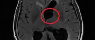
Diagnosis of brain cyst
Recognition of the presence of a cystic formation is carried out using the following diagnostic methods (tomography):
- magnetic resonance;
- computer
These methods are the most accurate in diagnosing the pathology in question, since they provide the most complete description of the course of the disease. Additional research methods include:
- USDG (ultrasound Doppler scanning);
- ECG, Echo-CG;
- blood pressure monitoring.
To differentiate a cyst from a tumor, a diagnostic procedure is performed that requires the introduction of a special contrast agent. In newborns, an ultrasound scan of the brain is prescribed in the first week of life, which excludes the presence of congenital pathological formations.
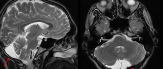
The following photo clearly shows the location of the retrocerebellar cyst:
Preparation rules
During a routine examination, doctors recommend following a few simple recommendations:
- If it is necessary to administer contrast, you must come to the clinic on an empty stomach, that is, do not eat food for 7-8 hours.
- If you are afraid of research or confined spaces, be sure to consult with a specialist. Your doctor may prescribe you sedatives to make the examination process more comfortable.
- Do not bring any metal products inside the tomograph. For this reason, doctors recommend removing metal accessories in advance.
- Prepare a change of clothes that do not have metal or metal-containing accessories.
- The duration of the diagnostic test is about 40 minutes. Empty your bladder in advance to avoid discomfort during the procedure.
Principles of treatment
Treatment of a brain cyst is a rather complex procedure that requires an integrated approach. For non-progressive forms of the cyst, dynamic observation by a neurologist is prescribed and drug therapy is recommended:
- Antibacterial or antiviral therapy. In this case, the indication is the presence of an inflammatory focus of an infectious or autoimmune nature.
- Taking immunomodulators. The goal is to increase the body’s immune barrier, stopping the aggressive effects of the autoimmune environment.
Tumors, cysts and hematomas can be a consequence of severe brain contusion.
Read our article about what to do if you have a brain injury. Also learn how to provide first aid for a fracture of the base of the skull from this article.You can learn about how headaches are associated with chronic intracranial hypertension at https://gidmed.com/bolezni-nevrologii/vnutrecherepnoe-davlenie/gipertenziya.html
Drug treatment of cerebrovascular disorders consists of three areas:
- Reducing the concentration of cholesterol and blood clotting - antiplatelet agents (regular aspirin, ticlopidine, pentoxifylline).
- Normalization of blood pressure indicators - Enalapril, Capoten.
- Resorption of adhesions - anticoagulants.
- Improving the flow of oxygen and glucose into brain cells - a group of nootropic drugs (Cerebramin, Cerebrolysin, Nootropil, Vinpotropil, Vinpocetine)
- Increasing the resistance of the cellular composition of the brain to increases in intracranial pressure - antioxidants.
If the severity of the disease is high, the surgeon will decide how to treat the retrocerebellar cyst of the brain.
Maybe:
- endoscopic intervention;
- cystocystrenostomy;
- cavitary cyst shunting;
- excision of the tumor.
Video of an operation to remove a retrocerebellar arachnoid cyst of the brain:
How is an MRI performed?
To ensure that the procedure is as informative as possible and that the images are clear, experts recommend performing magnetic resonance imaging in closed-type tomographs. Before starting the study, the patient must change into clothes without metal structures, take off his shoes and lie down on a retractable table. If the doctor has prescribed additional contrast enhancement, a special drug is injected into a vein, which is distributed throughout the circulatory system in just a few minutes. Then the table moves inside the tomograph, where the research process takes place.
Maintaining immobility is the main requirement for the patient! Remember that even minimal movements can lead to a decrease in the information content of the procedure.
It is also worth noting that when performing magnetic resonance imaging, a specific loud noise is observed. If this scares you, you can use earplugs.
Patients generally tolerate MRI well. Inside the tomograph there is good lighting and ventilation, there is a loudspeaker, as well as a panic button for emergency stop of the study.