Ventricular system of the brain
The ventricles of the brain are several interconnected collectors in which the formation and distribution of liquor fluid occurs.
Liquor washes the brain and spinal cord. Normally, there is always a certain amount of cerebrospinal fluid in the ventricles. Two large collectors of cerebrospinal fluid are located on either side of the corpus callosum. Both ventricles are connected to each other. On the left side is the first ventricle, and on the right is the second. They consist of horns and a body. The lateral ventricles are connected through a system of small holes to the 3rd ventricle.
In the distal part of the brain, between the cerebellum and the medulla oblongata, there is the 4th ventricle. It is quite large in size. The fourth ventricle is diamond-shaped. At the very bottom there is a hole called the diamond-shaped fossa.
Proper functioning of the ventricles allows cerebrospinal fluid to enter the subarachnoid space when necessary. This zone is located between the dura mater and the arachnoid membrane of the brain. This ability allows you to maintain the required volume of cerebrospinal fluid in various pathological conditions.
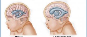
In newborn babies, dilatation of the lateral ventricles is often observed. In this condition, the horns of the ventricles are enlarged, and increased accumulation of fluid in the area of their bodies may also be observed. This condition often causes both left and right ventricle enlargement. In differential diagnosis, asymmetry in the area of the main brain collectors is excluded.
Danger of disease
At an early stage, the disease contributes to the development of the above symptoms, which, moreover, like some delay in movements, disappear forever with age. The appearance of such severe effects, for example, cerebral palsy, does not occur in this case. If the pressure inside the skull is already high, and the cerebrospinal fluid continues to accumulate, then the human brain will inevitably be damaged. With the tireless increase in the size of the ventricles, the very properties of the brain change, reducing nervous regulation. The child’s brain bones are elastic and not so rigidly connected to each other, so the pressure will always be slightly lower.
Indicators of normal sizes
In the human body, the ventricular system consists of several cavities that anastomize with each other. They communicate with the subarachnoid space, as well as the spinal cord canal. A special liquid, cerebrospinal fluid, moves directly inside the cavities. With its help, tissues receive nutrients and oxygen molecules.
The largest intracerebral hollow formations are, of course, the lateral ventricles. They are localized below the corpus callosum - on both sides of the midline of the brain, symmetrical relative to each other. In each, it is customary to distinguish several sections - the anterior and lower, as well as the posterior horns and the body itself. The shape resembles the English S.
The normal size of the ventricles is assessed taking into account individual anatomical characteristics - there are no uniform standards. Experts focus on average parameters. It is important to know these sizes for babies under one year old - for the purpose of early diagnosis of hydrocephalus.
- What is heart dilatation
Normal values for children:
| Anatomical unit | Newborns, mm | 3 months, mm | 6 months – 9 months, mm | 12 months, mm |
| Lateral ventricle | 23.5-/+ 6.8 | 36.2 -/+3.9 | 60.8-/+6.7 | 64.7-/+12.7 |
For adults, the parameters should be in the range - the anterior horn of the lateral ventricle is less than 12 mm in people under 40 years of age, while its body is 18–21 mm in people under 60 years of age. Exceeding the age-related size of the brain ventricles by more than 10% requires additional research to identify and eliminate the root cause.
How can the disease be cured?
To get rid of the diagnosis, you should contact neurosurgeons and neuropathologists. To avoid the possibility of developing side effects, the child is constantly monitored by attending physicians. The main methods of combating asymmetry include the use of diuretics to reduce the amount of cerebrospinal fluid produced and the use of neurometabolic stimulants (nootropics) for better blood supply to the brain. In addition, sedatives are also added here. Additionally, special gymnastics and therapeutic massage are prescribed. Babies up to six months of age are treated on an outpatient basis. Older children are treated taking into account the disease that caused the asymmetry. For example, if it is an infection, then antiviral drugs and antibiotics are added. Surgeries are performed to treat head trauma and tumors. The course of treatment is long and takes up to several months.
Causes of dilatation
Enlargement or dilatation of the lateral ventricles of the brain occurs due to increased production of cerebrospinal fluid. This leads to the fact that it cannot be excreted normally.
This, in turn, leads to disruption of the flow of cerebrospinal fluid. This disease most often occurs in premature babies, but is observed in people of any age.
What causes the disorder in newborns?
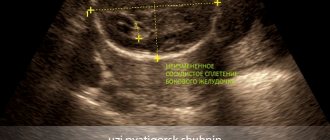
This is how dilatation of the lateral ventricles looks schematically
Dilatation of the lateral ventricles of the brain in infants is often a sign of hydrocephalus, and can also be caused by a number of other reasons.
In newborns, asymmetry is caused by trauma or space-occupying lesions in the brain. Regardless of the possible cause, urgent consultation with a neurosurgeon is required.
Mild asymmetry may be a congenital disorder that does not cause symptoms. In this case, only constant monitoring is required so that the difference between the ventricles does not change.
- What is dilatation of the left atrium cavity: causes, symptoms, treatment and prognosis
The main causes of dilatation include:
- viral and other diseases of a woman during pregnancy;
- oxygen starvation of the fetus;
- premature birth;
- birth injuries;
- malformations of the central nervous system.
Ventricular asymmetry can also result from hemorrhage. This pathology occurs due to compression of one of the ventricles by an additional volume of blood. Due to hemorrhage, the ventricles of the brain in an infant may be enlarged for the following reasons:
- various maternal diseases, for example, type I diabetes or heart defects;
- intrauterine infections;
- a long time between the water breaking and the baby being born.
The most common cause of dilatation is hypoxia. Other causes account for less than 1% of cases. It is hypoxia that leads to the accumulation of cerebrospinal fluid, which, in turn, increases intracranial pressure. This leads to expansion of the cavity of the lateral ventricles.
Risk zone for adult patients
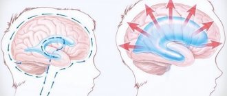
A change in the size of the lateral ventricles leads to disruption of the circulation of cerebrospinal fluid. Asymmetry of the lateral ventricles of the brain in adults occurs for the following reasons:
- difficulty in the outflow of cerebrospinal fluid;
- excessive production of cerebrospinal fluid;
- neuroinfections;
- cysts and neoplasms;
- skull injuries;
- hematomas;
- stroke;
- hydrocephalus;
- vascular thrombosis.
What are the typical symptoms?
Depending on the level of intracranial pressure, symptoms change accordingly. The very first thing is discomfort. Indeed, what disease can proceed comfortably for the patient? Regurgitation appears, but not always. Subsequently, throwing back the head is added - postural reflexes. Another beacon will be simply an enlargement of the head. In fact, the disease often does not manifest itself, so it can only be detected by ultrasound.
A short list of symptoms:
- Decreased muscle tone.
- Complete refusal to breastfeed.
- Constant worry and crying.
- Hand trembling.
- Weakening of swallowing and grasping reflexes.
- Dehiscence of the sagittal suture.
- Swelling and tension of the fontanel.
- On examination, the iris of the eye is partially hidden behind the lower eyelid - rising sun syndrome.
- Swelling of the optic discs.
Diagnostics
If a specialist observes signs of a malfunction in the circulation of cerebrospinal fluid through the cerebral ventricles, or the patient has complaints of deterioration in health, then instrumental confirmation of dilatation of the brain cavities is required.
It is possible to identify signs of slight dilation of the lateral ventricles using such a modern diagnostic examination method as magnetic resonance imaging. On the resulting images of brain structures, you can see in detail the area of expansion, the area of damage, and the involvement of neighboring brain tissues in the process.
Increased intracranial pressure will also be diagnosed using the following procedures:
- echoencephaloscopy;
- electroencephalography;
- ophthalmoscopy;
- examination of cerebrospinal fluid - identification of previous neuroinfections;
- blood tests - general, biochemical, for autoimmune processes.
Only after careful comparison of all information from diagnostic procedures, a neurologist will be able to assess the severity of dilatation of the lateral ventricles, establish the root cause of the pathological condition and select optimal therapeutic measures.
- The cardiac cycle consists of 3 phases
Classification
The main criteria for separating dilatations of the lateral ventricles in the brain are the size of the cavities, the etiology of the dilation, the age of the patient, and the localization of pathological changes.
Each neurologist chooses the optimal classification of the disorder for himself. However, most doctors adhere to the average principles of diagnosis:
- According to the time of expected appearance of the lesion in the brain:
- prenatal period;
- detection of enlarged cerebral ventricles in newborns;
- expansion of brain cavities in adults.
- By localization:
- left ventricle enlargement;
- right-sided lesion;
- bilateral defeat.
- By etiology:
- post-infectious ventricular dilatation;
- post-traumatic changes;
- toxic expansion;
- tumor focus in the brain;
- vascular diseases.
- By severity:
- slightly enlarged ventricles of the brain in infants;
- moderate dilatation;
- severe changes in the ventricles.
Additionally, the specialist can indicate in the diagnosis whether complications are present - for example, hydrocephalus or intellectual/neurological problems.
Materials and methods
Of the 16,839 patients included in the “Registry of Coronary Angiography Operations” [5] from 1998 to 2013, patients with stenosis of >75% of the lumen of at least one coronary artery, without acute or anamnestic myocardial infarction, without congenital and acquired heart defects: 75 patients with RV dilatation and 1134 without it. The transverse size of the pancreas in the parasternal position was 26 mm or less [6]. For a clearer division of the groups, patients with pancreatic dilatation included patients with a pancreatic size of 30 mm or more; patients with a slight enlargement of the pancreas (>26 and <30 mm) were not included in the study. A clinical, comprehensive echocardiographic examination was performed (one-dimensional, two-dimensional, Doppler echocardiography using Imagepoint NX ultrasound devices, Agilente Technologies, Phillips, USA; Vivid 3, 4, 7, 9 Systems, Vingmed-General Electric - Horten, Norway), determination of lipid composition of blood serum, Holter monitoring, selective coronary angiography according to Judkins (1967) (angiographic complexes Diagnost ARC A, Poly Diagnost C, Integris Allura, Phillips, Holland). Linear echocardiography (EchoCG) parameters were indexed to height; myocardial mass, calculated using the Devereux formula, to the body surface area [7]. Echocardiographic syndromes were diagnosed according to standard criteria: the LV was considered dilated when its end-diastolic diameter index was more than 32 mm/m2 in women and more than 33 mm/m2 in men [2], LV systolic function was reduced when the LV ejection fraction was less than 50%, hemodynamically significant mitral regurgitation (≥II degree) - with an effective area of the regurgitant orifice ≥0.2 cm2, regurgitant volume ≥30 ml [8].
Statistical processing of data was carried out using a package of applied statistical programs (SPSS Inc., version 17.0). Indicators are presented in the form M
±
SD
_
The distribution of variables was determined using the Kolmogorov–Smirnov test. To compare values with their normal distribution, t
; for a non-normal distribution, the nonparametric Mann-Whitney test was used.
When analyzing qualitative indicators, Pearson's χ2 test was used. Differences at p
<0.05 were assessed as statistically significant. Multivariate analysis was performed - binary logistic regression analysis with calculation of odds ratio (OR).
Treatment tactics
In itself, the expansion of the size of the ventricles of the brain does not require intervention - if there are no clinical signs of intracranial pressure failure. Whereas in the case of a violation of liquorodynamics and symptoms of deterioration of well-being formed against this background, doctors will recommend conservative therapy:
- diuretics – removing swelling from brain tissue;
- neuroprotectors – correction of the conduction of nerve impulses;
- vasoactive agents – improving brain nutrition;
- nootropics – improvement of local blood circulation;
- sedative medications – normalization of the psychosomatic background;
- anti-inflammatory/antibacterial drugs – if the disorder is caused by an infectious process.
Neurosurgical intervention will be required if ventricular dilatation is caused by brain tumors or thromboembolism of cerebral vessels. If necessary, ventriculostomy is performed to create a new connection between the cavities of the brain.
Symptoms
At the initial stage of formation of dilated ventricles of the brain in an infant, any special clinical signs may not be detected - the child behaves according to the age norm. After all, adaptation mechanisms are capable of combating overproduction of cerebrospinal fluid.
However, as the expansion of the lateral ventricles of the child’s brain increases, he begins to worry about the consequences of hydrocephalus - pathological pressure on the brain structures due to tissue swelling. The main signs of intracranial hypertension:
- frequent attacks of headache;
- slow growth of fontanelles;
- swelling of tissue between the sutures of the skull;
- nausea and vomiting without improvement of well-being;
- decreased appetite, frequent regurgitation;
- worsening sleep;
- throwing the head back;
- muscle hypertonicity;
- lack of interest in current events, apathy;
- tendency to epilepsy.
In adult patients, a violation of the outflow of cerebrospinal fluid from the lateral ventricles is manifested by a feeling of constant distension inside the head, persistent dizziness with nausea. A person’s ability to work decreases, and he develops anxiety-phobic conditions. At the same time, taking standard analgesics does not improve well-being.
With persistent hypertensive-hydrocephalic syndrome, people develop paresis/paralysis, as well as serious difficulties with speech, vision, hearing, and decreased intellectual capabilities.